Downloaded 363 times
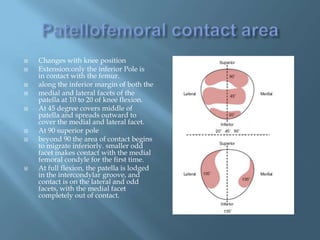
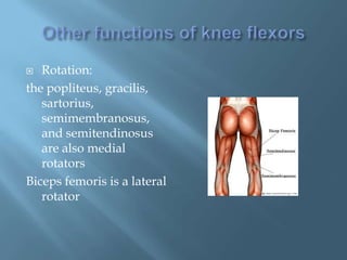
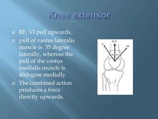
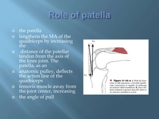
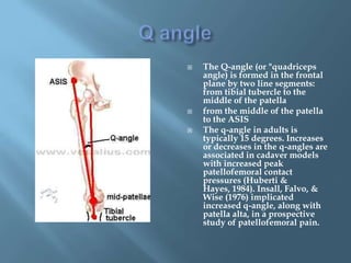
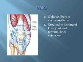
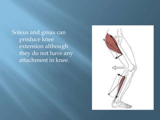

The document summarizes the anatomy and function of the knee joint. It describes the articulating surfaces of the patella and femur, the muscles that act on the knee joint as flexors, extensors, and rotators, and how the angle of pull of the quadriceps femoris muscle is increased by the patella acting as a pulley. It also discusses the Q-angle and how increased or decreased angles can impact patellofemoral contact pressures.


