Range of motion (ROM) measurements are performed to evaluate joint impairment, develop treatment goals, assess progress, and modify treatment. ROM is described in 3 planes and axes and measured using a goniometer. Active ROM is voluntary motion while passive ROM uses external assistance. Several factors determine ROM including joint integrity, scarring, age, gender, joint shape, and health of surrounding tissues. Common causes of limited ROM include contractures, arthritis, and pain. Precise positioning and stabilization are needed to reliably measure ROM of various joints like the shoulder, spine, and knee. Standardized testing procedures and documentation of measurements are important.
this PPT contain detailed kinetics & kinematics of ankle joint & all joints of foot complex, muscles of ankle & foot complex, plantar arches & weight distribution during standing.
Goniometry is the measuring of angles created by the bones of the body at the joints.1, 2, 3
The term goniometry is derived from two Greek words, gonia meaning angle and metron, meaning measure. 1, 2, 3, 4, 5,
System to measure the joint ranges in each plane of the joint is termed goniometry. 4
These measurements are done with instrument such as goniometer, a tape measure, inclinometers or by visual estimate.
Elbow complex is designed to serve hand.
They provide MOBILITY for Hand in space by apparent shortening and Lengthening of upper extremity.
They provide Stability for skillful and forceful movements
GONIOMETRY FOR UPPER LIMB DISCUSSES IN CONCISE THE DIFFERENT TYPES OF GONIOMETERS AVAILABLE FOR MEASURING VARIOUS JOINT ROM, PRINCIPLES OF GONIOMETRY AND PLACEMENT OF GONIOMETER FOR MEASURING RANGE OF MOTION IN UPPER LIMB (SHOULDER, ELBOW, FOREARM AND WRIST JOINT).
Short wave diathermy (s.w.d) electro therapyÂbhìšhék Singh
Electrotherapy topic shot wave diathermy ppt (physics)
Bachelor of physiotherapy topic swd . Swd introduction, and range of swd , indications and contraindications of swd
A chronicle on muscle strengthening:
MMT is a procedure for the evaluation of strength of individual
muscle or muscles group, based upon the effective performance of a movement in relation to the forces of gravity or manual resistance through the available ROM.
GONIOMETRY FOR THE LOWERLIMB DISCUSSES IN CONCISE THE DIFFERENT TYPES OF GONIOMETERS AVAILABLE FOR MEASURING VARIOUS JOINT ROM, PRINCIPLES OF GONIOMETRY, AND PLACEMENT OF GONIOMETER FOR MEASURING RANGE OF MOTION IN THE LOWER LIMB (HIP, KNEE, ANKLE).
A goniometer is a device used to measure angles, typically in the field of physiotherapy, occupational therapy, and biomechanics. It consists of a flat, circular, or semi-circular protractor-like instrument with an adjustable arm or arms. The primary purpose of a goniometer is to measure the range of motion at a joint in the body.
Here's a basic overview of how a goniometer is used and some key points about its features
A goniometer is an instrument that measures the available range of motion at a joint. The art and science of measuring the joint ranges in each plane of the joint are called goniometry. ... The term goniometry is derived from two Greek words, gonia, meaning "angle" and metron, meaning "measurement".
Physiotherapy aims at correcting movements. But how do we correct them if we don't know the measurement of these movements?
Movements occur in a particular range that is measured in degrees with the help of GONIOMETER.
So in this presentation, we are going to discuss about goniometry!
Title: Sense of Smell
Presenter: Dr. Faiza, Assistant Professor of Physiology
Qualifications:
MBBS (Best Graduate, AIMC Lahore)
FCPS Physiology
ICMT, CHPE, DHPE (STMU)
MPH (GC University, Faisalabad)
MBA (Virtual University of Pakistan)
Learning Objectives:
Describe the primary categories of smells and the concept of odor blindness.
Explain the structure and location of the olfactory membrane and mucosa, including the types and roles of cells involved in olfaction.
Describe the pathway and mechanisms of olfactory signal transmission from the olfactory receptors to the brain.
Illustrate the biochemical cascade triggered by odorant binding to olfactory receptors, including the role of G-proteins and second messengers in generating an action potential.
Identify different types of olfactory disorders such as anosmia, hyposmia, hyperosmia, and dysosmia, including their potential causes.
Key Topics:
Olfactory Genes:
3% of the human genome accounts for olfactory genes.
400 genes for odorant receptors.
Olfactory Membrane:
Located in the superior part of the nasal cavity.
Medially: Folds downward along the superior septum.
Laterally: Folds over the superior turbinate and upper surface of the middle turbinate.
Total surface area: 5-10 square centimeters.
Olfactory Mucosa:
Olfactory Cells: Bipolar nerve cells derived from the CNS (100 million), with 4-25 olfactory cilia per cell.
Sustentacular Cells: Produce mucus and maintain ionic and molecular environment.
Basal Cells: Replace worn-out olfactory cells with an average lifespan of 1-2 months.
Bowman’s Gland: Secretes mucus.
Stimulation of Olfactory Cells:
Odorant dissolves in mucus and attaches to receptors on olfactory cilia.
Involves a cascade effect through G-proteins and second messengers, leading to depolarization and action potential generation in the olfactory nerve.
Quality of a Good Odorant:
Small (3-20 Carbon atoms), volatile, water-soluble, and lipid-soluble.
Facilitated by odorant-binding proteins in mucus.
Membrane Potential and Action Potential:
Resting membrane potential: -55mV.
Action potential frequency in the olfactory nerve increases with odorant strength.
Adaptation Towards the Sense of Smell:
Rapid adaptation within the first second, with further slow adaptation.
Psychological adaptation greater than receptor adaptation, involving feedback inhibition from the central nervous system.
Primary Sensations of Smell:
Camphoraceous, Musky, Floral, Pepperminty, Ethereal, Pungent, Putrid.
Odor Detection Threshold:
Examples: Hydrogen sulfide (0.0005 ppm), Methyl-mercaptan (0.002 ppm).
Some toxic substances are odorless at lethal concentrations.
Characteristics of Smell:
Odor blindness for single substances due to lack of appropriate receptor protein.
Behavioral and emotional influences of smell.
Transmission of Olfactory Signals:
From olfactory cells to glomeruli in the olfactory bulb, involving lateral inhibition.
Primitive, less old, and new olfactory systems with different path
Pulmonary Thromboembolism - etilogy, types, medical- Surgical and nursing man...VarunMahajani
Disruption of blood supply to lung alveoli due to blockage of one or more pulmonary blood vessels is called as Pulmonary thromboembolism. In this presentation we will discuss its causes, types and its management in depth.
micro teaching on communication m.sc nursing.pdfAnurag Sharma
Microteaching is a unique model of practice teaching. It is a viable instrument for the. desired change in the teaching behavior or the behavior potential which, in specified types of real. classroom situations, tends to facilitate the achievement of specified types of objectives.
ARTIFICIAL INTELLIGENCE IN HEALTHCARE.pdfAnujkumaranit
Artificial intelligence (AI) refers to the simulation of human intelligence processes by machines, especially computer systems. It encompasses tasks such as learning, reasoning, problem-solving, perception, and language understanding. AI technologies are revolutionizing various fields, from healthcare to finance, by enabling machines to perform tasks that typically require human intelligence.
These simplified slides by Dr. Sidra Arshad present an overview of the non-respiratory functions of the respiratory tract.
Learning objectives:
1. Enlist the non-respiratory functions of the respiratory tract
2. Briefly explain how these functions are carried out
3. Discuss the significance of dead space
4. Differentiate between minute ventilation and alveolar ventilation
5. Describe the cough and sneeze reflexes
Study Resources:
1. Chapter 39, Guyton and Hall Textbook of Medical Physiology, 14th edition
2. Chapter 34, Ganong’s Review of Medical Physiology, 26th edition
3. Chapter 17, Human Physiology by Lauralee Sherwood, 9th edition
4. Non-respiratory functions of the lungs https://academic.oup.com/bjaed/article/13/3/98/278874
Knee anatomy and clinical tests 2024.pdfvimalpl1234
This includes all relevant anatomy and clinical tests compiled from standard textbooks, Campbell,netter etc..It is comprehensive and best suited for orthopaedicians and orthopaedic residents.
HOT NEW PRODUCT! BIG SALES FAST SHIPPING NOW FROM CHINA!! EU KU DB BK substit...GL Anaacs
Contact us if you are interested:
Email / Skype : kefaya1771@gmail.com
Threema: PXHY5PDH
New BATCH Ku !!! MUCH IN DEMAND FAST SALE EVERY BATCH HAPPY GOOD EFFECT BIG BATCH !
Contact me on Threema or skype to start big business!!
Hot-sale products:
NEW HOT EUTYLONE WHITE CRYSTAL!!
5cl-adba precursor (semi finished )
5cl-adba raw materials
ADBB precursor (semi finished )
ADBB raw materials
APVP powder
5fadb/4f-adb
Jwh018 / Jwh210
Eutylone crystal
Protonitazene (hydrochloride) CAS: 119276-01-6
Flubrotizolam CAS: 57801-95-3
Metonitazene CAS: 14680-51-4
Payment terms: Western Union,MoneyGram,Bitcoin or USDT.
Deliver Time: Usually 7-15days
Shipping method: FedEx, TNT, DHL,UPS etc.Our deliveries are 100% safe, fast, reliable and discreet.
Samples will be sent for your evaluation!If you are interested in, please contact me, let's talk details.
We specializes in exporting high quality Research chemical, medical intermediate, Pharmaceutical chemicals and so on. Products are exported to USA, Canada, France, Korea, Japan,Russia, Southeast Asia and other countries.
Lung Cancer: Artificial Intelligence, Synergetics, Complex System Analysis, S...Oleg Kshivets
RESULTS: Overall life span (LS) was 2252.1±1742.5 days and cumulative 5-year survival (5YS) reached 73.2%, 10 years – 64.8%, 20 years – 42.5%. 513 LCP lived more than 5 years (LS=3124.6±1525.6 days), 148 LCP – more than 10 years (LS=5054.4±1504.1 days).199 LCP died because of LC (LS=562.7±374.5 days). 5YS of LCP after bi/lobectomies was significantly superior in comparison with LCP after pneumonectomies (78.1% vs.63.7%, P=0.00001 by log-rank test). AT significantly improved 5YS (66.3% vs. 34.8%) (P=0.00000 by log-rank test) only for LCP with N1-2. Cox modeling displayed that 5YS of LCP significantly depended on: phase transition (PT) early-invasive LC in terms of synergetics, PT N0—N12, cell ratio factors (ratio between cancer cells- CC and blood cells subpopulations), G1-3, histology, glucose, AT, blood cell circuit, prothrombin index, heparin tolerance, recalcification time (P=0.000-0.038). Neural networks, genetic algorithm selection and bootstrap simulation revealed relationships between 5YS and PT early-invasive LC (rank=1), PT N0—N12 (rank=2), thrombocytes/CC (3), erythrocytes/CC (4), eosinophils/CC (5), healthy cells/CC (6), lymphocytes/CC (7), segmented neutrophils/CC (8), stick neutrophils/CC (9), monocytes/CC (10); leucocytes/CC (11). Correct prediction of 5YS was 100% by neural networks computing (area under ROC curve=1.0; error=0.0).
CONCLUSIONS: 5YS of LCP after radical procedures significantly depended on: 1) PT early-invasive cancer; 2) PT N0--N12; 3) cell ratio factors; 4) blood cell circuit; 5) biochemical factors; 6) hemostasis system; 7) AT; 8) LC characteristics; 9) LC cell dynamics; 10) surgery type: lobectomy/pneumonectomy; 11) anthropometric data. Optimal diagnosis and treatment strategies for LC are: 1) screening and early detection of LC; 2) availability of experienced thoracic surgeons because of complexity of radical procedures; 3) aggressive en block surgery and adequate lymph node dissection for completeness; 4) precise prediction; 5) adjuvant chemoimmunoradiotherapy for LCP with unfavorable prognosis.
NVBDCP.pptx Nation vector borne disease control programSapna Thakur
NVBDCP was launched in 2003-2004 . Vector-Borne Disease: Disease that results from an infection transmitted to humans and other animals by blood-feeding arthropods, such as mosquitoes, ticks, and fleas. Examples of vector-borne diseases include Dengue fever, West Nile Virus, Lyme disease, and malaria.
Flu Vaccine Alert in Bangalore Karnatakaaddon Scans
As flu season approaches, health officials in Bangalore, Karnataka, are urging residents to get their flu vaccinations. The seasonal flu, while common, can lead to severe health complications, particularly for vulnerable populations such as young children, the elderly, and those with underlying health conditions.
Dr. Vidisha Kumari, a leading epidemiologist in Bangalore, emphasizes the importance of getting vaccinated. "The flu vaccine is our best defense against the influenza virus. It not only protects individuals but also helps prevent the spread of the virus in our communities," he says.
This year, the flu season is expected to coincide with a potential increase in other respiratory illnesses. The Karnataka Health Department has launched an awareness campaign highlighting the significance of flu vaccinations. They have set up multiple vaccination centers across Bangalore, making it convenient for residents to receive their shots.
To encourage widespread vaccination, the government is also collaborating with local schools, workplaces, and community centers to facilitate vaccination drives. Special attention is being given to ensuring that the vaccine is accessible to all, including marginalized communities who may have limited access to healthcare.
Residents are reminded that the flu vaccine is safe and effective. Common side effects are mild and may include soreness at the injection site, mild fever, or muscle aches. These side effects are generally short-lived and far less severe than the flu itself.
Healthcare providers are also stressing the importance of continuing COVID-19 precautions. Wearing masks, practicing good hand hygiene, and maintaining social distancing are still crucial, especially in crowded places.
Protect yourself and your loved ones by getting vaccinated. Together, we can help keep Bangalore healthy and safe this flu season. For more information on vaccination centers and schedules, residents can visit the Karnataka Health Department’s official website or follow their social media pages.
Stay informed, stay safe, and get your flu shot today!
2. Why Is It Performed ?
• Determining the presence of joint
impairment
• Developing treatment goals.
• Evaluating progress or lack of progress.
• Modifying treatment.
• Motivating the subject.
• Research
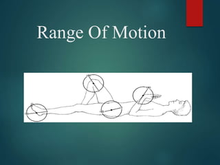
3. PLANES AND AXIS
• Osteo-kinematic motions are described to be taking
place in 3 cardinal planes and axis
4.
5. A frontal or coronal axis lies parallel to the transverse suture of the
skull. It is also horizontal and at right angle to the sagittal axis.
Movement about frontal axis occurs in a sagittal plane. Flexion and
extension (except of the thumb) occurs about a frontal axis and in a
sagittal plane.
A sagittal or antero-posterior axis lies parallel to the sagittal suture
of the skull, i.e., in an antero-posterior direction. Movement about this
axis occurs in a frontal plane. Abduction and adduction (except pf the
thumb) and side flexion movements are said to take about a sagittal
axis and in a frontal plane.
A vertical axis lies parallel to the line of gravity and movement about
it occurs in a horizontal plane. Rotation occurs about a vertical axis
and in a horizontal plane
6. Joint Ranges
Active ROM Passive ROM
• Active motion is the unassisted
voluntary movement of a joint.
(Quality of ROM)
• Passive motion is attained by the examiner
without the patient’s assistance.(Quantity of
ROM)
• Normally, PROM is slightly greater than
AROM because joints have a small amount
of motion at the end range that is not under
voluntary control.
7. MEASURING JOINT RANGE OF
MOTION
• Range Of Motion (ROM) is the arc of motion
that occurs at a joint or a series of joints.
• Three notation systems have been
used to define ROM :
1. The 0 to 180 degree system
2. The 180 to 0 degree system
3. The 360 degree system
Most commonly used is the 0 to 180
degree notation system
8. Prerequisite Knowledge For Measuring
ROM
a) Normal ROM’s (Range)
b) Joint Structure And Function
c) Recommended positioning for self and
patient
d) Bony landmarks related to each joint
e) Alignment of Goniometer
f) Normal end-feel
g) Factors that can alter normal ROM
9. FACTORS DETERMINING AMOUNT
OF ROM
Integrity
Of Joint
SurfaceRELIABILI
TY
Amount
Of
Scarring
Present
AG
E
GEND
ER
Shape Of
Articulati
ng
Surface
Healt
h Of
Joint
Various
diseases/
pathologic
al
conditions
Health Of
Surroundi
ng
Tissues
Mobilty &
Pliabilty Of
Soft Tissue
10. Common pathological causes of
ROM Restriction
• Skin/soft tissue contracture
• Arthritis
• Fracture
• Burns
• Muscle weakness/paralysis
• Pain
• Edema
• Spasticity
• Presence of foreign body in the
joint
11. Prerequisite Skills For Measuring
ROM
• The therapist should be skilled in
Correct positioning
Stabilization for measurement
Palpation
Alignment
Recording measurements accurately
Documentation
12. Testing Procedure
PLACE THE SUBJECT IN
TESTING POSITION
STABILIZE THE PROXIMAL JOINT SEGMENT
MOVE THE DISTAL JOINT SEGMENT TO ZERO STARTING POSITION.
SLOWLY MOVE THE DISTAL JOINT SEGMENT TO THE END OF PASSIVE ROM
AND DETERMINE END FEEL
MAKE VISUAL ESTIMATE OF THE ROM
RETURN THE DISTAL JOINT SEGMENT TO THE STARTING POSITION
PALPATE THE BONY ANATOMICAL
LANDMARKS ALIGN THE GONIOMETER
13. RECORD THE STARTING POSITION.
REMOVE THE GONIOMETER
STABILIZE THE PROXIMAL JOINT
SEGMENT
MOVE THE DISTAL
SEGMENT
THROUGH FULL
ROM
REALIGN THE GONIOMETER. PALPATE THE ANATOMICAL LAND
MARKS AGAIN IF NECESSARY
RECORD THE ROM
14. Documentation
• Hypo Mobility : A motion that does not start
with 0 degree or ends prematurely indicates
joint hypomobility
Example : if knee joint has 30 degree of
hypomobility in flexion, it would be recorded as 30
– 135 deg
• Hyper Mobility : Joint hypermobility at the
beginning of the range is noted by inclusion of a
zero between the starting & ending measurements
Example : if the elbow joint has 5 degree of
hypermobility in extension and 140 degree of
flexion , it would be recorded as 5 – 0 – 140 deg
15. What is Goniometry?
• The term goniometry is derived from two Greek words :
Gonia-metron
• Therefore, goniometry refers to the measurement of angles, in
particular the measurement of angles created at human joints
by the bones.
ANGL
E
MEASU
RE
16. Types of Goniometer
• Full Circle Manual Universal Goniometer (360)
• Half circle manual Goniometer (180)
• Gravity Goniometer :-
• a) Double Inclinometer (used for spine
goniometry)
• b) Pendulum Inclinometer
• c) BubbleGoniometer
• Electrogoniometer
• Digital Goniometer
• Tape Measurements
• Smartphone Devices
• Use of malleable wires/sheets (in cases of
deformities)
20. UNIVERSAL GONIOMETER
• A universal Goniometer may be
constructed of metal or plastic and it has 3
parts :-
1. Body of
Goniometer2. Stationary
arm
3. Movable arm
(placed over the Joint being
measured)
(aligned parallel with the longitudinal axis of
the fixed part)
(aligned parallel with the longitudinal axis of
the movable part)
21.
22. Precautions !!!
1. Joint irritability status
2. Presence of Pain
3. Instability
4. Recent trauma
5. Is it really important to assess accurate
ROM ??
23. END-FEEL
• The end of each motion at each joint is
limited from further movement by
particular anatomical structures.
• The type of structure that limits a joint
motion has a characteristic feel, which may
be detected by the therapist performing the
passive ROM.
• This feeling, which is experienced by the
therapist as resistance or a barrier to
further motion, is called the end-feel.
24. NORMAL END-FEEL DESCRIPTION EXAMPLE
Soft Soft Tissue Approximation Knee flexion (contact
between soft tissue of
posterior leg and
posterior thigh)
Firm Muscular stretch Hip flexion with knee
straight (passive
elastic tension of
hamstring muscles)
Capsular stretch Extension of
metacarpophalangeal
joints of fingers
Ligamentous stretch Forearm supination
(tension in the palmar
radioulnar ligament of the
inferior radioulnar joint)
Hard Bone contacting bone Elbow extension
(olecranon process of the
ulna and olecranon fossa
25. ABNORMAL END-FEEL DESCRIPTION EXAMPLES
Soft Occurs sooner or later in the Soft tissue edema
ROM than is usual or in a
joint
Synovitis
that normally has a firm or
hard end-feel . Feels boggy.
Firm Occurs sooner or later in the Increased muscular tonus
ROM than is usual or in a
joint
Capsular , muscular ,
that normally has a soft or ligamentous, and fascial
hard end-feel. shortening
Hard Occurs sooner or later in the Chondromalacia
ROM than is usual or in a
joint
Osteoarthritis
that normally has a soft or Loose bodies in joint
firm end-feel. A bony grating Myositis ossificans
or bony block is felt. Fracture
Empty No real end-feel because
pain
Acute joint inflammation
prevents reaching end of Bursitis
ROM. No resistance is felt Abscess
except for patient’s protective Fracture
26. Capsular & Non-capsular Pattern Of
Movement Restriction
• Cyriax proposed that pathological
conditions involving the entire joint capsule
cause a particular pattern of restriction
involving most of the passive motions of
the joint. This pattern is called as capsular
pattern
• Restriction caused by condition involving
structures other than the entire joint
capsule is called as non-capsular pattern
• Example – Adhesive Capsulitis Shoulder
27. Shoulder ROM
FLEXION:
Motion: 0-180º
Position: Subject supine with knees flexed or sitting. elbow
extended with the palm facing the body
Goniometer: Axis at the acromion process, laterally
through the head of the humerus.
Stationary arm is placed along the mid-axillary line of the
trunk
Moving arm place along the lateral mid-line of the
humerus in line with the lateral epicondyle.
28. EXTENSION:
Motion: 0-45º~60º from neutral position
Position: Subject prone or sitting , elbow in slight flexion
with the palm facing the body.
Goniometer: Axis at the acromion process, laterally through
the head of the humerus
Stationary Arm aligned with mid- axillary line of the trunk
Moving arm along the lateral mid-line of humerus in line
with lateral epicondyle
29.
30. ABDUCTION:
Motion:0-180º
Position: Supine, prone or sitting with the limb in anatomic
position
Goniometer: Axis at anterior portion of acromion process.
Stationary arm at lateral aspect of anterior surface of chest
parallel to midline of sternum.
Moving arm on anterior aspect of arm parallel to midline of
humerus and in line with medial epicondyle. OR Goniometer:
Axis at the posterior portion of the acromion process; Stationary
arm aligned parallel to spinous process of the vertebral colomn
Moving arm aligned with the midline of the humerus in line with
lateral epicondyle
ADDUCTION:
Motion: 0-30º
Aligment of goniometer is same as abduction.
31.
32. EXTERNAL ROTATION:
Motion: 0-90º
Position: Supine. Shoulder is abducted to 90º. Elbow flexed with
forearm in neutral and perpendicular to table top such that the palm is
facing the feet. Elbow not supported. Humerus is fully supported on
the table. Stabilize the distal humerus, thorax, and scapula.
Goniometer: Axis at olecranon process of the ulna.
Stationary arm placed parallel to the table top or perpendicular to the
floor.
Moving arm along the ulnar shaft aligned with the styloid process of
the ulna.
INTERNAL ROTATION:
Motion: 0-65~90º
Positioning and goniometer alignment is same as in external rotation
33.
34. Radio-ulnar ROM
Supination:
Motion: 0- 80º~ 90º
Position: Subject sitting or supine, with the elbow flexed to 90º. Shoulder in zero
degrees of its’ ROM. Position starts midway between Supination and Pronation.
Goniometer: Axis is medial to the ulnar styloid process.
Stationary arm is aligned parallel to the anterior midline of the humerus.
Moving arm across the ventral aspect of the wrist on a line between and
proximal to the styloid process of the radius and the ulna.
Pronation:
Motion: 0- 80º~ 90º
Position: same for supination.
Goniometer: Axis is lateral to the ulnar styloid process.
Stationary arm is aligned parallel to the anterior midline of the humerus.
Moving arm across the dorsum of the wrist on a line between and proximal to
the styloid process of the radius and the ulna.
35.
36. JOINT MOTION TESTING
POSITION
STABILIZATION MEASUREMENT
S
CERVICAL • FLEXION Sitting Shoulder &
chest
1 cm– 4.3 cm
• EXTENSION Shoulder &
chest to
prevent
extension of
thoracic &
lumbar spine
18.5 cm–22.4cm
• SIDE FLEXION To prevent
side flexion
of thoracic &
lumbar spine
10.7cm-12.9cm
• ROTATION To prevent
rotation of
thoracic &
lumbar
11cm-13.2cm
TAPE MEASUREMENTS OF THE
SPINE
37. JOINT MOTION TESTING
POSITION
STABILIZATION MEASUREMEN
TS
THORACIC • FLEXION STANDING PELVIS
To prevent
anterior
tilting
10 cms (4
inches)
• EXTENSION •If the subject
has balance
problems or
muscle
weakness in
the LE,
measurement
can be taken in
prone/side lying
To prevent
posterior
tilting
• LATERAL
FLEXION
To prevent lateral
tilting
15.9cm for rt LF
16.9cm for lt LF
• ROTATION SITTING To
prevent
rotation
45 degree
(universal
goniometer
)
38. JOINT MOTION TESTIN
G
POSITIO
N
STABILIZATIO
N
MEASUREMEN
TS
LUMBAR • FLEXION STANDING PELVIS
To prevent
anterior tilting
6.7cm in males
5.8cm in
females
Average
6.3cm-
6.9cm
(Modified
Schober
test)
•EXTENSION To prevent
posterior tilting
1.6cm (Modified
Schober Test)
•LATER
AL
FLEXIO
N
To prevent
lateral
tilting
25 – 30
degree by
AMA (double
inclinometer)