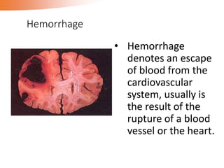
Hemodynamic Disorders
Nasuhi Engin AYDIN, M.D.
Department of Pathology
Aims and scope of the lecture
•Basic mechanisms and clinical relevance
with pathological aspects
Pathological aspects of normal
• Loss of vessel wall integrity : bleeding (hemorrhage)
•Changes in intravascular pressure or vascular
volüme : hyperemia, edema, ischemia
• Disorders in maintence of blood as a liquid :
thrombosis, embolism, infarction
Hemorrhage
• Hemorrhage
denotes an escape
of blood from the
cardiovascular
system, usually is
the result of the
rupture of a blood
vessel or the heart.
Hemorrhage
1-2 mm petechiae
3-5 mm purpura
1-2 cm ecchymose
Nomenculature according to site:
Hemothorax, hemopericardium,
hemoperitoneum, hemarthrosis
5
6
Petechial hemorrhages seen on the epicardium of the heart.
Petechiae are classically found when a coagulopathy is due
to a low platelet count. They can also appear following sudden hypoxia.
7
Petechial hemorrhages on the conjunctiva
8
The blotchy areas of hemorrhage in the skin are called ecchymoses , or also as areas of
purpura. Ecchymoses are larger than petechiae. They can appear with coagulation disorders.
9
Localized collection of blood outside the vascular system within tissues is known as a
hematoma. Here is a small hematoma under the toenail following trauma, which has a bluish
appearance from the deoxygenated blood within it.
10
This is hemopericardium as demonstrated by the dark blood in the pericardial sac opened
at autopsy. Massive blunt force trauma to the chest (often from the steering wheel) causes
a rupture of the myocardium and/or coronary arteries with bleeding into the pericardial
cavity. This leads to cardiac tamponade
11
A blood clot is seen over the external surface of the dura. Thus, this is an epidural
hematoma. Such a location for hemorrhage is virtually always the result of trauma
that causes a tear in the middle meningeal artery
12
This cerebral hemorrhage occurred in conjunction with an overdose of cocaine.
Such acute hemorrhages can occasionally be seen with cocaine use.
Hyperemia
•a local increase of blood volume and flow in
a particular tissue.
Normal blood flow
Hyperemia
Physiological : Exercise
Pathological : Inflammation
•Hyperemia is a increase of blood flow and volume
in a particular tissue due to arteriolar dilation
• Gross or macroscopic ( i.e., naked eye examination):
Larger, red, increased temperature, cut surface is full rich
in blood and wet
• Light microscopic: Dilation of arterioles and capillaries
Morphology of hyperemia
•Congestion is a local increase of blood volüme in
capillaries and venules resulting from impaired venous
return from a tissue.
Congestion
Normal blood fluid
Hyperemia
Congestion
Local increase of blood volume
Hyperemia : active process arteriolar dilatation
Congestion : passive process venous stagnation
Cyanosis: accumulation of deoxygenated hemoglobin
giving the tissue a blue-red color
17
18
 Congestion
Systemic due to heart failure
•Etiology
-Left heart failure
-Right heart failure
Pulmonary cong.
Systemic cong.
1. External pressure
2. Internal occlusion
Local
Edema
Hemorrhage
Heart failure cells
Brown induration
Consequences of pulmonary congestion
Gross (macroscopic) :
Increased volume and
weight of the organ; blue-
red color (cyanosis),
wetness and excessive
blood on the cut surface.
Morphology
22
The lung has a red, hyperemic cut surface, reflecting congestion,
due to increased hydrostatic pressure, as seen in cases of left
heart failure. The transudate, mixed with air in the alveoli, gives
the cut surface a frothy appearance
Acute pulmonary congestion
Chronic pulmonary congestion
Consequences of liver congestion
Atrophy
Fatty change
Centrilobular necrosis
Fibrosis ‘cardiac cirrhosis’
25
A "nutmeg" liver seen with chronic congestion of the liver. The dark red congested regions
represent accumulation of erythrocytes in centrilobular regions.
 The central regions of the lobules become dark red
surrounded by a lighter zone of uncongested liver substance.
“ Nutmeg liver”
•Liver congestion
Central veins and hepatic sinuses of the
centrilobular regions are distended with
blood.
28
Microscopically, the nutmeg pattern results from congestion around
the central veins, as seen here. This is usually due to a "right sided" heart failure.
• Microscopic changes in ‘nutmeg’ liver:
1) Atrophy and/or necrosis of the liver cells in the
mainly in centrilobular regions
2) Fatty change of the liver cells in the peripheral
part of the lobules
‘Cardiac cirrhosis’ of the liver in the longstanding cases
F
F
F
Thrombosis and thrombus
• Thrombosis is the process of formation of
a solid mass of blood within blood vessels
or the heart in living body.
• The resultant mass is called a thrombus.
Normal hemostatic process
when a vessell is severed
normal hemostatic process
stops bleeding
VIRCHOW’S TRIAD,
i.e., factors effective in thrombus formation
• Endothelial injury (most important).
can induce thrombosis alone !
• Alterations in blood flow (stasis, turbulence)
• Hypercoagulability states
Antithrombotic activities of endothelial cells
Endothelial injury
• Trauma
• Radiation
• Vasculitis
• ‘cigarette smoke’ , Atherosclerosis
• Bacterial toxins
• Heart infarction
Alterations in normal blood flow
• Platelets are activated endothelial contact
• Slowed flow retards dilution of activated
clotting factors and also hepatic clearance
• Turbulence may also induce endothelial injury
Hypercoagulable states
Primary (genetic):
Factor V Leiden mutation
Antithrombin III deficiency
Protein C deficiency
Protein S deficiency
Hypercoagulable states
Secondary (acquired): High risk
Prolonged bed rest or immobilization.
Heart infarction,
Tissue damage
(surgery, fractures, burns),
Cardiac failure.
Cancer, leukemia
Hypercoagulable states
Secondary (acquired): Low risk
Smoking,
Hyperlipidemia
Late pregnancy/postdelivery
Oral contraceptives
Cardiomyopathy
Atrial fibrillation
Nephrotic syndrome
Sickle cell anemia
Thrombocytosis
Initiation and propagation of thrombus
White thrombus
Red thrombus
Mixed thrombus
Fibrin thrombus
Types of thrombus
Mural thrombus
in heart chamber
RED THROMBUS IN A VEIN
White thrombus
Site: heart valves , arteries
Components: Platelet, fibrin
F F
F
Mixed thrombus
Site: heart chambers, veins
Components: Platelet, fibrin, erythrocytes
E
E
F
P
F
Fibrin thrombi
are visible
within
capillaries
 Fibrin thrombi in a glomerulus
Organization and recanalization
Detachment, ‘embolism’
Fate of thrombus formation
Organization and recanalization of thrombus
Thrombus is dissolved and
blood could flow again.
Recanalization
Arterial thrombus
Congestion, ischemia
Occlusion of a distal vessel by emboli
(i.e., detached thrombi)
Effects of thrombus formation
Embolism and embolus
• Embolism is a partial or complete obstruction of some
part of the vascular system by any mass (solid, liquid
or even gas !) carried in the circulation.
• The transported material is called an embolus
• The etiology of embolism > 95 % is detached thrombi
Types of embolus
•Thrombi >95 %, i.e., thromboemboli
•Foreign body
•Fat embolism
•Air embolism
•Fluid (amniotic) embolism
Sources:
Fractures of long bones
Soft tissue trauma
Fat embolism
90% of individuals with severe skeletal injuries
10% with clinical findings(1-3 days)
Pulmonary insufficiency, neurologic symptoms
Fat embolism
Fat embolism
Etiology:
Intravenous therapeutic procedures
Obstetric procedures
Chest wall injury
Decompression sickness (nitrogen)
Air embolism
•Gas bubbles within the circulation can obstruct
vascular flow.
•A particular form of gas embolism called decompression
sickness occurs when individuals are exposed to sudden
changes in atmospheric pressure.
AMNIOTIC FLUID EMBOLISM
Incidence: 1/50 000 deliveries
Mortality rate: 80%
Clinical onset: Sudden severe dyspnea,
cyanosis, hypotensive shock, DIC
• Embolus from left heart cavity or arterial system
• Embolus from right heart cavity or venous system
• Embolus from portal veins
• Paradoxical embolism
• Retrograde embolism
Pathway of an embolus depends
on the part of the circulation:
PULMONARY THROMBOEMBOLISM
•Instantaneous death (>60%).
•Cardiovascular collapse.
•Right heart failure
1.Large emboli (5%):
2. Small emboli (60-80%):
•Clinical silent in patients without
cardiovascular failure.
• blood flow from bronchial arteries
(collateral vascular supply)
3. Between the extremes of large and small
emboli (10-15%): Pulmonary hemorrhage.
4. Multiple small emboli: Pulmonary
hypertension and vascular sclerosis.
Systemic (arteriel) embolism
I. 80-85% from heart, secondary to myocardial infarction.
II. 5-10% from auricular thrombi associated with rheumatic
heart disease and atrial fibrillation.
III. 5% from the dilated cardiac chamber of myocarditis /
cardiomyopathy.
VI. Less common sources: Debris from ulcerative
atheromata, or thrombi in aneurysms, infectious
endocarditis, prosthetic valves, paradoxical emboli.
INFARCTION
(INFARCT)
infarct/infarction
•An infarct is a localized area of ischemic
necrosis in a tissue or organ produced
by occlusion of either its arterial supply
or its venous drainage.
•The process whereby the infarct is
developed is known as infarction.
Intrinsic occlusion
for example, thrombosis,
embolism
expansion of atheroma
Vasospasm
Extrinsic compression
for example, twisting of the vessels
Etiology
INFARCTION
• Shape: Wedge-shaped
Segmental
Irregular
• Nature of necrosis
• Types: Red and white infarcts
Morphology of infarct
• LM:
1) Ischemic coagulative necrosis
2) Anemic infarct with few RBC
3) Hemorrhagic infarct has engorgement
and hemorrhage
4) The pathology changes secondary to
infarct such as hyperemia, hemorrhage,
infla., organization and so on.
•Myocardial infarct
The myocardial
cells shows
coagulative
necrosis with the
outline of the
myocardium. In
the margin of the
infarct there are
numerous
inflammatory
exudation and
connective tissue.
white infarct/anemic infarct
•arterial occlusions
•firm tissues
Venous occlusions
Loose tissues
Tissues with dual circulations
Tissues previously congested
Blood flow reestablished
Red infarct/hemorrhagic infarct
The alteration of blood in pulmonary embolism
•Hemorrhagic infarct of the lung
Hemodynamic disorders

Hemodynamic disorders

  • 1.
    Hemodynamic Disorders Nasuhi EnginAYDIN, M.D. Department of Pathology
  • 2.
    Aims and scopeof the lecture •Basic mechanisms and clinical relevance with pathological aspects
  • 3.
    Pathological aspects ofnormal • Loss of vessel wall integrity : bleeding (hemorrhage) •Changes in intravascular pressure or vascular volüme : hyperemia, edema, ischemia • Disorders in maintence of blood as a liquid : thrombosis, embolism, infarction
  • 4.
    Hemorrhage • Hemorrhage denotes anescape of blood from the cardiovascular system, usually is the result of the rupture of a blood vessel or the heart.
  • 5.
    Hemorrhage 1-2 mm petechiae 3-5mm purpura 1-2 cm ecchymose Nomenculature according to site: Hemothorax, hemopericardium, hemoperitoneum, hemarthrosis 5
  • 6.
    6 Petechial hemorrhages seenon the epicardium of the heart. Petechiae are classically found when a coagulopathy is due to a low platelet count. They can also appear following sudden hypoxia.
  • 7.
  • 8.
    8 The blotchy areasof hemorrhage in the skin are called ecchymoses , or also as areas of purpura. Ecchymoses are larger than petechiae. They can appear with coagulation disorders.
  • 9.
    9 Localized collection ofblood outside the vascular system within tissues is known as a hematoma. Here is a small hematoma under the toenail following trauma, which has a bluish appearance from the deoxygenated blood within it.
  • 10.
    10 This is hemopericardiumas demonstrated by the dark blood in the pericardial sac opened at autopsy. Massive blunt force trauma to the chest (often from the steering wheel) causes a rupture of the myocardium and/or coronary arteries with bleeding into the pericardial cavity. This leads to cardiac tamponade
  • 11.
    11 A blood clotis seen over the external surface of the dura. Thus, this is an epidural hematoma. Such a location for hemorrhage is virtually always the result of trauma that causes a tear in the middle meningeal artery
  • 12.
    12 This cerebral hemorrhageoccurred in conjunction with an overdose of cocaine. Such acute hemorrhages can occasionally be seen with cocaine use.
  • 13.
    Hyperemia •a local increaseof blood volume and flow in a particular tissue. Normal blood flow Hyperemia
  • 14.
    Physiological : Exercise Pathological: Inflammation •Hyperemia is a increase of blood flow and volume in a particular tissue due to arteriolar dilation
  • 15.
    • Gross ormacroscopic ( i.e., naked eye examination): Larger, red, increased temperature, cut surface is full rich in blood and wet • Light microscopic: Dilation of arterioles and capillaries Morphology of hyperemia
  • 16.
    •Congestion is alocal increase of blood volüme in capillaries and venules resulting from impaired venous return from a tissue. Congestion Normal blood fluid Hyperemia Congestion
  • 17.
    Local increase ofblood volume Hyperemia : active process arteriolar dilatation Congestion : passive process venous stagnation Cyanosis: accumulation of deoxygenated hemoglobin giving the tissue a blue-red color 17
  • 18.
  • 19.
     Congestion Systemic dueto heart failure •Etiology -Left heart failure -Right heart failure Pulmonary cong. Systemic cong. 1. External pressure 2. Internal occlusion Local
  • 20.
    Edema Hemorrhage Heart failure cells Browninduration Consequences of pulmonary congestion
  • 21.
    Gross (macroscopic) : Increasedvolume and weight of the organ; blue- red color (cyanosis), wetness and excessive blood on the cut surface. Morphology
  • 22.
    22 The lung hasa red, hyperemic cut surface, reflecting congestion, due to increased hydrostatic pressure, as seen in cases of left heart failure. The transudate, mixed with air in the alveoli, gives the cut surface a frothy appearance
  • 23.
  • 24.
    Consequences of livercongestion Atrophy Fatty change Centrilobular necrosis Fibrosis ‘cardiac cirrhosis’
  • 25.
    25 A "nutmeg" liverseen with chronic congestion of the liver. The dark red congested regions represent accumulation of erythrocytes in centrilobular regions.
  • 26.
     The centralregions of the lobules become dark red surrounded by a lighter zone of uncongested liver substance. “ Nutmeg liver”
  • 27.
    •Liver congestion Central veinsand hepatic sinuses of the centrilobular regions are distended with blood.
  • 28.
    28 Microscopically, the nutmegpattern results from congestion around the central veins, as seen here. This is usually due to a "right sided" heart failure.
  • 29.
    • Microscopic changesin ‘nutmeg’ liver: 1) Atrophy and/or necrosis of the liver cells in the mainly in centrilobular regions 2) Fatty change of the liver cells in the peripheral part of the lobules
  • 30.
    ‘Cardiac cirrhosis’ ofthe liver in the longstanding cases F F F
  • 31.
  • 32.
    Thrombosis and thrombus •Thrombosis is the process of formation of a solid mass of blood within blood vessels or the heart in living body. • The resultant mass is called a thrombus.
  • 33.
    Normal hemostatic process whena vessell is severed normal hemostatic process stops bleeding
  • 34.
    VIRCHOW’S TRIAD, i.e., factorseffective in thrombus formation • Endothelial injury (most important). can induce thrombosis alone ! • Alterations in blood flow (stasis, turbulence) • Hypercoagulability states
  • 35.
  • 36.
    Endothelial injury • Trauma •Radiation • Vasculitis • ‘cigarette smoke’ , Atherosclerosis • Bacterial toxins • Heart infarction
  • 37.
    Alterations in normalblood flow • Platelets are activated endothelial contact • Slowed flow retards dilution of activated clotting factors and also hepatic clearance • Turbulence may also induce endothelial injury
  • 38.
    Hypercoagulable states Primary (genetic): FactorV Leiden mutation Antithrombin III deficiency Protein C deficiency Protein S deficiency
  • 39.
    Hypercoagulable states Secondary (acquired):High risk Prolonged bed rest or immobilization. Heart infarction, Tissue damage (surgery, fractures, burns), Cardiac failure. Cancer, leukemia
  • 40.
    Hypercoagulable states Secondary (acquired):Low risk Smoking, Hyperlipidemia Late pregnancy/postdelivery Oral contraceptives Cardiomyopathy Atrial fibrillation Nephrotic syndrome Sickle cell anemia Thrombocytosis
  • 41.
  • 43.
    White thrombus Red thrombus Mixedthrombus Fibrin thrombus Types of thrombus
  • 44.
  • 45.
  • 46.
    White thrombus Site: heartvalves , arteries Components: Platelet, fibrin F F F
  • 47.
    Mixed thrombus Site: heartchambers, veins Components: Platelet, fibrin, erythrocytes E E F P F
  • 48.
  • 49.
    Organization and recanalization Detachment,‘embolism’ Fate of thrombus formation
  • 50.
    Organization and recanalizationof thrombus Thrombus is dissolved and blood could flow again.
  • 51.
  • 52.
    Congestion, ischemia Occlusion ofa distal vessel by emboli (i.e., detached thrombi) Effects of thrombus formation
  • 53.
  • 54.
    • Embolism isa partial or complete obstruction of some part of the vascular system by any mass (solid, liquid or even gas !) carried in the circulation. • The transported material is called an embolus • The etiology of embolism > 95 % is detached thrombi
  • 55.
    Types of embolus •Thrombi>95 %, i.e., thromboemboli •Foreign body •Fat embolism •Air embolism •Fluid (amniotic) embolism
  • 56.
    Sources: Fractures of longbones Soft tissue trauma Fat embolism
  • 57.
    90% of individualswith severe skeletal injuries 10% with clinical findings(1-3 days) Pulmonary insufficiency, neurologic symptoms Fat embolism
  • 58.
  • 60.
    Etiology: Intravenous therapeutic procedures Obstetricprocedures Chest wall injury Decompression sickness (nitrogen) Air embolism •Gas bubbles within the circulation can obstruct vascular flow. •A particular form of gas embolism called decompression sickness occurs when individuals are exposed to sudden changes in atmospheric pressure.
  • 61.
    AMNIOTIC FLUID EMBOLISM Incidence:1/50 000 deliveries Mortality rate: 80% Clinical onset: Sudden severe dyspnea, cyanosis, hypotensive shock, DIC
  • 63.
    • Embolus fromleft heart cavity or arterial system • Embolus from right heart cavity or venous system • Embolus from portal veins • Paradoxical embolism • Retrograde embolism Pathway of an embolus depends on the part of the circulation:
  • 65.
    PULMONARY THROMBOEMBOLISM •Instantaneous death(>60%). •Cardiovascular collapse. •Right heart failure 1.Large emboli (5%):
  • 67.
    2. Small emboli(60-80%): •Clinical silent in patients without cardiovascular failure. • blood flow from bronchial arteries (collateral vascular supply)
  • 68.
    3. Between theextremes of large and small emboli (10-15%): Pulmonary hemorrhage. 4. Multiple small emboli: Pulmonary hypertension and vascular sclerosis.
  • 69.
    Systemic (arteriel) embolism I.80-85% from heart, secondary to myocardial infarction. II. 5-10% from auricular thrombi associated with rheumatic heart disease and atrial fibrillation. III. 5% from the dilated cardiac chamber of myocarditis / cardiomyopathy. VI. Less common sources: Debris from ulcerative atheromata, or thrombi in aneurysms, infectious endocarditis, prosthetic valves, paradoxical emboli.
  • 70.
  • 71.
    infarct/infarction •An infarct isa localized area of ischemic necrosis in a tissue or organ produced by occlusion of either its arterial supply or its venous drainage. •The process whereby the infarct is developed is known as infarction.
  • 72.
    Intrinsic occlusion for example,thrombosis, embolism expansion of atheroma Vasospasm Extrinsic compression for example, twisting of the vessels Etiology
  • 73.
    INFARCTION • Shape: Wedge-shaped Segmental Irregular •Nature of necrosis • Types: Red and white infarcts Morphology of infarct
  • 77.
    • LM: 1) Ischemiccoagulative necrosis 2) Anemic infarct with few RBC 3) Hemorrhagic infarct has engorgement and hemorrhage 4) The pathology changes secondary to infarct such as hyperemia, hemorrhage, infla., organization and so on.
  • 78.
    •Myocardial infarct The myocardial cellsshows coagulative necrosis with the outline of the myocardium. In the margin of the infarct there are numerous inflammatory exudation and connective tissue.
  • 79.
    white infarct/anemic infarct •arterialocclusions •firm tissues
  • 83.
    Venous occlusions Loose tissues Tissueswith dual circulations Tissues previously congested Blood flow reestablished Red infarct/hemorrhagic infarct
  • 84.
    The alteration ofblood in pulmonary embolism
  • 85.