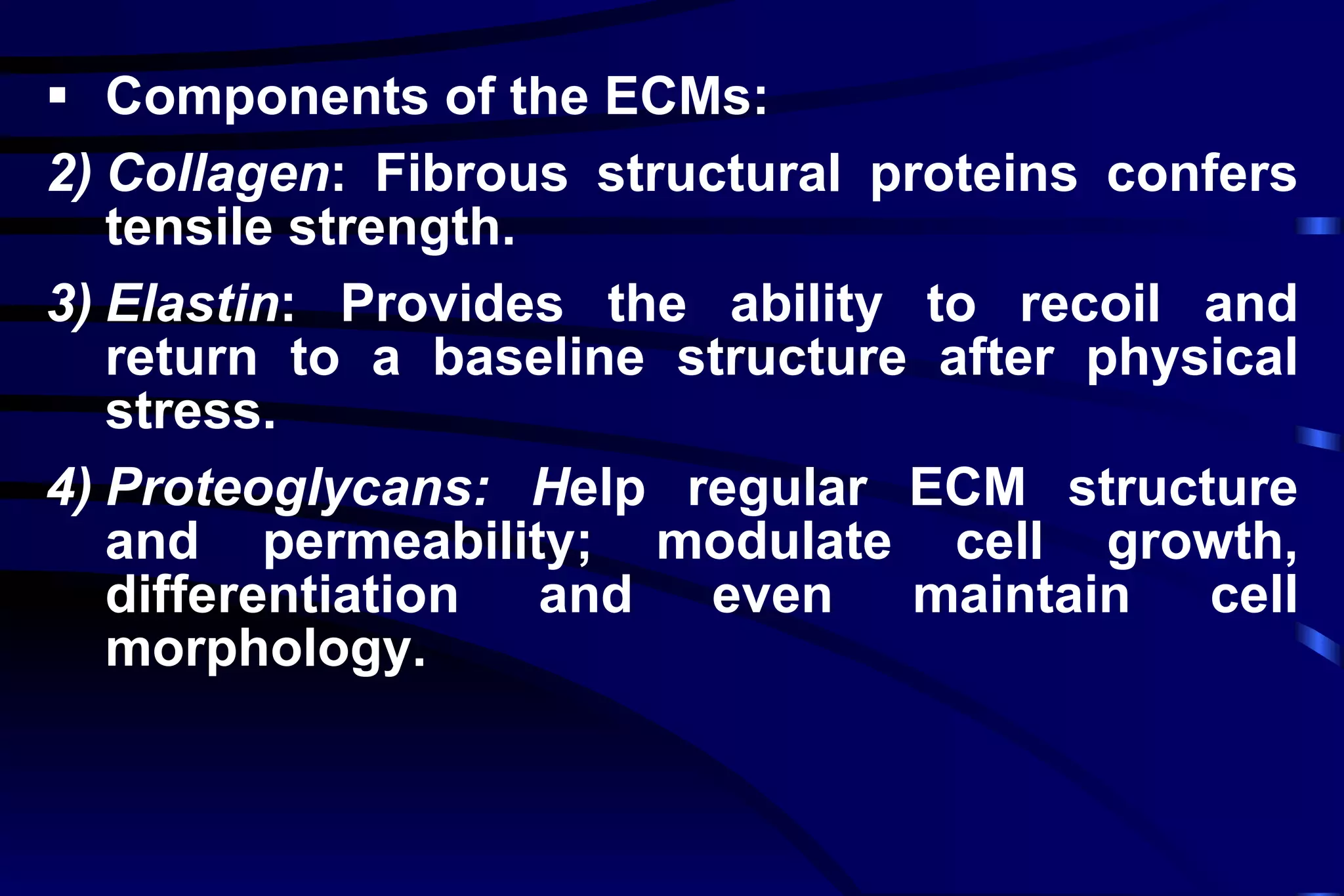
Repair : Regeneration of injured tissue by parenchymal cells of the same type or replacement by connective tissue. Chapter Three Repair Section A
Repair Completely regeneration: Regeneration of injured tissue by parenchymal cells of the same type. Fibrous repair: Replacement by connective tissue In  other words Regeneration Scar
 
Cell population and cell cycle phases
 
Proliferative Potential Labile cells  - continuously dividing  Epidermis, mucosal epithelium, GI tract epithelium etc Stable cells  - low level of replication Hepatocytes, renal tubular epithelium, pancreatic acini Permanent cells  - never divide Nerve cells, cardiac myocytes, skeletal mm


Regeneration   of  e pithelial  t issues   Skin regeneration  :  BM not breached, repaired by the proliferation of epithelial cells. .
Regeneration of  renal tubular cells and hepatocytes : Renal tubular cells: repaired by surviving renal tubular epithelial cells.  If the basic framework is not intact, massive scar tissue is formed.   Hepatocytes  are analogous to the above.
Regeneration of  c onnective  t issue   connective tissue includes: inactive fibroblasts(fibrocyte), activated fibroblasts  extracellular matrix Fibroblasts produce collagen, elastic, and reticular fibers and amorphous material.
Regeneration of cartilage and bone  Cartilage regeneration:  weak of repair capability  Bone tissue: a strong regenerative ability perichondrial cells chondrocytes with new cartilage matrix the quiescent cells and embed in the increased  matrix or the wall of lacunae.
 
 
Angiogenesis:   by two processes:  Vasculogenesis: from angioblasts Angiogenesis: capillary sprouts
 
Fig 4-15
Muscle   Cardiac muscle fibe r s and s keletal  muscle : scar tissue.   skeletal muscle: Repair may be possible only when sarcolemma keeps alive and portion of myofibrils destroy in muscle fiber.  V ascular smooth muscle:  a limited replicative  protential,  new  small  vessels can be formed.  Sarcolemma: a coating of BM-like material adhering to the plasma membrane.
Neural tissue   the central nervous system: scar formation.  the peripheral nervous system: axonal regeneration.
Nerve fiber regeneration Normal nerve fiber Regeneration of Schwann cells and axon filaments at site of nerve lesion or section New axon extending into original channel.
1. Cell-cell interaction Conditioned medium Contact inhibition 2. Growth factors 3. Extracellular matrix Laminin:    Epithelia    Fibroblasts Fibronectin :    Epithelia  Fibroblasts Factors influencing regeneration
1. Chalon and  c ontact inhibition Chalones   is a number of growth inhibitory signals.  A ny different tissues can produce and release inhibitors to control its own proliferation.   Contact inhibition:  Cells continue dividing until they touch one another.
Cells anchor to dish surface and divide. When cells have formed a complete single layer, they stop dividing. If some cells are scraped away, the remaining cells divide to fill the dish with a single layer and then stop.
2. Growth factor s Present in serum or produced locally Exert pleiotropic effects: proliferation, cell migration, differentiation, tissue remodeling Regulate growth of cells by controlling expression of genes that regulate cell proliferation


The common growth factor s: Platelet-derived growth factor (PDGF) : activates fibroblasts, smooth muscle cells, and monocytes for their proliferation and migration; promotes mitosis of gliacytes. Fibroblast growth factor (FGF) : mitogenic for most mesenchymal cells and induces endothelial cell to release proteolytic enzyme.
Epidermal growth factor (EGF) : mitogenic for epithelial cells ,  fibroblasts ,  glial cells and SMC.   Tr ansforming  growth factors(TGF) :  T wo types :   alpha TGF shares homology with EGF .  Beta TGF acts as either a growth stimulator or a growth inhibitor.
Vascular endothelial growth factor (VEGF) : a central role in the growth of new blood.  Cytokines:   IL-1 and TNF induce fibroblast proliferation and collagen synthesis. TNF can also stimulate angiogenesis.
 
Extracellular matrix( ECM ) ECM provides turgor to soft tissue and rigidity to bone, and supplies a substratum for cell adhesion and critically regulates the growth, movement, and differentiation of the cells living within it.
 
Components of the ECMs: Collagen :  Fibrous structural proteins confers tensile strength.  Elastin :   Provides the ability to recoil and return to a baseline structure after physical stress.  Proteoglycans: H elp regular ECM structure and permeability; modulate cell growth, differentiation and even maintain cell morphology.
Adhesive glycoproteins: Include fibronectin, laminin and so on. Link ECM components and  link ECM to cells via cell surface integrins.  Integrins: A family of cell surface receptors mediating adhesion of cells to ECMs.
 
Section B Fibrous Repair Granulation tissues: Newly formed capillaries Fibroblasts Inflammatory cells
Repair by connective tissue Occurs when repair by parenchymal regeneration alone cannot be accomplished Involves production of  Granulation Tissue replacement of parenchymal cells with proliferating fibroblasts and vascular endothelial cells
Granulation tissue Gross: soft, pink, and granular.  LM: fibroblasts, new thin-walled capillaries and inflammatory cells in a loose ECM with edema.
Fibroblasts -- divide and secrete collagen.  Eeventually results in fibrosis with connective tissue matrix.
Granulation tissue : with numerous new blood vessels,  fibroblasts and inflammatory cells.
 
Granulation tissue
Components of the process of fibrosis Angiogenesis - New vessels budding from old Fibrosis, consisting of emigration and proliferation of fibroblasts and deposition of ECM Scar remodeling, tightly regulated by proteases and protease inhibitors
Roles of granulation tissue: Anti-infection and protecting the wound surface from further injure.  Filling incision or wound and any defect of tissue.  Replacing necrosis tissue, effusion and other foreign body.
Fig 4-14
Scar  f ormation (Fibrosis) Angiogenesis - New vessels budding from old Fibrosis: emigration and proliferation of fibroblasts and deposition of ECM. Scar remodeling: tightly regulated by proteases and protease inhibitors
Scar tissue  a pale, avascular scar with largely inactive fibroblasts, dense collagen, fragments of elastic tissue, and other ECM components.  may undergo a reduction in size of 90 percent.
Scar tissue: dense collagen, fibrocytes, scattered fibroblasts and sparse vessels.
Advantage of scar:  provides a resilient permanent patch  provides a  tensile strength and can keep the reparative site solid.
 
Fig 4-19
Healing by first intention Healing by second intention Healing under scab Section C Wound healing
Wound healing Induction of acute inflammatory response by an initial injury Parenchymal cell regeneration Migration and proliferation of parenchymal and connective tissue cells
Wound healing (cont’d) Synthesis of ECM proteins Remodeling of parenchymal elements to restore tissue function Remodeling of connective tissue to achieve wound strength
Healing by First Intention Focal Disruption of Basement Membrane and loss of only a few epithelial cells e.g. Surgical Incision
Healing by  Second Intention Larger injury, abscess, infarction Process is similar but Results in much larger Scar and then CONTRACTION
Wound Strength After sutures are removed at one week, wound strength is only 10% of unwounded skin (Walker’ Law) By 3-4 months, wound strength is about 80% of unwounded skin (Walker’s Law)
Healing  Skin wound
Healing - Skin Scar
Factors affecting Healing: Systemic Age  Nutrition Vitamin def. Immune status Other diseases Local Infection Size or extent. apposition  Blood supply Mobility Foreign body
Fig 4-18
Summary: Healing – Proliferation & Differentiation. Labile, Stabe & Permanent cells Stages of Healing: 1-2-3-4…. Healing by First or Second intention. Skin wound healing - bone healing. Factors affecting healing – Local / Systemic

Tissue Repair

  • 1.
    Repair : Regenerationof injured tissue by parenchymal cells of the same type or replacement by connective tissue. Chapter Three Repair Section A
  • 2.
    Repair Completely regeneration:Regeneration of injured tissue by parenchymal cells of the same type. Fibrous repair: Replacement by connective tissue In other words Regeneration Scar
  • 3.
  • 4.
    Cell population andcell cycle phases
  • 5.
  • 6.
    Proliferative Potential Labilecells - continuously dividing Epidermis, mucosal epithelium, GI tract epithelium etc Stable cells - low level of replication Hepatocytes, renal tubular epithelium, pancreatic acini Permanent cells - never divide Nerve cells, cardiac myocytes, skeletal mm
  • 7.
  • 8.
  • 9.
  • 10.
  • 11.
  • 12.
    Regeneration of e pithelial t issues Skin regeneration : BM not breached, repaired by the proliferation of epithelial cells. .
  • 13.
    Regeneration of renal tubular cells and hepatocytes : Renal tubular cells: repaired by surviving renal tubular epithelial cells. If the basic framework is not intact, massive scar tissue is formed. Hepatocytes are analogous to the above.
  • 14.
    Regeneration of c onnective t issue connective tissue includes: inactive fibroblasts(fibrocyte), activated fibroblasts extracellular matrix Fibroblasts produce collagen, elastic, and reticular fibers and amorphous material.
  • 15.
    Regeneration of cartilageand bone Cartilage regeneration: weak of repair capability Bone tissue: a strong regenerative ability perichondrial cells chondrocytes with new cartilage matrix the quiescent cells and embed in the increased matrix or the wall of lacunae.
  • 16.
  • 17.
  • 18.
    Angiogenesis: by two processes: Vasculogenesis: from angioblasts Angiogenesis: capillary sprouts
  • 19.
  • 20.
  • 21.
    Muscle Cardiac muscle fibe r s and s keletal muscle : scar tissue. skeletal muscle: Repair may be possible only when sarcolemma keeps alive and portion of myofibrils destroy in muscle fiber. V ascular smooth muscle: a limited replicative protential, new small vessels can be formed. Sarcolemma: a coating of BM-like material adhering to the plasma membrane.
  • 22.
    Neural tissue the central nervous system: scar formation. the peripheral nervous system: axonal regeneration.
  • 23.
    Nerve fiber regenerationNormal nerve fiber Regeneration of Schwann cells and axon filaments at site of nerve lesion or section New axon extending into original channel.
  • 24.
    1. Cell-cell interactionConditioned medium Contact inhibition 2. Growth factors 3. Extracellular matrix Laminin:  Epithelia  Fibroblasts Fibronectin :  Epithelia  Fibroblasts Factors influencing regeneration
  • 25.
    1. Chalon and c ontact inhibition Chalones is a number of growth inhibitory signals. A ny different tissues can produce and release inhibitors to control its own proliferation. Contact inhibition: Cells continue dividing until they touch one another.
  • 26.
    Cells anchor todish surface and divide. When cells have formed a complete single layer, they stop dividing. If some cells are scraped away, the remaining cells divide to fill the dish with a single layer and then stop.
  • 27.
    2. Growth factors Present in serum or produced locally Exert pleiotropic effects: proliferation, cell migration, differentiation, tissue remodeling Regulate growth of cells by controlling expression of genes that regulate cell proliferation
  • 28.
  • 29.
  • 30.
  • 31.
  • 32.
    The common growthfactor s: Platelet-derived growth factor (PDGF) : activates fibroblasts, smooth muscle cells, and monocytes for their proliferation and migration; promotes mitosis of gliacytes. Fibroblast growth factor (FGF) : mitogenic for most mesenchymal cells and induces endothelial cell to release proteolytic enzyme.
  • 33.
    Epidermal growth factor(EGF) : mitogenic for epithelial cells , fibroblasts , glial cells and SMC. Tr ansforming growth factors(TGF) : T wo types : alpha TGF shares homology with EGF . Beta TGF acts as either a growth stimulator or a growth inhibitor.
  • 34.
    Vascular endothelial growthfactor (VEGF) : a central role in the growth of new blood. Cytokines: IL-1 and TNF induce fibroblast proliferation and collagen synthesis. TNF can also stimulate angiogenesis.
  • 35.
  • 36.
    Extracellular matrix( ECM) ECM provides turgor to soft tissue and rigidity to bone, and supplies a substratum for cell adhesion and critically regulates the growth, movement, and differentiation of the cells living within it.
  • 37.
  • 38.
    Components of theECMs: Collagen : Fibrous structural proteins confers tensile strength. Elastin : Provides the ability to recoil and return to a baseline structure after physical stress. Proteoglycans: H elp regular ECM structure and permeability; modulate cell growth, differentiation and even maintain cell morphology.
  • 39.
    Adhesive glycoproteins: Includefibronectin, laminin and so on. Link ECM components and link ECM to cells via cell surface integrins. Integrins: A family of cell surface receptors mediating adhesion of cells to ECMs.
  • 40.
  • 41.
    Section B FibrousRepair Granulation tissues: Newly formed capillaries Fibroblasts Inflammatory cells
  • 42.
    Repair by connectivetissue Occurs when repair by parenchymal regeneration alone cannot be accomplished Involves production of Granulation Tissue replacement of parenchymal cells with proliferating fibroblasts and vascular endothelial cells
  • 43.
    Granulation tissue Gross:soft, pink, and granular. LM: fibroblasts, new thin-walled capillaries and inflammatory cells in a loose ECM with edema.
  • 44.
    Fibroblasts -- divideand secrete collagen. Eeventually results in fibrosis with connective tissue matrix.
  • 45.
    Granulation tissue :with numerous new blood vessels, fibroblasts and inflammatory cells.
  • 46.
  • 47.
  • 48.
    Components of theprocess of fibrosis Angiogenesis - New vessels budding from old Fibrosis, consisting of emigration and proliferation of fibroblasts and deposition of ECM Scar remodeling, tightly regulated by proteases and protease inhibitors
  • 49.
    Roles of granulationtissue: Anti-infection and protecting the wound surface from further injure. Filling incision or wound and any defect of tissue. Replacing necrosis tissue, effusion and other foreign body.
  • 50.
  • 51.
    Scar formation (Fibrosis) Angiogenesis - New vessels budding from old Fibrosis: emigration and proliferation of fibroblasts and deposition of ECM. Scar remodeling: tightly regulated by proteases and protease inhibitors
  • 52.
    Scar tissue a pale, avascular scar with largely inactive fibroblasts, dense collagen, fragments of elastic tissue, and other ECM components. may undergo a reduction in size of 90 percent.
  • 53.
    Scar tissue: densecollagen, fibrocytes, scattered fibroblasts and sparse vessels.
  • 54.
    Advantage of scar: provides a resilient permanent patch provides a tensile strength and can keep the reparative site solid.
  • 55.
  • 56.
  • 57.
    Healing by firstintention Healing by second intention Healing under scab Section C Wound healing
  • 58.
    Wound healing Inductionof acute inflammatory response by an initial injury Parenchymal cell regeneration Migration and proliferation of parenchymal and connective tissue cells
  • 59.
    Wound healing (cont’d)Synthesis of ECM proteins Remodeling of parenchymal elements to restore tissue function Remodeling of connective tissue to achieve wound strength
  • 60.
    Healing by FirstIntention Focal Disruption of Basement Membrane and loss of only a few epithelial cells e.g. Surgical Incision
  • 61.
    Healing by Second Intention Larger injury, abscess, infarction Process is similar but Results in much larger Scar and then CONTRACTION
  • 62.
    Wound Strength Aftersutures are removed at one week, wound strength is only 10% of unwounded skin (Walker’ Law) By 3-4 months, wound strength is about 80% of unwounded skin (Walker’s Law)
  • 63.
  • 64.
  • 65.
    Factors affecting Healing:Systemic Age Nutrition Vitamin def. Immune status Other diseases Local Infection Size or extent. apposition Blood supply Mobility Foreign body
  • 66.
  • 67.
    Summary: Healing –Proliferation & Differentiation. Labile, Stabe & Permanent cells Stages of Healing: 1-2-3-4…. Healing by First or Second intention. Skin wound healing - bone healing. Factors affecting healing – Local / Systemic

Editor's Notes