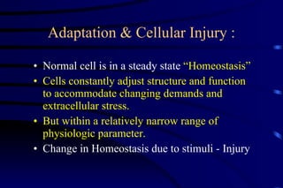
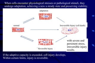
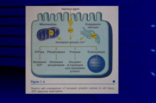
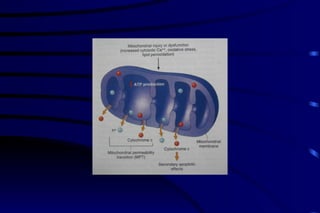
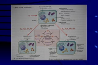
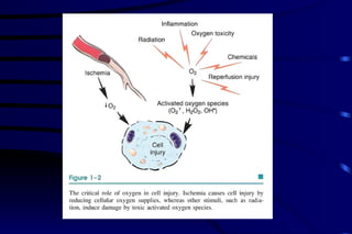
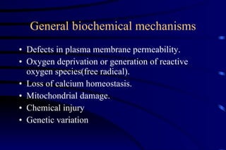
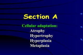
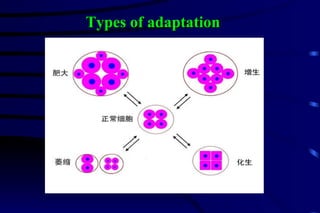
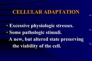
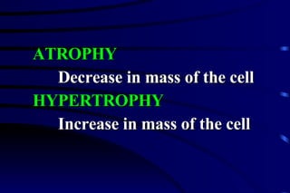
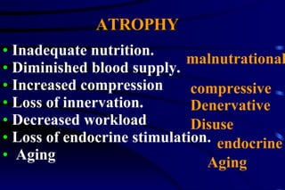
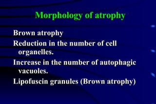
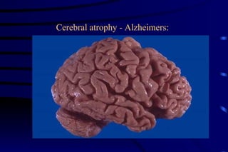
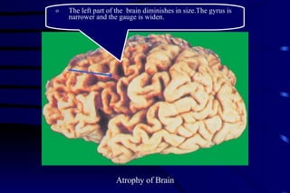
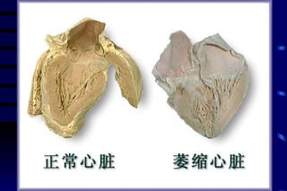
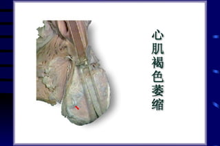
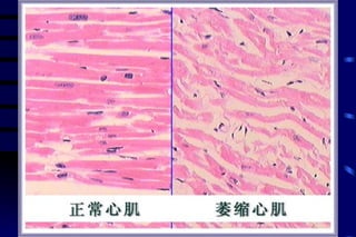
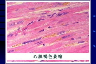
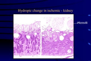
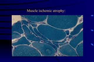
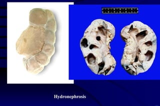
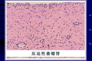
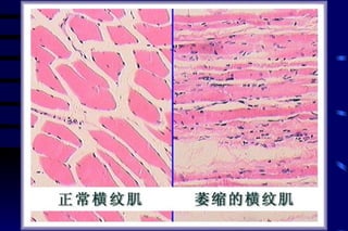
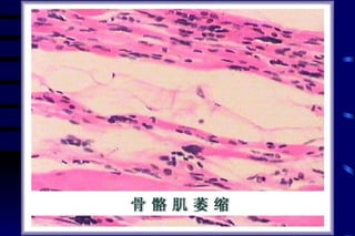
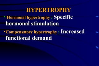
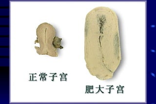
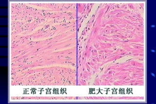
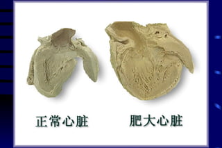
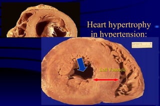
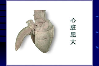
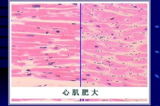
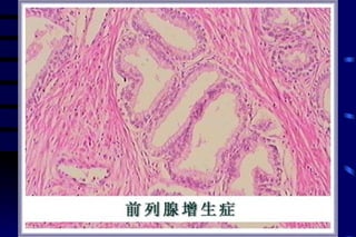
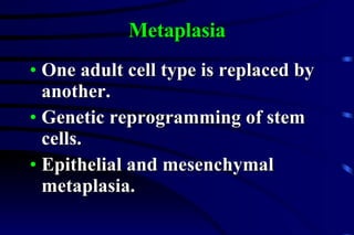
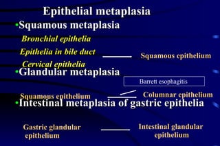
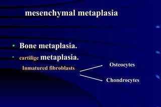
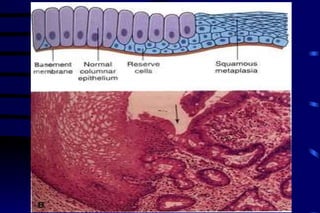
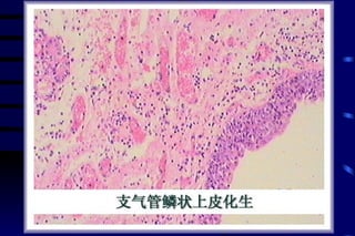
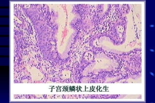
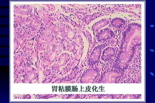
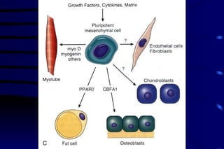
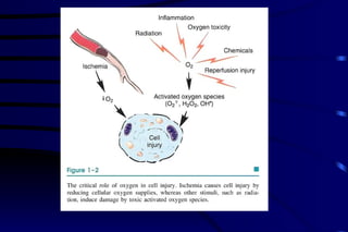
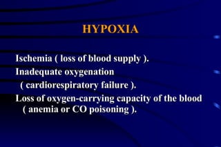
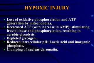
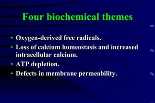
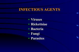
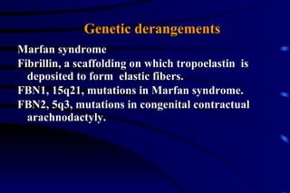
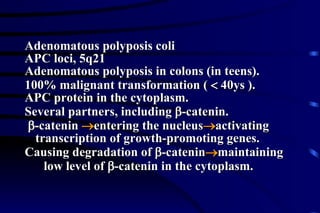
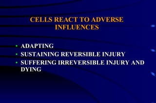
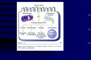
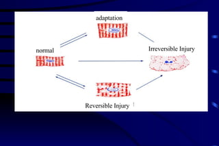
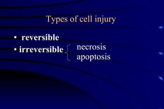
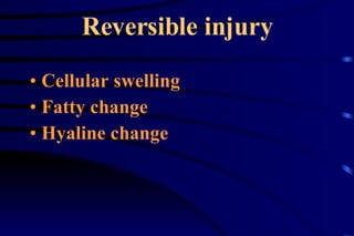
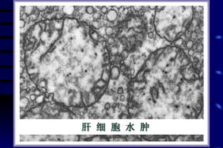
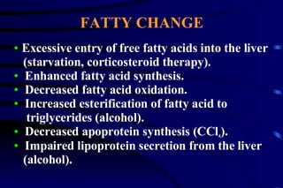
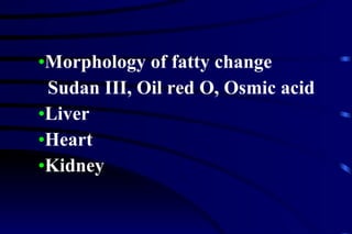
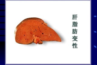
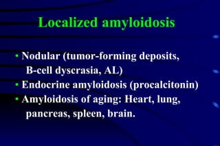
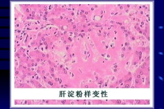
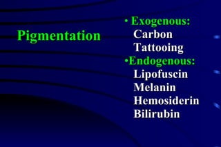
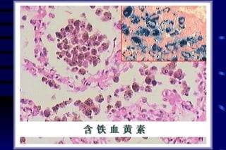
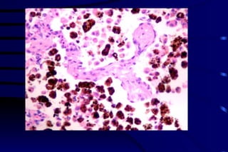
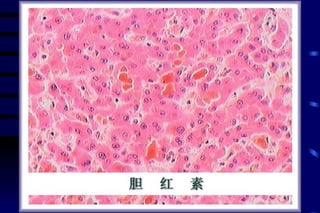
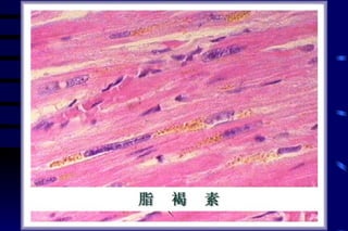
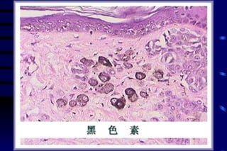
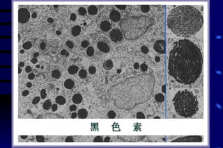
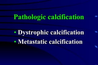
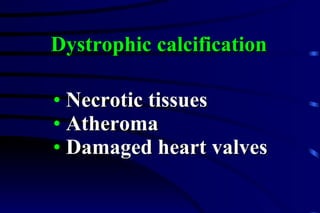
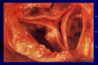
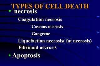
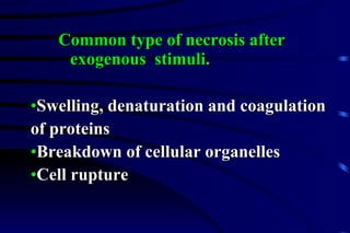
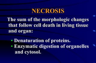
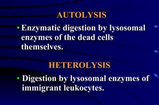
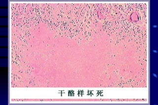
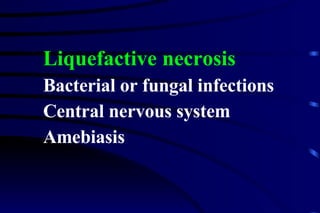
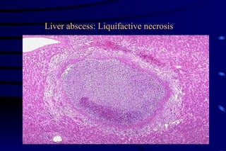
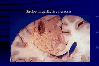
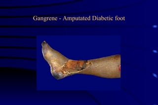
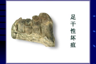
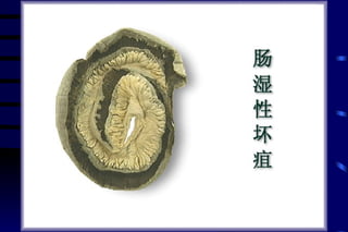
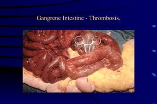
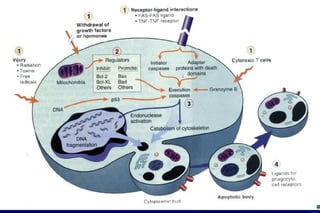
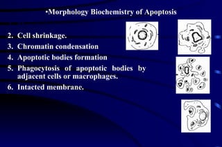
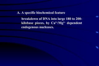
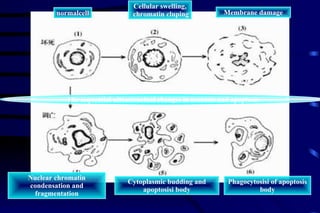
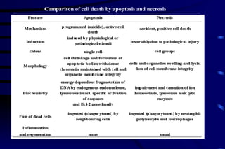
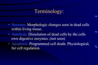
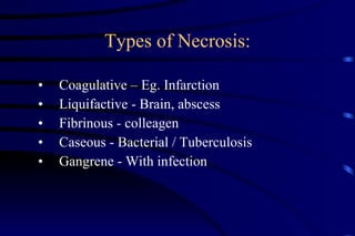
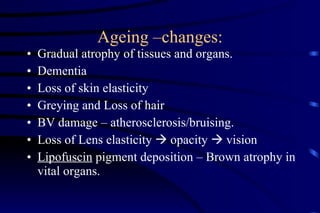
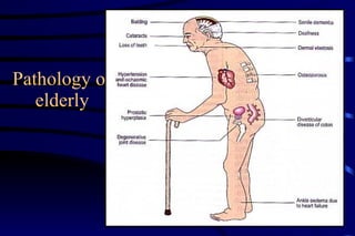
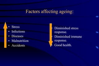
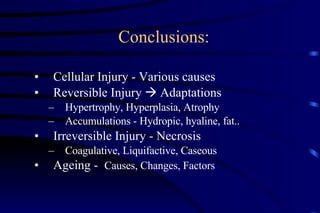
Chapter Two  adaptation and injury
Adaptation & Cellular Injury : Normal cell is in a steady state  “Homeostasis” Cells constantly adjust structure and function to accommodate changing demands and extracellular stress. But within a relatively narrow range of physiologic parameter. Change in Homeostasis due to stimuli - Injury
Irreversible Injury   (cell death)   Reversible Injury   adaptation  normal   When cells encounter physiological stresses or pathological stimuli, they undergo adaptation, achieving a new a steady state and preserving viability.  If the adaptive capacity is exceeded, cell injury develops.  Within certain limits, injury is reversible. with severe and persistent stress, irreversible injury results.
adaptation A state that between normal, unstressed cell  and the injured, overstressed cell.
General principles 1. The cellular response to injurious stimuli depends on the type of injury.
Incomplete occlusion of coronary artery  Complete or prolonged occlusion hypertesion Prolonged starvation
General principles 2.The consequences of an injurious stimulus depend on the type, status, adaptability, and genetic makeup of injured cell.
Skeletal muscle accommodates complete ischemia for 2 to 3 hours without irreversible injury. cardiac muscle dies after 20 to 30 minutes. Neuron dies after a few minutes.
General principles 3.Cellular function is far before cell death occurs, and the morphologic changes of cell injury(or death) lag far behind both.
 
General principles Cell membrane integrity, critical to cellular ionic and osmotic homeostasis; ATP generation, largely via mitochondrial aerobic respiration; Protein synthesis;  Intergrity of the genetic apparatus. 4. four intracellular systems are particularly vulnerable.


General biochemical mechanisms Defects in plasma membrane permeability. Oxygen deprivation or generation of reactive oxygen species(free radical). Loss of calcium homeostasis. Mitochondrial damage. Chemical injury Genetic variation
Cellular adaptation: Atrophy Hypertrophy Hyperplasia Metaplasia Section A
Types of adaptation
CELLULAR ADAPTATION Excessive physiologic stresses. Some pathologic stimuli. A new, but altered state preserving  the viability of the cell.
ATROPHY Decrease in mass of the cell HYPERTROPHY  Increase in mass of the cell
ATROPHY  Inadequate nutrition. Diminished blood supply. Increased compression Loss of innervation. Decreased workload Loss of endocrine stimulation. Aging  malnutrational Denervative endocrine Disuse compressive Aging
Morphology of atrophy Brown atrophy Reduction in the number of cell organelles. Increase in the number of autophagic vacuoles. Lipofuscin granules (Brown atrophy)
Fig 2-5
Cerebral atrophy - Alzheimers:
Atrophy of Brain The left part of the  brain diminishes in size.The gyrus is narrower and the gauge is widen.


Hydropic change in ischemic - kidney Microvilli
Muscle ischemic atrophy:


Hydronephrosis


Hormonal hypertrophy  :  Specific hormonal stimulation   Compensatory hypertrophy  :  Increased functional demand HYPERTROPHY
Fig 2-6
 
 
Heart hypertrophy in hypertension: Left Ventricle
 
 
HYPERPLASIA
HYPERPLASIA Physiologic hyperplasia: Hormonal hyperplasia Compensatory hyperplasia Pathologic hyperplasia: Excessive hormonal stimulation. Effects of locally produced GFs on target cells.  female breast and uterus at puberty and pregnancy.
Fig 2-1
PARTIAL HEPATECTOMY Priming Proliferation Growth lnhibition GROWTH FACTORS AND CYTOKINES HGF  TGF-  EGF TNF-  IL-6 Others ADJUVANTS Norepinephrine Insulin Glucagon Thyroid hormone GROWTH INHIBITORS TGF-    Others Growth factors Adjuvanis Matrix degradation
 
 
Metaplasia One adult cell type is replaced by another. Genetic reprogramming of stem cells. Epithelial and mesenchymal metaplasia.
Intestinal glandular  epithelium Squamous metaplasia Glandular metaplasia Intestinal metaplasia of gastric epithelia Bronchial epithelia Epithelia in bile duct Cervical epithelia Epithelial metaplasia  Columnar epithelium   Squamous epithelium Squamous epithelium Gastric glandular epithelium Barrett esophagitis
Connective tissue metaplasia Bone metaplasia. cartilige  metaplasia. Inmatured fibroblasts Osteocytes Chondrocytes mesenchymal metaplasia


significance of metaplasia A two-edged sword An undesirable change Survive but some important protective mechanism is lost. The influences that predispose to such squamous metaplasia, if persistent, may promote cancer transformation in metaplastic epithelium.
Cell  injury Section B
Causes of cell injury and disease Oxygen deprivation ( hypoxia, ischemia) Nutritional imbalances Physical agents Chemical agents and drugs Infectious agents Immunologic reactions Genetic derangements
 
HYPOXIA Ischemia ( loss of blood supply ). Inadequate oxygenation ( cardiorespiratory failure ). Loss of oxygen-carrying capacity of the blood ( anemia or CO poisoning ).
HYPOXIC INJURY Loss of oxidative phosphorylation and ATP generation by mitochondria. Decreased ATP (with increase in AMP): stimulating fructokinase and phosphorylation, resulting in aerobic glycolysis. Depleted glycogen. Reduced intracellular pH: Lactic acid and inorganic phosphate. Clumping of nuclear chromatin.
Four biochemical themes Oxygen-derived free radicals. Loss of calcium homeostasis and increased intracellular calcium. ATP depletion. Defects in membrane permeability.
PHYSICAL AGENTS Trauma Heat Cold Radiation Electric shock
CHEMICAL AGENTS AND DRUGS Endogenous products: urea Exogenous agents: Therapeutic drugs: hormones Nontherapeutic agents:  lead or alcohol
MECHANISMS OF CHEMICAL INJURY Directly:  Mercury of mercuric chloride binds to SH groups of cell membrane proteins, causing increased permeability and inhibition of ATPase-dependent transport.
By conversion to reactive toxic metabolites which in turn cause cell injury either by direct covalent binding to membrane protein and lipid, or more commonly by the formation of free radicals. MECHANISMS OF CHEMICAL INJURY
CCl 4  in SER of liver cell (P-450) – CCl 3 .  – lipid peroxidation and autocatalytic reactions – swelling and breakdown of ER, dissociation of ribosome, and decreased hepatic protein synthesis ( loss of lipid acceptor protein – fatty change of liver cell) – progressive cellular swelling, plasma membrane damage, and cell death.
FREE RADICAL INITIATION Absorption of energy (UV light and x-rays) Oxidative metabolic reactions Enzymatic conversion of exogenous chemicals or drugs (CCl 4 >CCl 3 . ) Oxygen-derived radicals Superoxide
Cell injury caused by free radicals through Peroxidation of lipids. Cross linking proteins by the formation of disulfide bonds. Induction of DNA damage that has been implicated both in cell killing and malignant transformation.
INFECTIOUS AGENTS Viruses Rickettsiae Bacteria Fungi Parasites
Marfan syndrome Fibrillin, a scaffolding on which tropoelastin   is deposited to form  elastic fibers. FBN1, 15q21, mutations in Marfan syndrome. FBN2, 5q3, mutations in congenital contractual arachnodactyly.  Genetic derangements
Adenomatous polyposis coli APC loci, 5q21 Adenomatous polyposis in colons (in teens). 100% malignant transformation (    40ys ). APC protein in the cytoplasm. Several partners, including   -catenin.  -catenin   entering the nucleus  activating transcription of growth-promoting genes. Causing degradation of   -catenin  maintaining low level of   -catenin in the cytoplasm.
CELLS REACT TO ADVERSE INFLUENCES ADAPTING SUSTAINING REVERSIBLE INJURY SUFFERING IRREVERSIBLE INJURY AND DYING
CELL INJURY AND NECROSIS General mechanisms: Maintenance of the integrity of cell membranes.  Aerobic respiration and production of ATP.  Synthesis of enzymes and structure proteins.  Preservation of the integrity of the genetic apparatus.
 
Irreversible Injury   Reversible Injury   adaptation   normal
Types of cell injury  reversible irreversible necrosis apoptosis
Reversible injury Cellular swelling Fatty change Hyaline change
Cellular swelling
 
Excessive entry of free fatty acids into the liver (starvation, corticosteroid therapy). Enhanced fatty acid synthesis. Decreased fatty acid oxidation. Increased esterification of fatty acid to  triglycerides (alcohol). Decreased apoprotein synthesis (CCl 4 ). Impaired lipoprotein secretion from the liver  (alcohol). FATTY CHANGE
Morphology of fatty change Sudan III, Oil red O, Osmic acid Liver Heart Kidney
 
 
Intracellular hyaline changes Hyaline degeneration of arterioles Hyaline degeneration of connective tissue Hyaline changes (degeneration)
Absorption of protein causing hyaline  droplets in proximal epithelial cells in the kidney. Russel bodies in plasma cells. Viral inclusions in the cytoplasm or the nucleus. Masses of altered intermediate filaments  (Mallory bodies). Intracellular hyaline changes


Hyaline change of the central artery of the spleen (spleen of hypertension disease) The narrowing of the lumina with thickened vessel wall.  Homogeneous pink hyaline material deposits under the intima.
Mucoid Degeneration mucopolysaccharide deposition in the stroma of connective tissue.
mucoid degeneration
A heterogeneous group of pathogenic  fibrillar proteins accumulating in tissues and organs. Excess synthesis Resistance to catabolism AMYLOIDOSIS
 
Chemical nature of amyloid fibrils Two major forms: AL (amyloid light chain protein) AA (amyloid-associated protein):   Derived from serum AA (12kd)  synthesized in liver and elevated in  inflammatory states.
Minor forms of amyloid fibrils: Transthyretin (TTR): A mutant form of a  serum protein in familial amyloid  polyneuropathy. A variant of TTR in aging. Beta-2-microglobulin (the component of  class I MHC molecules) in long-term hemidialysis.
Minor forms of amyloid fibrils:   Beta-2-amyloid protein forms the core of  cerebral plaques and deposits within  cerebral vessel walls in Alzheimer disease, deriving from a transmembrane glycoprotein precursor.
primary (B-cell dyscrasia, AL) Secondary or reactive (AA):  Collagen diseases, bronchiectasis, chronic  osteomyelitis. Hemodialysis-related: Beta-2-microglobulin  deposition. Hereditary (AA) Clinical forms of amyloidosis Systemic  amyloidosis:
Nodular (tumor-forming deposits,  B-cell dyscrasia, AL)  Endocrine amyloidosis (procalcitonin) Amyloidosis of aging: Heart, lung,  pancreas, spleen, brain. Localized amyloidosis
 
Exogenous:   Carbon Tattooing Endogenous: Lipofuscin Melanin Hemosiderin Bilirubin Pigmentation


Dystrophic calcification Metastatic calcification Pathologic calcification
Necrotic tissues Atheroma Damaged heart valves Dystrophic calcification
Fig 2-13
 
Increased secretion of parathyroid  hormone Destruction of bone tissue Vitamin D-related disorders:  Sarcoidosis Renal failure Metastatic calcification Hypercalcimia
Metastatic calcification Affecting Interstitial tissue of gastric mucosa Kidneys Lungs Pulmonary veins  Systemic arteries
Section C CELL DEATH
TYPES OF CELL DEATH necrosis Coagulation necrosis Caseous necrosis Gangrene Liquefaction necrosis( fat necrosis) Fibrinoid necrosis Apoptosis
Swelling, denaturation and coagulation  of proteins Breakdown of cellular organelles Cell rupture Common type of necrosis after exogenous  stimuli.
NECROSIS The sum of the morphologic changes that follow cell death in living tissue and organ: Denaturation of proteins. Enzymatic digestion of organelles and cytosol.
Enzymatic digestion by lysosomal enzymes of the dead cells themselves. AUTOLYSIS HETEROLYSIS Digestion by lysosomal enzymes of immigrant leukocytes.
Nucleus changes : The hallmarks of cell death in three patterns: Basic Pathologic Change of Necrosis Normal cell Chromatin margination karyorrhexis pyknosis karyolysis Nuclear Alteration of Necrosis
1)Three pattern of nuclear changes Karyolysis (DNase activity) Pyknosis (DNA condensation) Karyorrhexis (fragmentation of pyknotic nucleus)
Pyknosis : nuclear shrinkage and increased basophilia, and the DNA apparently condenses into a solid, shrunken basophilic mass.  Karyorrhexis:  nucleus undergoes fragmentation, scattered about the cytoplasm. Karyolysis:  the basophilia of the chromatin may fade and the nucleus disappears.
 
Cytoplasm change:   increased eosinophilium and a more glassy homogeneous appearance and even vacuolated cytoplasm. The Necrosis of heptocytes
Types of Necrosis Coagulative Necrosis Liquefactive Necrosis Fibrinoid Necrosis Necrosis Gangrenous Necrosis
Morphologic appearance of necrosis Increased eosinophilia: Loss of RNA in the cytoplasm  Increased binding of eosin to denatured  cytoplasmic   protein  More glassy homogeneous appearance  Loss of glycogen particles Vacuolated and moth-eaten cytoplasm Calcification of necrotic cells
Coagulation necrosis Denatures of both structural and  enzymatic proteins by injury or the  subsequent increasing intracellular  acidosis.
Renal Infarction - Coagulative
Splenic Infarction - Coagulative necrosis
Infarction -  Adrenal gland:
 
Caseous necrosis A subtype of coagulation necrosis White and cheesy Tuberculosis Completely obliterated tissue  architecture
Caseous necrosis of kidney The necrosis area is soft, white-yellow cheesy appearance.
 
Extensive  Caseous necrosis Tuberculosis
 
Liquefactive necrosis Bacterial or fungal infections Central nervous system Amebiasis
Liver abscess: Liquifactive necrosis
Stroke- Liquifactive necrosis
 
 
Fat necrosis Traumatic Activated pancreatic lipases
Fat necrosis(Steatonecrosis) Only shadowy outlines of necrotic fat cells may be seen, with basophilic calcium deposits and a surrounding inflammatory reaction.
Fibrinoid degeneration Deeply eosinophilic Collagen diseases Necrotic vasculitis Malignant hypertension
Fibrinoid Necrosis homogeneous, deeply eosinophilic in necrosis.
Gangrene A subtype of coagulation necrosis Dry gangrene Wet gangrene Gas gangrene
Caseous necrosis - Tuberculosis
Gangrene - Amputated Diabetic foot
 
 
Gangrene Intestine - Thrombosis.
 
Absorption Discharge: Erosion  Ulcer Sinus  Fistula  Cavitation Organization Encapsulation Calcification Fates of necrosis


Section D APOPTOSIS
APOPTOSIS  (Programmed cell death)   Programmed destruction of cells during  embryogenesis. Hormone dependent involution of tissues  in the adult. Cell deletion in proliferating cell popula-  tions (intestinal crypt epithelium),  tumors, and lymphoid organs.
Pathologic atrophy in parenchymal  organs after duct obstruction. Cell death by cytotoxic T cells. Cell injury in certain viral diseases. Cell death produced by a variety of  injurious stimuli given in low doses (e.g. mild thermal injury).
MORPHOLOGICAL FEATURES OF APOPTOSIS Cell shrinkage Chromatin condensation and fragmentation. Formation of cytoplasmic blebs and apoptotic bodies. Phagocytosis of apoptotic bodies by adjacent  healthy cells or macrophages. Lack of inflammation.
Necrosis   Apoptosis Stimuli   Hypoxia  Physical Toxins  Pathological Histology   Cell swelling  Single cell  Coagulation N  Chromatin Disruption of  condensation organelles  Apoptotic bodies DNA   Random  Internucleosomal breakdown   Diffuse
Necrosis   Apoptosis Mechanism  ATP depletion  Gene activation Membrane  Endonuclease injury Free radicals Tissue  Inflammation  No inflammation   reaction   Phagocytosis of  apoptotic bodies
Fig 1-18
 
Biochemical features of apoptosis 1.PROTEIN CLEAVAGE:   Caspases (cysteine protease) Nuclear scaffold Cytoskeletal protein 2.PROTEIN CROSS-LINKING:   Transglutaminase Cytoplasmic protein  shrunken shalls  apoptotic bodies Biochemical features of apoptosis
3. DNA breakdown:   50-300 kb pieces Ca2+, Mg2+ dependent endonucleases DNA oligonucleosomes DNA ladders (also seen in necrosis) 4. PHAGOCYTIC RECOGNITION   Receptors on macrophages for the  surface  components  (phosphatidylserine, thrombospondin)  on apoptotic bodies.
Fig 1-19
Apoptosis-associated genes bcl-2, c-myc, p53
Fig 1-20
Occuring conditions during embryogenesis and development; as a homeostatic mechanism to maintain normal cell populations of tissue in the face of cell turnover; as a defense mechanism such as in immune reactions; when cells are damaged by diseases or noxious agents, such as injury, tumors and inflammation;  reduction cell during atrophy; in aging.
Morphologic features   Cell shrinkage.  Chromatin condensation Apoptotic bodies formation  Phagocytosis of apoptotic bodies by adjacent cells or macrophages. Intacted membrane. Morphology Biochemistry of Apoptosis
A specific biochemical feature breakdown of DNA into large 180 to 200-kilobase pieces, by Ca 2+ /Mg 2+  dependent endogenous nucleases.
normalcell Cellular swelling, chromatin cluping Membrane damage Nuclear chromatin condensation and fragmentation Cytoplasmic budding and apoptosisi body Phagocytosisi of apoptosis body The sequential ultrastructual changes in necrosis and apoptosis
Apotosis of hepatocytes
Comparison of cell death by apoptosis and necrosis
Terminology: Necrosis:  Morphologic changes seen in dead cells within living tissue. Autolysis:  Dissolution of dead cells by the cells own digestive enzymes. (not seen) Apoptosis:  Programmed cell death. Physiological, for cell regulation.
Types of Necrosis: Coagulative – Eg. Infarction Liquifactive - Brain, abscess Fibrinous - colleagen Caseous - Bacterial / Tuberculosis Gangrene - With infection
Ageing: “ Progressive time related loss of structural and functional capacity of cells leading to death” Senescence, Senility, Senile changes. Ageing of a person is intimately related to cellular ageing.
Factors affecting Ageing: Genetic – Clock genes,  (fibroblasts) Diet – malnutrition, obesity etc. Social conditions -  Diseases – Atherosclerosis, diabetes etc. Werner’s syndrome.
Cellular mechanisms of ageing Cross linking proteins & DNA. Accumulation of toxic by-products. Ageing genes. Loss of repair mechanism. Free radicle injury Telomerase shortening.
Telomerase in ageing: Germ Cells Somatic Cells
Ageing –changes: Gradual atrophy of tissues and organs. Dementia Loss of skin elasticity Greying and Loss of hair BV damage – atherosclerosis/bruising. Loss of Lens elasticity    opacity    vision Lipofuscin  pigment deposition – Brown atrophy in vital organs.
Pathology of elderly
Factors affecting ageing: Stress Infections Diseases Malnutrition Accidents Diminished stress response. Diminished immune response. Good health.
Conclusions: Cellular Injury - Various causes Reversible Injury    Adaptations Hypertrophy, Hyperplasia, Atrophy Accumulations - Hydropic, hyaline, fat.. Irreversible Injury - Necrosis Coagulative, Liquifactive, Caseous Ageing -  Causes, Changes, Factors

Tissue Injury

  • 1.
    Chapter Two adaptation and injury
  • 2.
    Adaptation & CellularInjury : Normal cell is in a steady state “Homeostasis” Cells constantly adjust structure and function to accommodate changing demands and extracellular stress. But within a relatively narrow range of physiologic parameter. Change in Homeostasis due to stimuli - Injury
  • 3.
    Irreversible Injury (cell death) Reversible Injury adaptation normal When cells encounter physiological stresses or pathological stimuli, they undergo adaptation, achieving a new a steady state and preserving viability. If the adaptive capacity is exceeded, cell injury develops. Within certain limits, injury is reversible. with severe and persistent stress, irreversible injury results.
  • 4.
    adaptation A statethat between normal, unstressed cell and the injured, overstressed cell.
  • 5.
    General principles 1.The cellular response to injurious stimuli depends on the type of injury.
  • 6.
    Incomplete occlusion ofcoronary artery Complete or prolonged occlusion hypertesion Prolonged starvation
  • 7.
    General principles 2.Theconsequences of an injurious stimulus depend on the type, status, adaptability, and genetic makeup of injured cell.
  • 8.
    Skeletal muscle accommodatescomplete ischemia for 2 to 3 hours without irreversible injury. cardiac muscle dies after 20 to 30 minutes. Neuron dies after a few minutes.
  • 9.
    General principles 3.Cellularfunction is far before cell death occurs, and the morphologic changes of cell injury(or death) lag far behind both.
  • 10.
  • 11.
    General principles Cellmembrane integrity, critical to cellular ionic and osmotic homeostasis; ATP generation, largely via mitochondrial aerobic respiration; Protein synthesis; Intergrity of the genetic apparatus. 4. four intracellular systems are particularly vulnerable.
  • 12.
  • 13.
  • 14.
  • 15.
  • 16.
    General biochemical mechanismsDefects in plasma membrane permeability. Oxygen deprivation or generation of reactive oxygen species(free radical). Loss of calcium homeostasis. Mitochondrial damage. Chemical injury Genetic variation
  • 17.
    Cellular adaptation: AtrophyHypertrophy Hyperplasia Metaplasia Section A
  • 18.
  • 19.
    CELLULAR ADAPTATION Excessivephysiologic stresses. Some pathologic stimuli. A new, but altered state preserving the viability of the cell.
  • 20.
    ATROPHY Decrease inmass of the cell HYPERTROPHY Increase in mass of the cell
  • 21.
    ATROPHY Inadequatenutrition. Diminished blood supply. Increased compression Loss of innervation. Decreased workload Loss of endocrine stimulation. Aging malnutrational Denervative endocrine Disuse compressive Aging
  • 22.
    Morphology of atrophyBrown atrophy Reduction in the number of cell organelles. Increase in the number of autophagic vacuoles. Lipofuscin granules (Brown atrophy)
  • 23.
  • 24.
    Cerebral atrophy -Alzheimers:
  • 25.
    Atrophy of BrainThe left part of the brain diminishes in size.The gyrus is narrower and the gauge is widen.
  • 26.
  • 27.
  • 28.
  • 29.
  • 30.
    Hydropic change inischemic - kidney Microvilli
  • 31.
  • 32.
  • 33.
  • 34.
  • 35.
  • 36.
  • 37.
  • 38.
  • 39.
  • 40.
    Hormonal hypertrophy : Specific hormonal stimulation Compensatory hypertrophy : Increased functional demand HYPERTROPHY
  • 41.
  • 42.
  • 43.
  • 44.
    Heart hypertrophy inhypertension: Left Ventricle
  • 45.
  • 46.
  • 47.
  • 48.
    HYPERPLASIA Physiologic hyperplasia:Hormonal hyperplasia Compensatory hyperplasia Pathologic hyperplasia: Excessive hormonal stimulation. Effects of locally produced GFs on target cells. female breast and uterus at puberty and pregnancy.
  • 49.
  • 50.
    PARTIAL HEPATECTOMY PrimingProliferation Growth lnhibition GROWTH FACTORS AND CYTOKINES HGF TGF-  EGF TNF-  IL-6 Others ADJUVANTS Norepinephrine Insulin Glucagon Thyroid hormone GROWTH INHIBITORS TGF-  Others Growth factors Adjuvanis Matrix degradation
  • 51.
  • 52.
  • 53.
    Metaplasia One adultcell type is replaced by another. Genetic reprogramming of stem cells. Epithelial and mesenchymal metaplasia.
  • 54.
    Intestinal glandular epithelium Squamous metaplasia Glandular metaplasia Intestinal metaplasia of gastric epithelia Bronchial epithelia Epithelia in bile duct Cervical epithelia Epithelial metaplasia Columnar epithelium Squamous epithelium Squamous epithelium Gastric glandular epithelium Barrett esophagitis
  • 55.
    Connective tissue metaplasiaBone metaplasia. cartilige metaplasia. Inmatured fibroblasts Osteocytes Chondrocytes mesenchymal metaplasia
  • 56.
  • 57.
  • 58.
  • 59.
  • 60.
  • 61.
    significance of metaplasiaA two-edged sword An undesirable change Survive but some important protective mechanism is lost. The influences that predispose to such squamous metaplasia, if persistent, may promote cancer transformation in metaplastic epithelium.
  • 62.
    Cell injurySection B
  • 63.
    Causes of cellinjury and disease Oxygen deprivation ( hypoxia, ischemia) Nutritional imbalances Physical agents Chemical agents and drugs Infectious agents Immunologic reactions Genetic derangements
  • 64.
  • 65.
    HYPOXIA Ischemia (loss of blood supply ). Inadequate oxygenation ( cardiorespiratory failure ). Loss of oxygen-carrying capacity of the blood ( anemia or CO poisoning ).
  • 66.
    HYPOXIC INJURY Lossof oxidative phosphorylation and ATP generation by mitochondria. Decreased ATP (with increase in AMP): stimulating fructokinase and phosphorylation, resulting in aerobic glycolysis. Depleted glycogen. Reduced intracellular pH: Lactic acid and inorganic phosphate. Clumping of nuclear chromatin.
  • 67.
    Four biochemical themesOxygen-derived free radicals. Loss of calcium homeostasis and increased intracellular calcium. ATP depletion. Defects in membrane permeability.
  • 68.
    PHYSICAL AGENTS TraumaHeat Cold Radiation Electric shock
  • 69.
    CHEMICAL AGENTS ANDDRUGS Endogenous products: urea Exogenous agents: Therapeutic drugs: hormones Nontherapeutic agents: lead or alcohol
  • 70.
    MECHANISMS OF CHEMICALINJURY Directly: Mercury of mercuric chloride binds to SH groups of cell membrane proteins, causing increased permeability and inhibition of ATPase-dependent transport.
  • 71.
    By conversion toreactive toxic metabolites which in turn cause cell injury either by direct covalent binding to membrane protein and lipid, or more commonly by the formation of free radicals. MECHANISMS OF CHEMICAL INJURY
  • 72.
    CCl 4 in SER of liver cell (P-450) – CCl 3 . – lipid peroxidation and autocatalytic reactions – swelling and breakdown of ER, dissociation of ribosome, and decreased hepatic protein synthesis ( loss of lipid acceptor protein – fatty change of liver cell) – progressive cellular swelling, plasma membrane damage, and cell death.
  • 73.
    FREE RADICAL INITIATIONAbsorption of energy (UV light and x-rays) Oxidative metabolic reactions Enzymatic conversion of exogenous chemicals or drugs (CCl 4 >CCl 3 . ) Oxygen-derived radicals Superoxide
  • 74.
    Cell injury causedby free radicals through Peroxidation of lipids. Cross linking proteins by the formation of disulfide bonds. Induction of DNA damage that has been implicated both in cell killing and malignant transformation.
  • 75.
    INFECTIOUS AGENTS VirusesRickettsiae Bacteria Fungi Parasites
  • 76.
    Marfan syndrome Fibrillin,a scaffolding on which tropoelastin is deposited to form elastic fibers. FBN1, 15q21, mutations in Marfan syndrome. FBN2, 5q3, mutations in congenital contractual arachnodactyly. Genetic derangements
  • 77.
    Adenomatous polyposis coliAPC loci, 5q21 Adenomatous polyposis in colons (in teens). 100% malignant transformation (  40ys ). APC protein in the cytoplasm. Several partners, including  -catenin.  -catenin  entering the nucleus  activating transcription of growth-promoting genes. Causing degradation of  -catenin  maintaining low level of  -catenin in the cytoplasm.
  • 78.
    CELLS REACT TOADVERSE INFLUENCES ADAPTING SUSTAINING REVERSIBLE INJURY SUFFERING IRREVERSIBLE INJURY AND DYING
  • 79.
    CELL INJURY ANDNECROSIS General mechanisms: Maintenance of the integrity of cell membranes. Aerobic respiration and production of ATP. Synthesis of enzymes and structure proteins. Preservation of the integrity of the genetic apparatus.
  • 80.
  • 81.
    Irreversible Injury Reversible Injury adaptation normal
  • 82.
    Types of cellinjury reversible irreversible necrosis apoptosis
  • 83.
    Reversible injury Cellularswelling Fatty change Hyaline change
  • 84.
  • 85.
  • 86.
    Excessive entry offree fatty acids into the liver (starvation, corticosteroid therapy). Enhanced fatty acid synthesis. Decreased fatty acid oxidation. Increased esterification of fatty acid to triglycerides (alcohol). Decreased apoprotein synthesis (CCl 4 ). Impaired lipoprotein secretion from the liver (alcohol). FATTY CHANGE
  • 87.
    Morphology of fattychange Sudan III, Oil red O, Osmic acid Liver Heart Kidney
  • 88.
  • 89.
  • 90.
    Intracellular hyaline changesHyaline degeneration of arterioles Hyaline degeneration of connective tissue Hyaline changes (degeneration)
  • 91.
    Absorption of proteincausing hyaline droplets in proximal epithelial cells in the kidney. Russel bodies in plasma cells. Viral inclusions in the cytoplasm or the nucleus. Masses of altered intermediate filaments (Mallory bodies). Intracellular hyaline changes
  • 92.
  • 93.
  • 94.
  • 95.
  • 96.
    Hyaline change ofthe central artery of the spleen (spleen of hypertension disease) The narrowing of the lumina with thickened vessel wall. Homogeneous pink hyaline material deposits under the intima.
  • 97.
    Mucoid Degeneration mucopolysaccharidedeposition in the stroma of connective tissue.
  • 98.
  • 99.
    A heterogeneous groupof pathogenic fibrillar proteins accumulating in tissues and organs. Excess synthesis Resistance to catabolism AMYLOIDOSIS
  • 100.
  • 101.
    Chemical nature ofamyloid fibrils Two major forms: AL (amyloid light chain protein) AA (amyloid-associated protein): Derived from serum AA (12kd) synthesized in liver and elevated in inflammatory states.
  • 102.
    Minor forms ofamyloid fibrils: Transthyretin (TTR): A mutant form of a serum protein in familial amyloid polyneuropathy. A variant of TTR in aging. Beta-2-microglobulin (the component of class I MHC molecules) in long-term hemidialysis.
  • 103.
    Minor forms ofamyloid fibrils: Beta-2-amyloid protein forms the core of cerebral plaques and deposits within cerebral vessel walls in Alzheimer disease, deriving from a transmembrane glycoprotein precursor.
  • 104.
    Minor forms ofamyloid fibrils: Transthyretin (TTR): A mutant form of a serum protein in familial amyloid polyneuropathy. A variant of TTR in aging. Beta-2-microglobulin (the component of class I MHC molecules) in long-term hemidialysis.
  • 105.
    Minor forms ofamyloid fibrils: Beta-2-amyloid protein forms the core of cerebral plaques and deposits within cerebral vessel walls in Alzheimer disease, deriving from a transmembrane glycoprotein precursor.
  • 106.
    primary (B-cell dyscrasia,AL) Secondary or reactive (AA): Collagen diseases, bronchiectasis, chronic osteomyelitis. Hemodialysis-related: Beta-2-microglobulin deposition. Hereditary (AA) Clinical forms of amyloidosis Systemic amyloidosis:
  • 107.
    Nodular (tumor-forming deposits, B-cell dyscrasia, AL) Endocrine amyloidosis (procalcitonin) Amyloidosis of aging: Heart, lung, pancreas, spleen, brain. Localized amyloidosis
  • 108.
  • 109.
    Exogenous: Carbon Tattooing Endogenous: Lipofuscin Melanin Hemosiderin Bilirubin Pigmentation
  • 110.
  • 111.
  • 112.
  • 113.
  • 114.
  • 115.
  • 116.
    Dystrophic calcification Metastaticcalcification Pathologic calcification
  • 117.
    Necrotic tissues AtheromaDamaged heart valves Dystrophic calcification
  • 118.
  • 119.
  • 120.
    Increased secretion ofparathyroid hormone Destruction of bone tissue Vitamin D-related disorders: Sarcoidosis Renal failure Metastatic calcification Hypercalcimia
  • 121.
    Metastatic calcification AffectingInterstitial tissue of gastric mucosa Kidneys Lungs Pulmonary veins Systemic arteries
  • 122.
  • 123.
    TYPES OF CELLDEATH necrosis Coagulation necrosis Caseous necrosis Gangrene Liquefaction necrosis( fat necrosis) Fibrinoid necrosis Apoptosis
  • 124.
    Swelling, denaturation andcoagulation of proteins Breakdown of cellular organelles Cell rupture Common type of necrosis after exogenous stimuli.
  • 125.
    NECROSIS The sumof the morphologic changes that follow cell death in living tissue and organ: Denaturation of proteins. Enzymatic digestion of organelles and cytosol.
  • 126.
    Enzymatic digestion bylysosomal enzymes of the dead cells themselves. AUTOLYSIS HETEROLYSIS Digestion by lysosomal enzymes of immigrant leukocytes.
  • 127.
    Nucleus changes :The hallmarks of cell death in three patterns: Basic Pathologic Change of Necrosis Normal cell Chromatin margination karyorrhexis pyknosis karyolysis Nuclear Alteration of Necrosis
  • 128.
    1)Three pattern ofnuclear changes Karyolysis (DNase activity) Pyknosis (DNA condensation) Karyorrhexis (fragmentation of pyknotic nucleus)
  • 129.
    Pyknosis : nuclearshrinkage and increased basophilia, and the DNA apparently condenses into a solid, shrunken basophilic mass.  Karyorrhexis: nucleus undergoes fragmentation, scattered about the cytoplasm. Karyolysis: the basophilia of the chromatin may fade and the nucleus disappears.
  • 130.
  • 131.
    Cytoplasm change: increased eosinophilium and a more glassy homogeneous appearance and even vacuolated cytoplasm. The Necrosis of heptocytes
  • 132.
    Types of NecrosisCoagulative Necrosis Liquefactive Necrosis Fibrinoid Necrosis Necrosis Gangrenous Necrosis
  • 133.
    Morphologic appearance ofnecrosis Increased eosinophilia: Loss of RNA in the cytoplasm Increased binding of eosin to denatured cytoplasmic protein More glassy homogeneous appearance Loss of glycogen particles Vacuolated and moth-eaten cytoplasm Calcification of necrotic cells
  • 134.
    Coagulation necrosis Denaturesof both structural and enzymatic proteins by injury or the subsequent increasing intracellular acidosis.
  • 135.
    Renal Infarction -Coagulative
  • 136.
    Splenic Infarction -Coagulative necrosis
  • 137.
    Infarction - Adrenal gland:
  • 138.
  • 139.
    Caseous necrosis Asubtype of coagulation necrosis White and cheesy Tuberculosis Completely obliterated tissue architecture
  • 140.
    Caseous necrosis ofkidney The necrosis area is soft, white-yellow cheesy appearance.
  • 141.
  • 142.
    Extensive Caseousnecrosis Tuberculosis
  • 143.
  • 144.
    Liquefactive necrosis Bacterialor fungal infections Central nervous system Amebiasis
  • 145.
  • 146.
  • 147.
  • 148.
  • 149.
    Fat necrosis TraumaticActivated pancreatic lipases
  • 150.
    Fat necrosis(Steatonecrosis) Onlyshadowy outlines of necrotic fat cells may be seen, with basophilic calcium deposits and a surrounding inflammatory reaction.
  • 151.
    Fibrinoid degeneration Deeplyeosinophilic Collagen diseases Necrotic vasculitis Malignant hypertension
  • 152.
    Fibrinoid Necrosis homogeneous,deeply eosinophilic in necrosis.
  • 153.
    Gangrene A subtypeof coagulation necrosis Dry gangrene Wet gangrene Gas gangrene
  • 154.
    Caseous necrosis -Tuberculosis
  • 155.
    Gangrene - AmputatedDiabetic foot
  • 156.
  • 157.
  • 158.
  • 159.
  • 160.
    Absorption Discharge: Erosion Ulcer Sinus Fistula Cavitation Organization Encapsulation Calcification Fates of necrosis
  • 161.
  • 162.
  • 163.
  • 164.
  • 165.
  • 166.
  • 167.
  • 168.
    APOPTOSIS (Programmedcell death) Programmed destruction of cells during embryogenesis. Hormone dependent involution of tissues in the adult. Cell deletion in proliferating cell popula- tions (intestinal crypt epithelium), tumors, and lymphoid organs.
  • 169.
    Pathologic atrophy inparenchymal organs after duct obstruction. Cell death by cytotoxic T cells. Cell injury in certain viral diseases. Cell death produced by a variety of injurious stimuli given in low doses (e.g. mild thermal injury).
  • 170.
    MORPHOLOGICAL FEATURES OFAPOPTOSIS Cell shrinkage Chromatin condensation and fragmentation. Formation of cytoplasmic blebs and apoptotic bodies. Phagocytosis of apoptotic bodies by adjacent healthy cells or macrophages. Lack of inflammation.
  • 171.
    Necrosis Apoptosis Stimuli Hypoxia Physical Toxins Pathological Histology Cell swelling Single cell Coagulation N Chromatin Disruption of condensation organelles Apoptotic bodies DNA Random Internucleosomal breakdown Diffuse
  • 172.
    Necrosis Apoptosis Mechanism ATP depletion Gene activation Membrane Endonuclease injury Free radicals Tissue Inflammation No inflammation reaction Phagocytosis of apoptotic bodies
  • 173.
  • 174.
  • 175.
    Biochemical features ofapoptosis 1.PROTEIN CLEAVAGE: Caspases (cysteine protease) Nuclear scaffold Cytoskeletal protein 2.PROTEIN CROSS-LINKING: Transglutaminase Cytoplasmic protein  shrunken shalls  apoptotic bodies Biochemical features of apoptosis
  • 176.
    3. DNA breakdown: 50-300 kb pieces Ca2+, Mg2+ dependent endonucleases DNA oligonucleosomes DNA ladders (also seen in necrosis) 4. PHAGOCYTIC RECOGNITION Receptors on macrophages for the surface components (phosphatidylserine, thrombospondin) on apoptotic bodies.
  • 177.
  • 178.
  • 179.
  • 180.
    Occuring conditions duringembryogenesis and development; as a homeostatic mechanism to maintain normal cell populations of tissue in the face of cell turnover; as a defense mechanism such as in immune reactions; when cells are damaged by diseases or noxious agents, such as injury, tumors and inflammation; reduction cell during atrophy; in aging.
  • 181.
    Morphologic features Cell shrinkage. Chromatin condensation Apoptotic bodies formation Phagocytosis of apoptotic bodies by adjacent cells or macrophages. Intacted membrane. Morphology Biochemistry of Apoptosis
  • 182.
    A specific biochemicalfeature breakdown of DNA into large 180 to 200-kilobase pieces, by Ca 2+ /Mg 2+ dependent endogenous nucleases.
  • 183.
    normalcell Cellular swelling,chromatin cluping Membrane damage Nuclear chromatin condensation and fragmentation Cytoplasmic budding and apoptosisi body Phagocytosisi of apoptosis body The sequential ultrastructual changes in necrosis and apoptosis
  • 184.
  • 185.
    Comparison of celldeath by apoptosis and necrosis
  • 186.
    Terminology: Necrosis: Morphologic changes seen in dead cells within living tissue. Autolysis: Dissolution of dead cells by the cells own digestive enzymes. (not seen) Apoptosis: Programmed cell death. Physiological, for cell regulation.
  • 187.
    Types of Necrosis:Coagulative – Eg. Infarction Liquifactive - Brain, abscess Fibrinous - colleagen Caseous - Bacterial / Tuberculosis Gangrene - With infection
  • 188.
    Ageing: “ Progressivetime related loss of structural and functional capacity of cells leading to death” Senescence, Senility, Senile changes. Ageing of a person is intimately related to cellular ageing.
  • 189.
    Factors affecting Ageing:Genetic – Clock genes, (fibroblasts) Diet – malnutrition, obesity etc. Social conditions - Diseases – Atherosclerosis, diabetes etc. Werner’s syndrome.
  • 190.
    Cellular mechanisms ofageing Cross linking proteins & DNA. Accumulation of toxic by-products. Ageing genes. Loss of repair mechanism. Free radicle injury Telomerase shortening.
  • 191.
    Telomerase in ageing:Germ Cells Somatic Cells
  • 192.
    Ageing –changes: Gradualatrophy of tissues and organs. Dementia Loss of skin elasticity Greying and Loss of hair BV damage – atherosclerosis/bruising. Loss of Lens elasticity  opacity  vision Lipofuscin pigment deposition – Brown atrophy in vital organs.
  • 193.
  • 194.
    Factors affecting ageing:Stress Infections Diseases Malnutrition Accidents Diminished stress response. Diminished immune response. Good health.
  • 195.
    Conclusions: Cellular Injury- Various causes Reversible Injury  Adaptations Hypertrophy, Hyperplasia, Atrophy Accumulations - Hydropic, hyaline, fat.. Irreversible Injury - Necrosis Coagulative, Liquifactive, Caseous Ageing - Causes, Changes, Factors