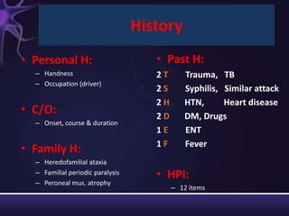
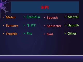
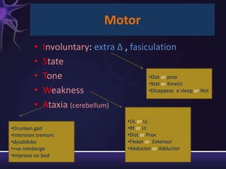
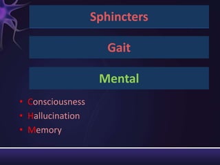
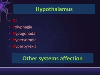
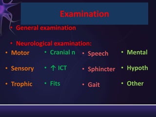
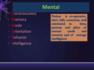
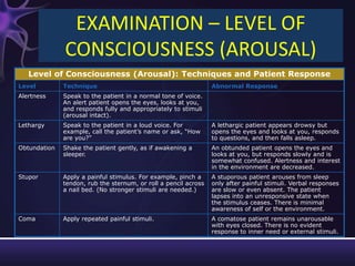
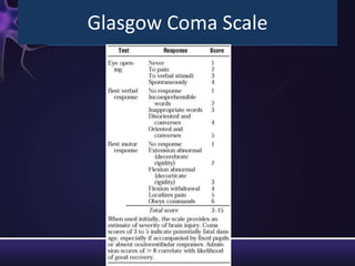
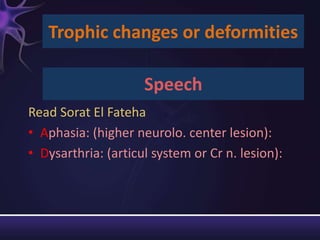
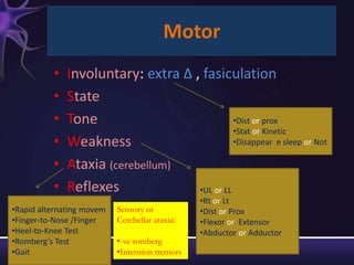
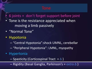
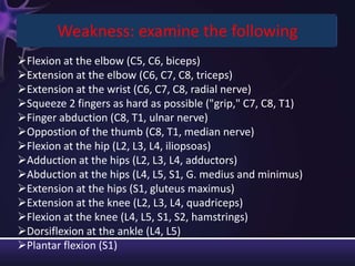
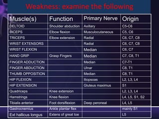
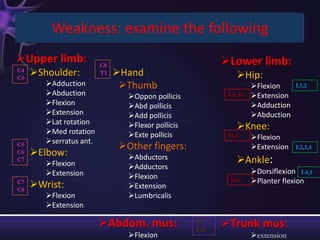
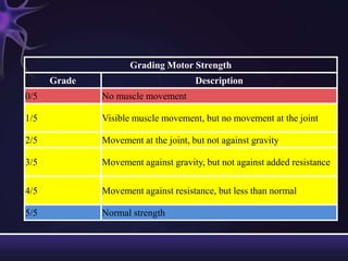
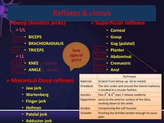
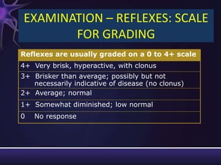
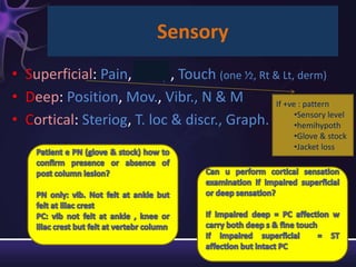
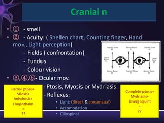
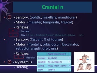
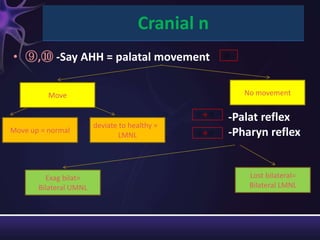
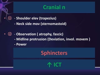
This neurological examination document provides details on:
1) The history, presenting complaints, family history, and review of systems to obtain for a neurological exam.
2) The components of the neurological exam including motor function, sensory function, cranial nerves, reflexes, and mental status.
3) Descriptions of how to examine each system, what abnormalities to look for, and what they may indicate. Rating scales are provided for things like strength, reflexes, and level of consciousness.
4) Common gait patterns and their potential causes are listed at the end.
Upper motor neurons originate in the brain and convey signals for voluntary movement through descending pathways to lower motor neurons. Lower motor neurons originate in the spinal cord and brainstem and directly innervate skeletal muscles. An upper motor neuron lesion causes spastic weakness without atrophy while a lower motor neuron lesion causes flaccid paralysis with atrophy.
The Babinski sign is an abnormal extensor plantar reflex elicited by stroking the sole of the foot. It indicates damage to the upper motor neurons. Joseph Babinski discovered this sign in 1896. A positive Babinski sign (big toe extending upward with fanning of other toes) suggests lesions in the corticospinal tract above the spinal cord. It is seen in conditions like stroke, spinal cord injury, etc. and in infants under 1 year of age. There are different types of Babinski responses based on the characteristics.
This document outlines the components of a general neurological assessment, including subjective and objective assessments. The subjective assessment involves taking a neurological history and headache/present complaint. The objective assessment examines various neurological functions like speech, mental status, sensory function, motor function, coordination, gait, and functional status. Key parts of the neurological exam include tests of consciousness, cranial nerves, reflexes, muscle tone/strength, and higher cognitive functions. The assessment aims to identify impairments, activities, and participation based on the ICF model of functioning.
This document provides an overview of how to examine a patient's sensory system. It discusses the anatomy of sensation, primary and combined sensations to be tested, and how to properly examine touch, pain, temperature, and proprioceptive senses. The examination should determine if there is a sensory loss, identify the level and pattern of loss. Tests include light touch with cotton, pinprick for pain, temperature with warm and cool water, and joint position sense. Care must be taken to avoid tiring the patient or leading responses.
This document provides information on low back pain, including its definition, prevalence, costs, causes, examination, diagnosis, and treatment options. Some key points:
- Low back pain is very common, affecting 60-80% of adults at some point. It costs the US over $90 billion annually in direct medical expenses and lost work.
- Causes can be non-spinal (e.g. hernia, infection) or spinal (e.g. arthritis, herniated disc, stenosis).
- Examination involves assessing gait, range of motion, motor strength, sensation, and reflexes. Common diagnostic tests are x-rays, MRI, CT.
- Treatment depends on cause but
The document discusses the sensory system and how it processes and transmits sensory information from peripheral receptors to the sensory cortex. It describes how different sensory modalities like pain, temperature, vibration and proprioception are carried by different nerve fiber types and pathways in the body. It provides details on testing various sensory modalities and how the patterns of sensory loss can help localize lesions to different parts of the central or peripheral nervous system.
Neck pain can have many causes, but is generally due to soft tissue injuries or abnormalities of the cervical spine structures. The cervical spine is comprised of 7 vertebrae, intervertebral discs, and supporting ligaments and muscles. Common causes of neck pain include muscle strains, disc injuries such as herniations or protrusions, arthritis, and fractures or dislocations. Symptoms vary depending on the underlying cause but often include neck pain and tenderness as well as reduced range of motion. Treatment involves physical therapy, medications, and sometimes surgery or injections depending on the severity and nature of the condition causing neck pain.
This document discusses the vestibular system and causes of vertigo. It describes the three main stabilizing sensory systems - vestibular, visual, and somatosensory. Vertigo can be caused by physiological or pathological issues in any of these systems. Common tests to evaluate vertigo include Dix-Hallpike maneuver, caloric testing, electronystagmography, and rotational chair testing. Positional maneuvers like Epley maneuver, Semont maneuver, and canalith repositioning therapy can be used to treat benign paroxysmal positional vertigo.
Cauda equina syndrome is a surgical emergency that occurs when the spinal canal is significantly narrowed, compressing the spinal cord and nerves below. It causes a variety of symptoms like leg and bladder problems. Early diagnosis and treatment are crucial, as waiting over 24 hours or symptoms worsening requires immediate surgery to decompress the spine. Prognosis depends on the severity and extent of symptoms, with bilateral leg pain or complete groin numbness indicating a poorer prognosis.
Transverse myelitis is a rare neurological condition where the spinal cord becomes inflamed across its width. It is often caused by an autoimmune response following a viral infection. Symptoms depend on the level of spinal cord involvement and may include sensory changes, motor weakness, and sphincter disturbances. Diagnosis involves ruling out other causes and showing signs of spinal cord inflammation. The goals of physiotherapy are to improve strength, mobility, and independence through exercises and management of issues like spasticity and skin care.
Part 1: Neurological history and physicaltschmitt2002
The document discusses assessment of the neurological system in adults. It outlines important information to gather prior to assessment, such as the location and effect of any lesions. The assessment examines level of consciousness, cranial nerves, motor function, reflexes, and more. Tests include the Glascow Coma Scale and evaluating responses of the central nervous system, peripheral nervous system, and specific areas like the cerebral cortex. The goal is to identify any abnormal signs or symptoms and select proper interventions.
This document outlines the components of a neurological history. It discusses establishing the main complaint, performing a detailed symptom analysis and chronological history, and conducting a systematic neurological examination. Specific symptoms related to areas like headaches, seizures, motor and sensory function are described. Taking a full history of the patient's medical, social, and family histories is also emphasized to understand the illness from their perspective. The goal is to understand the biomedical and contextual aspects of the neurological problem.
Meralgia Paresthetica (MP) is a condition caused by impingement of the lateral femoral cutaneous nerve, causing numbness and pain along the front of the thigh. It is often caused by entrapment of the nerve under the inguinal ligament. Diagnosis involves history, physical exam including the pelvic compression test, and may include imaging or nerve blocks. Treatment options include removing any underlying causes, medications, physical therapy, injections, or surgery.
Coma is a common medical emergency that requires careful management to prevent further brain damage. Emergency treatment focuses on maintaining oxygenation, circulation, controlling seizures, reducing intracranial pressure, and maintaining normal body temperature. Ongoing care involves monitoring vital signs, providing nutrition, preventing infection, and stimulating the patient as appropriate. The cause of coma must be determined through examination, investigations, and monitoring the patient's response to treatment.
This document provides information about examining reflexes during a neurological examination. It discusses the purpose of examining reflexes and describes techniques for testing several upper and lower limb tendon reflexes as well as other reflexes. Abnormalities of deep tendon reflexes like pendular movements and hung-up reflexes are explained. A grading scale for reflexes from 0 to 4 is provided. The document also briefly summarizes neonatal reflexes and references several sources for more information.
This document discusses cervical rib, including its structure, symptoms, diagnosis, treatment options, and physiotherapy management. Cervical rib is an extra rib that grows from the base of the neck above the collarbone. It can compress nearby blood vessels and nerves, causing pain, numbness, and reduced circulation in the arm. Diagnosis involves imaging tests and physical exams like Adson's test. Treatment may include anti-inflammatory drugs, surgery to remove the rib, or physiotherapy focused on exercises to improve strength and mobility.
This document discusses osteoarthritis of the knee (OA). It defines OA as a degenerative joint disease characterized by destruction of cartilage and bone growth at joint surfaces. It can be primary, from general wear and tear, or secondary, due to injury, infection or other predisposing causes. Risk factors include obesity, previous knee injuries, and high impact activities. Symptoms include pain, swelling, stiffness and decreased mobility. Diagnosis involves x-rays, MRI or CT scan to detect cartilage loss, bone spurs and other changes. Treatment aims to reduce pain and disability through patient education, exercises, modalities like cryotherapy and ultrasound, and rehabilitation.
Scoliosis is an abnormal curvature of the spine that occurs most often during periods of rapid growth before puberty. Mild cases of scoliosis may require only monitoring, but more severe curves can impair lung function. The Cobb angle measurement is used to assess the degree of spinal curvature, with treatment such as bracing or surgery considered for curves over 20-40 degrees.
This document provides an overview of the 12 cranial nerves, including their functions, methods of testing, and common causes of lesions or damage. It describes the cranial nerves as carrying sensory or motor functions, or both, and innervating specific muscles or regions of the head and neck. For each nerve, it outlines the key functions and sensory regions, how examination is performed, and common pathological conditions that may impact the nerve.
This document provides information about motor neuron disease (MND), including its types, clinical features, pathology, diagnosis, and treatment. It discusses four main types of MND: amyotrophic lateral sclerosis, progressive muscular atrophy, progressive bulbar palsy, and primary lateral sclerosis. The pathology of MND involves degeneration of motor neurons in the brain and spinal cord. Diagnosis is based on involvement of upper and lower motor neurons and ruling out other conditions. Treatment focuses on exercises, positioning, bracing, and psychological support to maintain function and prevent complications as the disease progresses.
The document discusses cerebellar ataxia, a disorder caused by damage to the cerebellum that controls coordination. It causes loss of coordination, balance problems, and slurred speech. The cerebellum coordinates muscle movement and is located in the hindbrain. Causes include viruses, alcohol, tumors, and toxins. Symptoms are diagnosed through neurological exams, imaging scans, and lab tests. Treatment focuses on physical therapy, assistive devices, and treating any underlying causes to ease symptoms and improve quality of life. Prevention involves vaccinations to reduce risk of viral infections that can lead to cerebellar ataxia.
This document provides an overview of the 12 cranial nerves, including their functions, methods of testing, and common causes of lesions. It discusses each cranial nerve individually, describing the purpose of testing, functional components, and how to examine sensory and motor functions. The cranial nerves control important functions like smell, vision, eye movement, facial expression, hearing, taste, swallowing and neck movement. Testing of the cranial nerves provides insight into neurological impairments localized to specific brain regions or cranial nerves.
This document discusses various superficial reflexes and how to elicit them, including:
1. Plantar reflexes can be elicited by stroking the sole of the foot and will result in either flexion or extension responses depending on neurological conditions.
2. Other reflexes discussed include corneal, conjunctival, pharyngeal, scapular, abdominal, cremasteric, bulbocavernosus, and anal reflexes.
3. Babinski's sign, which is an extensor plantar response, can occur in conditions that involve upper motor neuron lesions or alterations in consciousness.
This document provides guidance on performing a neurological examination, outlining the assessment of each cranial nerve and other relevant systems. The examination begins with inspection of general appearance and higher mental functions, followed by testing of the 12 cranial nerves, with details provided on assessing the visual system, facial muscles, hearing, swallowing and other functions. The motor and sensory systems are also evaluated, along with coordination and gait.
Frozen shoulder, also known as adhesive capsulitis, is a condition characterized by pain and stiffness in the shoulder joint that limits range of motion. It involves thickening and scarring of the shoulder joint capsule. Treatment involves conservative measures like oral anti-inflammatory drugs, corticosteroid injections into the joint, physical therapy including heat therapy and gentle range of motion exercises, and manipulation under anesthesia for refractory cases. Physical therapy aims to reduce pain and inflammation in the early stage and increase mobility in the stiffening stage through heat, passive range of motion, and home exercises.
Syringomyelia is a condition where a cyst, called a syrinx, develops in the spinal cord. It most commonly affects the lower cervical spine. It is often associated with abnormalities of the skull or spinal column. The majority of cases are linked to Chiari malformation type 1, where the cerebellar tonsils are displaced into the spinal canal. Symptoms vary depending on the location of the syrinx but can include pain, loss of sensation, muscle weakness or atrophy, and autonomic dysfunction. Diagnosis is made using imaging like MRI. Treatment involves surgery to decompress pressure on the spinal cord like laminectomy with the goal of resolving the syrinx.
Back pain is one of the most common health problems and is usually due to mechanical causes like muscle strains or disc issues. While the cause is often unknown, signs and symptoms like radiating leg pain help identify patients who may have a herniated disc compressing the nerve root. MRI is the best way to visualize disc damage, and treatment ranges from conservative options to surgery for severe or progressive cases. Most acute back pain resolves on its own, but recurrence after initial episodes is common.
The document provides guidance on performing a neurological examination. It outlines assessing the cranial nerves, reflexes, motor system, and sensory system. The cranial nerve examination involves testing each nerve individually. Reflex testing grades reflexes on a scale from 0 to 4. The motor exam evaluates muscle strength on a scale from 0 to 5. Finally, the sensory exam tests sensations like pain, touch, and position sense. The goal is to identify which parts of the neurological system may be affected.
The document discusses the neurological examination process. It describes the various components that are assessed which include levels of consciousness, mental status, cranial nerve function, motor skills, sensation, cerebellar function, and reflexes. Nurses play an important role in conducting and documenting the neurological examination to evaluate the presence of any disease in the nervous system.
The document provides information on performing a neurologic examination, including:
1) Obtaining a health history to understand the onset and progression of symptoms.
2) Assessing mental status, cranial nerves, motor function, sensory function, and reflexes.
3) Common clinical manifestations of neurologic diseases include pain, seizures, dizziness, visual disturbances, weakness, and abnormal sensation.
The document provides guidance on performing a neurological examination to systematically evaluate patients for neurological abnormalities. It discusses evaluating the patient's conscious state, cognition, cranial nerves, motor system, sensory system, and extrapyramidal signs. The examination aims to detect any neurological abnormalities, localize them within the nervous system, and determine the specific lesion.
This document provides an overview of the pediatric examination process. It discusses examining various body systems including:
1. Mental status and neurological assessment including cranial nerves, motor and sensory systems, and reflexes.
2. Assessment of consciousness, behavior, intelligence, memory, and speech.
3. Evaluation of muscle tone, power, involuntary movements, and coordination.
4. Sensory testing including superficial sensations, deep sensations, and cortical sensations.
5. Assessment of various reflexes including superficial, deep, and visceral reflexes.
The summary outlines the key areas addressed in a comprehensive pediatric examination.
This document provides guidance on conducting a general clinical examination (GCE). It defines a GCE as the direct observation and examination of a patient using inspection, palpation, percussion and auscultation. The objectives and significance of a GCE are explained, which is to gather clinical signs, analyze them, make diagnoses or differential diagnoses, and guide further testing and treatment. Steps for systematically conducting a GCE are outlined, including preparing the patient, examining different body regions like the head, neck, hands, lymph nodes and feet.
The patient presented with no acute distress. A physical examination found the patient's pupils, mucous membranes, lungs, heart, abdomen, extremities, and cranial nerves were normal. No abnormalities were noted.
This document provides guidance on performing an adult general physical examination. It outlines the initial steps, which include greeting the patient, introducing yourself, confirming patient identity, explaining the exam, ensuring a proper environment, obtaining consent, hand washing, draping the patient, and considering pain. It then describes aspects to examine such as handshake, body build, face, clothing, complexion, sounds, odors, posture, gait, and movements. Specific examination techniques are also mentioned, including examining the pallor, edema, nails, cyanosis, icterus, lymph nodes, and skin. Causes and characteristics of various physical findings are listed.
Part 1 function of brain and history taking of a neurological patientAtul Saswat
This document provides an overview of neurological assessment and the function of the brain and nervous system. It discusses the classification of the nervous system and outlines the key functions of different parts of the brain like the frontal lobe, parietal lobe, temporal lobe, occipital lobe, brainstem, and cerebellum. It also summarizes the roles of structures like the basal ganglia, thalamus, limbic system, and reticular formation. The document describes how to take a history from a neurological patient, including gathering demographic data, clarifying symptoms, and doing a systemic inquiry. It provides examples of findings from the initial impression and neurological exam.
General History taking and physical examinatinaneez103
This document provides information on performing a general history and physical examination. It discusses collecting a health history, which includes data on a patient's wellness, family history, and sociocultural background. The objectives of a health history are to identify patterns of health/illness, risk factors, and available resources. Physical examination involves inspection, palpation, percussion, and auscultation of the entire body from head to toe. Proper preparation, patient positioning, and use of appropriate instruments and techniques are emphasized. The document outlines examination of major body systems and common abnormal findings.
This document summarizes the examination of the 12 cranial nerves. It describes the function and tests for each nerve. For the first cranial nerve (olfactory nerve), it explains how smell tests are performed using familiar odors. For the second cranial nerve (optic nerve), it outlines tests for visual acuity, visual fields, color vision, and pupillary reaction. It then discusses examination of eye movements and analysis of diplopia for the third, fourth, and sixth cranial nerves. The document provides a high-level overview of testing sensation and motor function for the remaining cranial nerves.
Cervical cancer is the second most common cancer in women worldwide. The document discusses opportunities and challenges for cervical cancer prevention including new HPV vaccines and screening assays. It provides an overview of HPV vaccines, countries that have introduced them, and challenges to introduction. Monitoring vaccine coverage and impact is also discussed.
Part 2 general physical and mental examinationAtul Saswat
The document provides an overview of general physical and mental examinations for neurologic patients. It describes examinations of the head, face, skin, back, and eyes and checks for conditions like hydrocephalus, Parkinson's disease, and myasthenia gravis. It also outlines mental status examinations including tests of consciousness, memory, the Mini-Mental State Examination, and the Glasgow Coma Scale. Finally, it lists tests of lobular brain functions for the frontal, parietal, temporal, and occipital lobes.
02 1 principles of history taking and physical examinationsatyam mahaseth
This document outlines principles for taking a patient's history and conducting a physical examination. It emphasizes establishing rapport, listening to the patient, and obtaining a chronological history of present illness and symptoms. The physical exam should be thorough but respectful of the patient. Key signs and symptoms should be methodically examined and findings presented in an organized manner. Developing these clinical skills requires extensive practice over time.
This document provides information on methods for examining central nervous system reflexes through superficial and deep tendon reflex tests. It describes the components and techniques for assessing several key reflexes, including:
- Superficial reflexes like the corneal, palatal, abdominal, plantar, and cremasteric reflexes.
- Deep tendon reflexes like the biceps, triceps, knee jerk/patellar, and ankle reflexes.
For each reflex, it outlines the involved spinal roots and nerves, normal responses, and potential causes for absence of the reflex. Performing a full reflex examination can help evaluate the integrity of the spinal cord and peripheral nervous system.
Part 4 examination of motor and sensory systemAtul Saswat
This document summarizes the examination of the motor and sensory systems. It describes how to examine muscle bulk, tone, power, and involuntary movements. It also outlines how to test various sensory modalities like pain, touch, temperature, proprioception, vibration, and cortical sensations. Key points examined include muscle wasting, tone (loss or increase), power grading, reflexes, coordination, dermatomes, and signs for proprioception. Assessment methods are provided for each test with normal and abnormal findings.
This document provides an overview of squamous cell carcinoma of the head and neck (SCCHN), including its anatomical sites, incidence and mortality rates, risk factors, staging guidelines, and treatment approaches. It discusses the roles of surgery, radiation therapy, chemotherapy, concurrent chemoradiation, and targeted therapies like cetuximab in managing localized and advanced SCCHN. Concurrent chemoradiation is now standard for improving local control and organ preservation compared to radiation alone. The addition of cetuximab to radiation was shown to improve locoregional control and overall survival.
This document provides an overview of conducting a head, eyes, ears, nose, and throat (HEENT) examination. It lists the learning objectives, components to examine, anatomical landmarks, examination procedures, and how to record findings. The key areas covered are inspecting and palpating the head, eyes, ears, nose, mouth, neck, and lymph nodes. Procedures are described for assessing structures like the thyroid, trachea, and temporal arteries. The goal is to identify abnormalities, landmarks, exam techniques, and document examination findings.
This document provides information on conducting a health examination, including definitions, indications, techniques, equipment, positioning, preparing the patient and environment, and assessing different body systems. A health examination involves systematically assessing the general physical and mental condition of the body through the senses of inspection, palpation, percussion, and auscultation. It is important to prepare the patient and environment, use the proper equipment and techniques, and document examination findings.
This document provides details on performing a neurological examination, including:
1. Examining various parts of the patient's history, mental status, cranial nerves, motor system, sensory system, and gait.
2. Descriptions of how to assess level of consciousness, speech, reflexes, muscle strength, and sensory modalities.
3. Guidelines for evaluating abnormalities in tone, weakness, reflexes, and patterns of gait.
1. The document discusses various levels of impaired consciousness ranging from confusion to coma and defines them. It also describes the anatomy of the reticular activating system and two patterns of coma - diffuse cerebral injury or focal brainstem injury.
2. Specific causes of diffuse cerebral injury or focal brainstem injury that can lead to coma are provided. Evaluation of a comatose patient involves assessing level of consciousness, brainstem reflexes, and determining if focal neurological signs are present.
3. Management may involve giving thiamine, dextrose, naloxone and flumazenil in cases of diffuse injury without a known cause. For focal injuries, neuroimaging is important to identify structural
Dr. Hossam Ala'a provides a detailed guide for taking a history from a patient presenting with ischemia. The history includes gathering information on the patient's personal history, current complaint, history of present illness, past medical history, family history, and review of symptoms in other body systems. The physical examination involves inspecting and palpating the affected limb to evaluate changes in skin, muscles, and temperature that may indicate ischemia.
This document provides an overview of peripheral neuropathy (PN), including:
- PN most commonly presents as a length-dependent, symmetric sensorimotor polyneuropathy affecting the distal portions of limbs more than proximal.
- The clinical exam evaluates superficial sensation, deep sensation, motor function, and autonomic involvement. Sensory testing assesses patterns, distributions, and cortical sensation when possible.
- Common causes of PN include diabetes, paraproteinemias, alcoholism, renal failure, vitamin deficiencies, and some infectious diseases. A thorough history helps determine the temporal pattern and potential etiologies.
This document provides an overview of the neurological examination. It discusses examining various cranial nerves including visual acuity, eye movements, facial sensation, hearing, balance, and tongue and facial muscle function. It also covers assessing strength, tone, reflexes, sensory function, coordination, gait, and higher cognitive abilities. The exam evaluates multiple neurological domains to localize potential lesions in the brain or peripheral nervous system.
This document contains guidelines for performing a neurological examination. It begins with an overview of the developmental, psychosocial and cultural factors to consider. It then provides details on assessing various aspects of the neurological exam including mental status, cranial nerves, motor function, sensation and reflexes. Questions are provided to gather relevant history. The document comprehensively covers the steps and
The document provides guidance on performing a thorough neurological history. It outlines the key components of the history including personal details, complaint, family history, past medical history, and present history with a focus on specific questions to ask regarding motor function, sensation, cranial nerves, sphincters and more. As an example, it summarizes the history of a 37-year-old male patient who presented with acute onset right-sided weakness over 10 days, including pertinent details from his examination.
This document provides information about Baymax, who introduces himself as a personal healthcare provider. It then provides details on performing a physical exam, including preparing for the exam, establishing rapport with the patient, ensuring privacy and comfort, and explaining findings. Common symptoms that may warrant examination are listed. The physical exam components covered include vital signs, skin, head, eyes, ears, nose, mouth, neck, lungs, heart, abdomen, back, extremities, neurologic exam and mental status exam.
The document discusses the definition, classification, features, and pathophysiology of dystonia. It is classified based on age of onset, distribution, and etiology. Primary dystonias have no known underlying brain lesion and can be hereditary or idiopathic in nature, while secondary dystonias have an identifiable cause such as drugs, toxins, or other neurological conditions. The pathophysiology of primary dystonias involves subtle changes in neuronal signaling and communication in basal ganglia circuits that lead to abnormal patterns of muscle contraction.
This document discusses seizure disorders and epilepsy. It begins by differentiating between types of seizures, such as partial and generalized seizures. Diagnostic tests for seizures are outlined, along with the assessment and management of seizures. Various anti-seizure medications are discussed, along with goals of drug therapy and patient education. Nursing priorities for patients with seizures include maintaining a patent airway, providing oxygenation, obtaining vascular access, using seizure precautions, administering appropriate medications, and assessing therapeutic drug levels.
This document describes various tests used in neurological examinations to assess sensory and motor function. It discusses tests of vibration sense, joint position sense, tone, reflexes, and strength. Sensory tests include two-point discrimination, temperature, and pain sensation. Motor tests include assessment of strength, reflexes, and tone. Tests of the cranial nerves like corneal reflex and facial expression are mentioned. Cerebellar function is evaluated using finger-nose coordination, Romberg test, and heel-to-shin. Auditory and vestibular systems are examined using Rinne, Weber, and Schwabach tests as well as caloric testing and nystagmus observation. Interpretation of test results is provided to localize
This document summarizes the steps for examining a patient's motor and sensory systems. It outlines how to assess muscle tone, reflexes, strength, coordination, and sensation. The motor exam evaluates muscles for wasting, fasciculations, and abnormalities in tone. Reflexes like biceps, triceps, knee, and ankle jerks are tested. Strength is graded from 0-5. Coordination is tested using finger-nose, heel-shin, and rapid alternating movements. Sensation is assessed over dermatomes for pain, temperature, vibration and fine touch. The goal is to localize signs to upper or lower motor neuron lesions.
This document summarizes how to perform a motor and sensory examination. It describes how to test strength, tone, deep tendon reflexes, plantar responses, and involuntary movements. It also outlines how to evaluate primary sensory modalities like pain, temperature, vibration, and joint position. Higher cortical sensory functions and gait are also discussed. The document provides details on grading scales, distributions of findings, and classical abnormal patterns.
1. The neurological examination document outlines the process and components of examining a patient's nervous system, including terminology, indications, and aspects of the exam such as level of consciousness, cranial nerve function, motor function, and reflexes.
2. Nurses play an important role in conducting and documenting the neurological exam. This includes setting up equipment, assessing vital signs, performing tests of mental status, cranial nerves, motor skills, sensation, and reflexes, and communicating findings to doctors.
3. The goal of the neurological exam is to determine if there is any disease or abnormality present in the nervous system by thoroughly assessing multiple domains of neurological function.
This document provides an overview of seizures and epilepsy, including:
1. It defines seizures and describes different seizure types such as partial seizures, absence seizures, tonic-clonic seizures, myoclonic seizures, and neonatal/infantile seizures.
2. Etiologies and classifications of epilepsy are discussed, including focal vs generalized and age-specific syndromes.
3. Details are given on symptoms, EEG findings, and treatment responses for different seizure types like partial motor/sensory seizures and complex partial seizures.
4. Causes of seizures including genetic, structural, metabolic and other factors are briefly outlined.
This document provides an overview of cerebral palsy (CP), including its definition, causes, types, symptoms, diagnosis, and treatment. It begins by explaining that CP is a non-progressive brain injury occurring early in development that causes lifelong movement problems. The major types of CP are then summarized as sp
This document outlines the key components of a neurological examination. It discusses organizing the exam into six subsets (mental status, cranial nerves, motor, coordination, sensory, gait) and using a screening exam to check the entire neuroaxis. It provides details on assessing each cranial nerve and different neurological functions like strength, tone, reflexes, and sensory modalities. It also describes examining the cerebellum and observing different aspects of gait. The goal is to understand neurological deficits and localize them to different areas of the nervous system.
This document outlines the components of a neurological examination, including the six subsets (mental status, cranial nerves, motor, coordination, sensory, and gait) and how to evaluate each one. It discusses screening the entire neuroaxis through these subsets and expanding the exam based on history or abnormal findings. Key aspects covered for each subset include mental status testing, cranial nerve assessment, strength and tone evaluation, reflex testing, sensory modalities, coordination exams, and gait observation. The goal is to localize any abnormalities and differentiate between upper and lower motor neuron signs.
This document outlines the components of a neurological examination, including the six subsets (mental status, cranial nerves, motor, coordination, sensory, and gait) and how to evaluate each one. It discusses screening the entire neuroaxis through these subsets and expanding the exam based on history or exam findings. Key points covered include evaluating mental status, cranial nerves I-XII, strength, tone, reflexes, involuntary movements, primary and cortical sensory functions, cerebellar testing, and gait observation. The goal is to localize any abnormalities and differentiate between upper and lower motor neuron findings.
This document summarizes the examination of the sensory system and neurogenic bladder. It describes evaluating superficial, deep, and cortical sensations. It also discusses speech disorders like dysarthria and different types. Language examination and aphasia are mentioned. Neurogenic bladder is examined, including lesions affecting the reflex arc at different levels causing atonic, motor atonic, or autonomic bladders. Lesions above the reflex arc can cause retention, incontinence, or automatic bladder function.
2. History
• Personal H: • Past H:
– Handness 2T Trauma, TB
– Occupation (driver)
2S Syphilis, Similar attack
2H HTN, Heart disease
• C/O: 2D DM, Drugs
– Onset, course & duration
1E ENT
1F Fever
• Family H:
– Heredofamilial ataxia
– Familial periodic paralysis • HPI:
– Peroneal mus. atrophy
– 12 items
3. HPI
• Motor • Cranial n • Speech • Mental
• Sensory • ↑ ICT • Sphincter • Hypoth
• Trophic • Fits • Gait • Other
4. Motor
• Involuntary: extra ∆ , fasiculation
• State
• Tone •Dist or prox
•Stat or Kinetic
• Weakness •Disappear e sleep or Not
• Ataxia (cerebellum)
•UL or LL
•Drunken gait •Rt or Lt
•Intension tremors •Dist or Prox
•dysdidoko •Flexor or Extensor
•+ve romberge •Abductor or Adductor
•Improve on bed
5. Sensory
• Superficial: Pain, Temp, Touch
• Deep: Position, Mov., Vibr. If +ve : pattern
•Sensory level
• Cortical: Steriog, T. loc., T. discr. •hemihypoth
•Glove & stock
•Jacket loss
Trophic changes or deformities
• Ulcers: (N.B. : painless)
6. • ①: Cranial n
• Anosmia
• : • , :
• Sensory •
• ②: • Tast ant ⅔
Dysph (phar)
• Acuity • N. regur (palat)
• Motor
• Field • N. tone (palat)
• Eey clos.
• Mouth clos. • Hoarsn (lary)
• ③,④,⑥:
• Diplopia • : • :
• Ptosis • Deaf • Shoulder elev
• Squint • Tinitus • neck side mov
• Vertigo
• ⑤: • :
• Sensory • Tounge mov
• Pain,Temp
• Motor
• Masticat.
7. ↑ ICT
• Papilledema
• Headache
• Vomiting
Fits
• Aura
• Post effect
• Cons. Loss
• Gener. Or local
• March
8. Speech
• Aphasia: (higher neurolo. center lesion):
– Receptive(sensory):
• Spoken(Auditory)(aud recogn area lesion)
• Written(Visual)(visual recogn area lesion)
– Expressive(motor):
• Spoken (broca’s area lesion)
• Written(Agraphia)(exner’s area lesion)
• Dysarthria: (articul system lesion):
– ∆: bilateral→ slurred (psudobulbar)
– Extra ∆ → slow monotonus
– Cerebellar → stacatto
– Cr n → slurred (true bulbar)
13. EXAMINATION – LEVEL OF
CONSCIOUSNESS (AROUSAL)
Level of Consciousness (Arousal): Techniques and Patient Response
Level Technique Abnormal Response
Alertness Speak to the patient in a normal tone of voice.
An alert patient opens the eyes, looks at you,
and responds fully and appropriately to stimuli
(arousal intact).
Lethargy Speak to the patient in a loud voice. For A lethargic patient appears drowsy but
example, call the patient’s name or ask, “How opens the eyes and looks at you, responds
are you?” to questions, and then falls asleep.
Obtundation Shake the patient gently, as if awakening a An obtunded patient opens the eyes and
sleeper. looks at you, but responds slowly and is
somewhat confused. Alertness and interest
in the environment are decreased.
Stupor Apply a painful stimulus. For example, pinch a A stuporous patient arouses from sleep
tendon, rub the sternum, or roll a pencil across only after painful stimuli. Verbal responses
a nail bed. (No stronger stimuli are needed.) are slow or even absent. The patient
lapses into an unresponsive state when
the stimulus ceases. There is minimal
awareness of self or the environment.
Coma Apply repeated painful stimuli. A comatose patient remains unarousable
with eyes closed. There is no evident
response to inner need or external stimuli.
15. Trophic changes or deformities
Speech
Read Sorat El Fateha
• Aphasia: (higher neurolo. center lesion):
• Dysarthria: (articul system or Cr n. lesion):
16. Motor
• Involuntary: extra ∆ , fasiculation
• State
• Tone •Dist or prox
•Stat or Kinetic
• Weakness •Disappear e sleep or Not
• Ataxia (cerebellum)
• Reflexes •UL or LL
•Rt or Lt
•Rapid alternating movem
•Drunken gait Sensory or •Dist or Prox
•Finger-to-Nose /Finger
•Intension tremors Cerebellar ataxia: •Flexor or Extensor
•Heel-to-Knee
•dysdidoko Test •Abductor or Adductor
•Romberg’s Test
•+ve romberge •-ve romberg
•Gait
•Improve on bed •Intension tremors
17. Tone
• 6 joints + don’t forget support before joint
• Tone is the resistance appreciated when
moving a limb passively
• “Normal Tone”
• Hypotonia
– “Central Hypotonia”:shock UMNL, cerebellar
– “Peripheral Hypotonia”: LMNL, myopathy
• Hypertonia
– Spasticity (Corticospinal Tract = ∆ )
– Rigidity (Basal Ganglia, Parkinson’s = extra ∆ )
18. Weakness: examine the following
Flexion at the elbow (C5, C6, biceps)
Extension at the elbow (C6, C7, C8, triceps)
Extension at the wrist (C6, C7, C8, radial nerve)
Squeeze 2 fingers as hard as possible ("grip," C7, C8, T1)
Finger abduction (C8, T1, ulnar nerve)
Oppostion of the thumb (C8, T1, median nerve)
Flexion at the hip (L2, L3, L4, iliopsoas)
Adduction at the hips (L2, L3, L4, adductors)
Abduction at the hips (L4, L5, S1, G. medius and minimus)
Extension at the hips (S1, gluteus maximus)
Extension at the knee (L2, L3, L4, quadriceps)
Flexion at the knee (L4, L5, S1, S2, hamstrings)
Dorsiflexion at the ankle (L4, L5)
Plantar flexion (S1)
19. Weakness: examine the following
Muscle(s) Function Primary Nerve Origin
DELTOID Shoulder abduction Axillary C5-C6
BICEPS Elbow flexion Musculocutaneous C5, C6
TRICEPS Elbow extension Radial C6, C7, C8
WRIST EXTENSORS Radial C6, C7, C8
WRIST FLEXION Median C6, C7
HAND GRIP Grasp Fingers Median C7, C8, T1
FINGER ADDUCTION Median C7-T1
FINGER ABDUCTION Ulnar C8, T1
THUMB OPPOSITION Median C8, T1
HIP FLEXION Iliopsoas L2, L3, L4
HIP EXTENSION Gluteus maximus S1
Quadriceps Knee extension L2, L3, L4
Hamstrings Knee flexion L4, L5, S1, S2
Tibialis anterior Foot dorsiflexion Deep peroneal L4, L5
Gastrocnemius Ankle plantar flex mainly S1
Ext hallicus longus Extens of great toe L5
21. Grading Motor Strength
Grade Description
0/5 No muscle movement
1/5 Visible muscle movement, but no movement at the joint
2/5 Movement at the joint, but not against gravity
3/5 Movement against gravity, but not against added resistance
4/5 Movement against resistance, but less than normal
5/5 Normal strength
22. Reflexes & clonus
Deep (tendon jerks) Superficial reflexes
UL • Corneal
C5,6
• BICEPS • Grasp
• BRACHIORADIALIS • Gag (palatal)
C6,7 • TRICEPS S1,2 • Planter
Sure
LL signs of T6-12 • Abdominal
∆???? L1
L2,3,4 • KNEE + clonus • Cremastric
S1,2 • ANKLE + clonus S3,4,5 • Anal
Technique
Abnormal Deep reflexes Babiniski Scratsh From below up- lat to medial
Chaddock The skin under and around the lateral malleolus
• Jaw jerk is stroked in a circular fashion.
• Wartenberg Gonda’s
rd th
Flex 3 & 4 toes 7 release suddenly
Oppenheim press to the anterior surface of the tibia,
• Finger jerk stroking down to the ankle.
• Hofman Gordon Compressing the calf muscles
• Patelal jerk Schaefer Pinching the Achilles tendon enough to cause
pain.
• Adductor jerk
23. EXAMINATION – REFLEXES: SCALE
FOR GRADING
Reflexes are usually graded on a 0 to 4+ scale
4+ Very brisk, hyperactive, with clonus
3+ Brisker than average; possibly but not
necessarily indicative of disease (no clonus)
2+ Average; normal
1+ Somewhat diminished; low normal
0 No response
24. Sensory
• Superficial: Pain, Temp, Touch (one ⅟2, Rt & Lt, derm)
• Deep: Position, Mov., Vibr., N & M If +ve : pattern
•Sensory level
• Cortical: Steriog, T. loc & discr., Graph. •hemihypoth
•Glove & stock
•Jacket loss
26. Cranial n
• ⑤ - Sensory: (ophth., maxillary, mandibular)
- Motor: (massiter, temporalis, tregoid)
- Reflexes:
→
• Corneal
→ • Jaw : if +ve = bilateral ∆ lesion above pons (above nc.)
• - Sensory: (Tast ant ⅔ of tounge)
- Motor: (frontalis, orbic occul., buccinator,
retractor angulii, orbic oris)
- Reflexes: Rapid phase toward
→ • glabellar occular pendular H
• ⑧ - Nystagmus cerebel fix i.e. (lesion) H
vestib Away from (norm) H
- Hearing
stem vertical V
27. Cranial n
• ⑨,⑩ -Say AHH = palatal movement
Move No movement
→
-Palat reflex
deviate to healthy =
Move up = normal
LMNL → -Pharyn reflex
Exag bilat= Lost bilateral=
Bilateral UMNL Bilateral LMNL