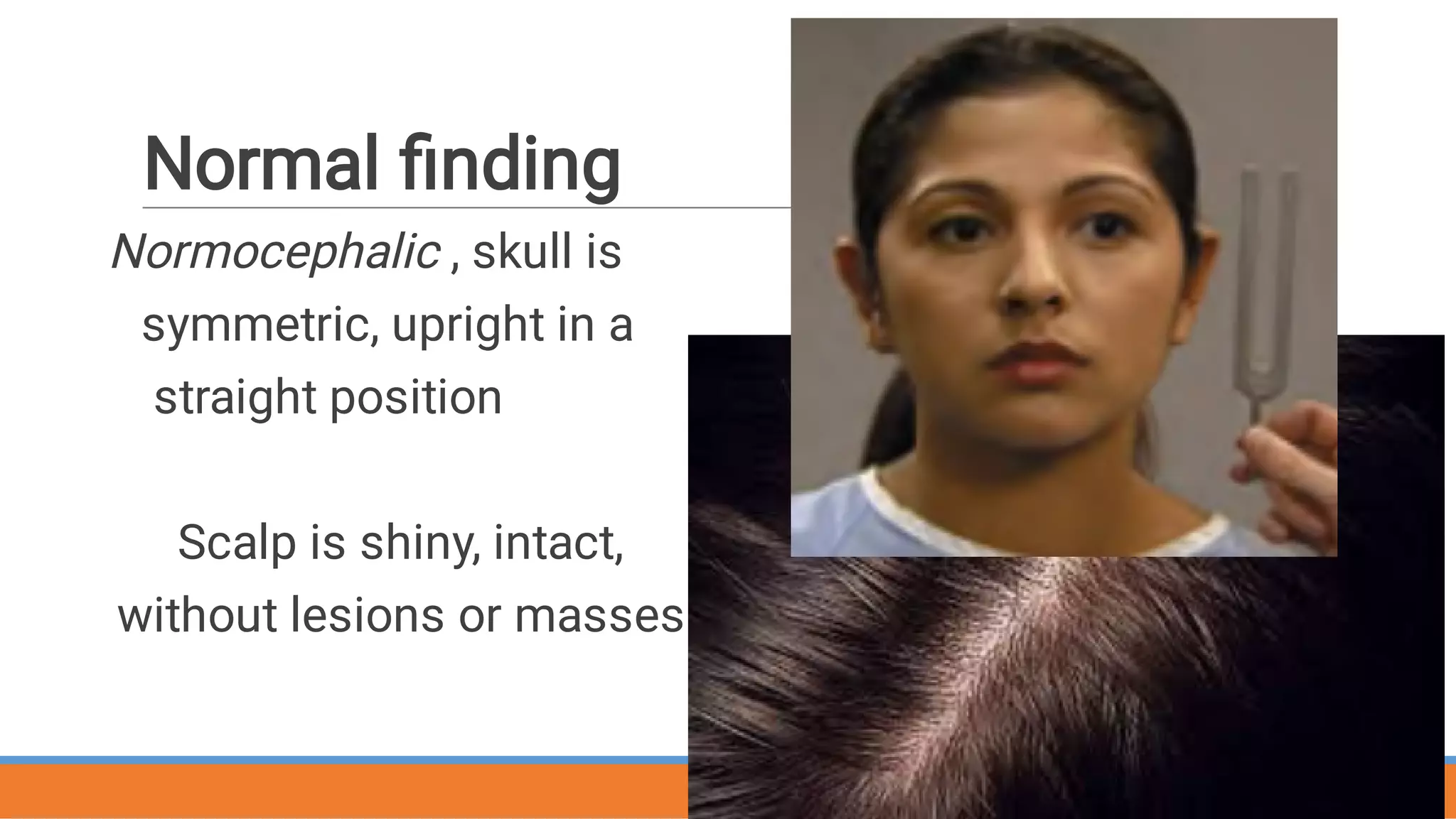
Head, mouth and
Neck Assessment
PREPARE BY. Rezq Mansour
Supervised By Dr
Supervised By Dr. Fawz Abu Algaith
Supervised By Dr
Supervised By Dr
Supervised By Dr. Fawz Abu Algaith
Supervised By Dr
Republic of Yemen
Ministry of Higher Education and
Scientific Research
Razi University
Master Program
Summary: Assessment of
Head, mouth & Neck
 Includes:
• Head
• Face
• Oral cavity
• Neck & lymph nodes
• Trachea
• Thyroid
Anatomy
THE HEAD
A skull is a rigid bony box that
protects the brain, sense organs
and upper spinal cord It includes :
The bones of the cranium
The bones of the face
The skull bone include
Six bones that form the skull and are
fused
together at sutures.
The skull is covered by scalp tissue,
which is
The skull face bones
Frontal bone
Parietal bone
Occipital bone
Temporal bone
Face bones :
consists of 14 bones that protect facial
structures, including :
The eyes, ears,nose, and mouth.
These structures are generally symmetric. Like
the skull.
•
•
These bones are immobile and are
fused at sutures, with the exception of
the mandible.
The mandible articulates with the
temporal bone of the skull at the
temporomandibular joint, allowing for
Facial muscles
The examination divided to
Subjective Data
post history
present history
Objective Data
Inspection
palpation
Head examination
Subjective Data-Head
Headache (HA)
  Injury
 Dizziness/Syncope
Hx. Head or Face Surgery
Objective Data-Head
Inspection and palpation head and face
size,
shape,
skin characteristics
Tenderness
Normal finding
Normocephalic , skull is
symmetric, upright in a
straight position
Scalp is shiny, intact,
without lesions or masses
Abnormal findings in Pediatric
Hydrocephalus
Down Syndrome
Craniosynostosis Depression
MASSES HEMATOMAS
LESIONS BELL’S PALSY
Cushing’s syndrome
Asymmetry
Vertical horizontal mixture
Mouth Anatomy
  • Hard and soft palates
•  Uvula
•  Tongue
Salivary glands
•  Parotid
•  Submandibular
•  Sublingual
Subjective Data- Oral cavity
• Sores/lesions
• Sore throat
• Bleeding gums
• Toothache
• Dysphagia
• Altered taste
• Tobacco
Objective Data- Oral Cavity Exam
• Inspect and palpate:
• Client opens and closes jaw- feel TMJ
(temporomandibular Joint)
• Note condition of teeth, gums and mucosa
• Parotid glands: if swelling
• Lips: (lumps, lesion, …etc)
• Open mouth, stick out tongue, move side to side
(tests CN XII)
• Say AAH …soft palate rises midline (tests CN X)
• With tongue blade inspect cheeks and underside lips
• With gloves and gauze- pull tongue L and R
Objective Data- Oral Cavity Exam
• Inspect uvula, palate, tonsils
• Uvula looks like hanging pendant if split in two-
bifid
• Palate: anterior hard palate, whitish while posterior
soft palate pinkish
• Tonsils- graded by enlargement:
• 1+ visible • 2+ near uvula
• 3+ touching uvula • 4+ touching together
Abnormalities
pharyngitis
Geographic Tongue
THE NECK EXAMMENATION







Structures of the neck include :
Upper portion of the spine,
Esophagus,
Trachea,
Thyroid gland,
Arteries,
Veins, and
Lymph nodes.


Neck anatomy
Structures of the neck
include :
Trachea,
Thyroid gland,
Arteries and Veins
Lymph nodes
- Preauricular
- Posterior
auricular
(mastoid)
- Occipital
- Submental
-Submandibular
Subjective Data-Neck
History of problems
 Neck pain  
Lumps
 Swelling
 Surgery
Neck Examination
Objective Data-Neck
Inspect- symmetry, midline, erect ,masses
and enlargement
 Note enlargement of lymph nodes
  Test for ROM, (naturally decreases with
age).
Palpate- carotid pulse, lymph nodes &
thyroid
Auscultate carotid arteries.
Range Of Motion
•Normal findings
–Full ROM (range of
motion), pain free,
symmetrical muscles, no
masses
•Abnormal findings
–Limited ROM, pain,
asymmetrical muscles,
masses Neck Mass
Trachea
Trachea normally centered;
(at the suprasternal notch)
Subjective Data- Trachea
 History of trachea problems?
Tracheostomy
 
Objective Data-Trachea
 Palpate trachea for :
Deviations
 Tenderness
Masses
The Mass push trachea away from affected
area, whereas atelectasis causes pull toward
affected area.
• The cartilages should be smooth, non
tender and move easily under examiner’s
fingers when the client swallow
• Palpation done by placing the thumb
and forefinger on each side of the
trachea
Thyroid Gland
Assessment by:
•Inspection
•Palpation
•Auscultation
Objective Data- Thyroid
Inspect: note deviations or bulges
 As person extends neck slightly and swallows
water, note upward, symmetrical movement of
trachea and other cartilage
Palpating Thyroid
 Ask person to tilt head slightly to side
and palpate lobes.
 Use fingers on opposite side to displace
gland in lateral direction, ask person to
swallow: isthmus rises
•
•
•
 Posterior approach
 Rest thumbs on back of
person’s neck,
Place fingers on cricoid
cartilage.
Displace gland & palpate
isthmus as person swallows,
thyroid rises
Palpating Thyroid
Palpating Thyroid
 Anterior approach
- Rest fingers around sternomastoid
muscle
- place thumbs on side of trachea.
- Displace gland & palpate isthmus
as person swallows, thyroid rises
 Normal thyroid finding as:
 
Thyroid palpated as symmetric,
without enlargement.
Symmetrical movement with
swallowing.
Texture consistent, with no masses,
nodules or tenderness.
Abnormal findings
Mass
Enlarged gland
Asymmetric
al
enlargement
Lymph nodes
Lymph nodes
- Preauricular
- Posterior
auricular
(mastoid)
- Occipital
- Submental
-Submandibular
•
•
•
Examination of lymphatic System :
Firstly inspection for
Enlarged lymph nodes,
Skin lesions
And edema
Examination of lymphatic System :
• Secondly palpating gently the lymph nodes areas using pads of "
2, 3, 4" fingers in gentle circular motion.
• Press lightly and then increasing pressure gradually.
• Move skin lightly over the under lying tissues & not moving the
examining fingers over the skin.
• Large nodes due to malignancy are generally not tender vary in
size, hard, asymmetrical
Lymph Nodes
•Normal findings
–Unable to palpate or see
nodes
•Abnormal findings
–Enlarged nodes
–Able to palpate or see
nodes
–Tenderness
–Firm, hard nodes
•Abnormal
–Enlarged
THANK YOU

Assessment head and neck(1).pdf