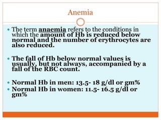
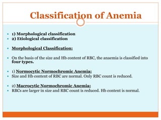
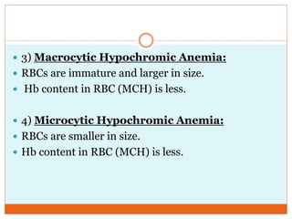
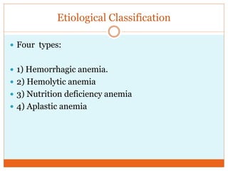
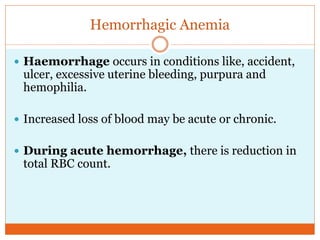
P R O F . D R S A B A K H A L I D
H O D P H Y S I O L O G Y
Anemias
Definition
 Anemia is defined as a reduction below the normal
limit in the concentration of hemoglobin or red
blood cells in the blood, for the age and sex of the
individual.
Anemia
 The term anaemia refers to the conditions in
which the amount of Hb is reduced below
normal and the number of erythrocytes are
also reduced.
 The fall of Hb below normal values is
usually, but not always, accompanied by a
fall of the RBC count.
 Normal Hb in men: 13.5- 18 g/dl or gm%
 Normal Hb in women: 11.5- 16.5 g/dl or
gm%
 A person is considered to be anaemic when his or her Hb level falls
bellow 13 g/dl for a man and 11.5 g/dl for a woman.
 Anaemia is called moderate when it falls below 9g/dL and
severe when it falls below 6g/dL.
 When fully oxygenated, each gram of Hg carries 1.39 ml of oxygen.
 Normal arterial blood with a Hb concentration of 15 g/dl carries
about 21 ml of oxygen, of which 4.5 ml are delivered to the tissues.
 In an anemic individual with a Hb concentration of only half-
normal, 7.5 g/dl, 100ml of blood can carry only about 10ml of
oxygen.
 Symptoms of Anemia:
 Anemia results in reduced oxygen carrying capacity of blood, resulting
in tissue hypoxia.
 The hypoxia causes symptoms like easy fatigability (due to muscle
hypoxia) and faintness (due to cerebral hypoxia) especially on exertion.
 Colour of skin, conjunctivae, pharyngeal mucous membrane, palm and
nail bed becomes pale.
 Headache, lack of concentration, restlessness, irritibility, muscles
becomes weak and the pt. gets fatigued quite often and quite easily.
 Anorexia, nausea, vomiting, abdominal discomfort and constipation.
 Skin becomes thin and dry, nails becomes
brittle and easily breakable.
 The hypoxia brings about several compensatory
responses.
 Breathlessness due to compensatory stimulation of
respiratory center and palpitation due to
compensatory increase in Cardiac output.
 Increase H.R and C.O. The velocity of blood flow
is increased.
 Signs include cardiac murmurs due to turbulence of
blood while passing through the cardiac valves-
another consequence of the raised C.O.
IRON TRANSPORT
Classification of Anemia
 1) Morphological classification
 2) Etiological classification
 Morphological Classification:
 On the basis of the size and Hb content of RBC, the anaemia is classified into
four types.
 1) Normocytic Normochromic Anemia:
 Size and Hb content of RBC are normal. Only RBC count is reduced.
 2) Macrocytic Normochromic Anemia:
 RBCs are larger in size and RBC count is reduced. Hb content is normal.
 3) Macrocytic Hypochromic Anemia:
 RBCs are immature and larger in size.
 Hb content in RBC (MCH) is less.
 4) Microcytic Hypochromic Anemia:
 RBCs are smaller in size.
 Hb content in RBC (MCH) is less.
Etiological Classification
 Four types:
 1) Hemorrhagic anemia.
 2) Hemolytic anemia
 3) Nutrition deficiency anemia
 4) Aplastic anemia
Hemorrhagic Anemia
 Haemorrhage occurs in conditions like, accident,
ulcer, excessive uterine bleeding, purpura and
hemophilia.
 Increased loss of blood may be acute or chronic.
 During acute hemorrhage, there is reduction in
total RBC count.
 Plasma portion of blood is replaced within 24 hours.
 Replacement of RBCs takes some time.
Hemodilution occurs with less no. of RBCs.
 This type of anaemia is called normocytic
normochromic anemia, as both the size and Hb
content of RBC are normal.
 This condition is corrected after 3-4 weeks, when
RBCs are produced due to the stimulation of bone
marrow.
 During chronic hemorrhage:
 Due to continuous loss of blood, a lot of iron is lost
from the body.
 Quantity of iron in diet is also limited.
 So the synthesis of Hb is affected.
 Cells do not contain adequate amount of Hb, so
this type of anaemia is known as hypochromic
and microcytic anemia.
Hemolytic Anemia
 Causes:
 1) Bacterial or chemical (industrial poisons) or by
malfunctioning of spleen, sickle cell anaemia,
congenital spherocytosis, thalassaemia.
 2) Presence of isoagglutinins like anti-Rh
agglutinins, when the recipient is Rh-
negative and the donor is Rh-positive.
 3) On the first blood transfusion nothing will happen
because plasma of recipient has no Anti-Rh agglutinins.
 If after sometime the transfusion is repeated between
the same donor and recipient, severe intravascular
agglutination will occur with consequent haemolysis
which may result in death.
 4) When the shape of RBCs is abnormal, the cells
becomes more fragile and hemolysis occurs easily.
Nutrition Deficiency Anemia
 Iron deficiency anaemia:
 RBCs are smaller in size and contain less Hb than
normal.
 MCV, MCH and MCHC% are all decreased.
 Present in bottle fed babies or in women with poor
diet and excessive menstrual bleeding and in
chronic blood loss due to internal bleeding.
IRON DEFICIENCY ANEMIA –
PERIPHEPRAL BLOOD PICTURE
Pernicious Anemia
 Is caused by an inability to form erythrocyte stroma
(structure), due to lack of absorption of vitamin B12
or destruction of gastric mucosa.
 This results in diminution on the number of RBCs.
 RBCs are large in size i.e; MCV is increased, but MCH is
normal (synthesis of Hb is normal) and MCHC% is also
about normal.
 The cells are macrocytic and normochromic.
Megaloblastic anemia
 Causes:
 Loss of any of these factors such as vit.B12, folic acid and intrinsic factor
from the stomach mucosa (parietal cell) can lead to slow reproduction of
erythroblasts in the bone marrow.
 Folic acid deficiency is usually dietary in origin and occurs in pregnancy.
 Loss of entire stomach (total gastrectomy).
 Intestinal sprue: folic acid, vit.B12 and other vit.B compounds are poorly
absorbed.
 As a result, these grow too large, with odd shapes and fragile membranes and
are called megaloblasts. These cells rupture easily and the cells are normocytic
and normochromic.
Aplastic anemia
 Bone marrow aplasia means lack of a functioning bone
marrow.
 Causes:
 Nuclear bomb blast
 Excessive X-ray treatment
 Certain industrial chemicals.
 Red bone marrow is reduced and replaced by fatty tissues.
 The cells are normocytic and normochromic.
Effects of Anemia on Function of the
Circulatory System
 In severe anemia viscosity may fall to as low as 1.5
times that of water (normal value:3).
 Increase blood flow through peripheral blood vessels
to tissues, increase venous return and increase
cardiac output.
 C.O increases 3 to 4 times than normal.
 Major effects of anemia is greatly increased C.O and
increased pumping workload on the heart (hypoxia results
in vasodilatation and increase in V.R and C.O).
 During exercise, the heart is not capable of pumping much
greater quantities of blood then it is already pumping.
 During exercise, tissue demand for oxygen increases.
Extreme tissue hypoxia results, and acute cardiac failure
results.

Anaemias

  • 1.
    P R OF . D R S A B A K H A L I D H O D P H Y S I O L O G Y Anemias
  • 2.
    Definition  Anemia isdefined as a reduction below the normal limit in the concentration of hemoglobin or red blood cells in the blood, for the age and sex of the individual.
  • 3.
    Anemia  The termanaemia refers to the conditions in which the amount of Hb is reduced below normal and the number of erythrocytes are also reduced.  The fall of Hb below normal values is usually, but not always, accompanied by a fall of the RBC count.  Normal Hb in men: 13.5- 18 g/dl or gm%  Normal Hb in women: 11.5- 16.5 g/dl or gm%
  • 4.
     A personis considered to be anaemic when his or her Hb level falls bellow 13 g/dl for a man and 11.5 g/dl for a woman.  Anaemia is called moderate when it falls below 9g/dL and severe when it falls below 6g/dL.  When fully oxygenated, each gram of Hg carries 1.39 ml of oxygen.  Normal arterial blood with a Hb concentration of 15 g/dl carries about 21 ml of oxygen, of which 4.5 ml are delivered to the tissues.  In an anemic individual with a Hb concentration of only half- normal, 7.5 g/dl, 100ml of blood can carry only about 10ml of oxygen.
  • 5.
     Symptoms ofAnemia:  Anemia results in reduced oxygen carrying capacity of blood, resulting in tissue hypoxia.  The hypoxia causes symptoms like easy fatigability (due to muscle hypoxia) and faintness (due to cerebral hypoxia) especially on exertion.  Colour of skin, conjunctivae, pharyngeal mucous membrane, palm and nail bed becomes pale.  Headache, lack of concentration, restlessness, irritibility, muscles becomes weak and the pt. gets fatigued quite often and quite easily.  Anorexia, nausea, vomiting, abdominal discomfort and constipation.
  • 6.
     Skin becomesthin and dry, nails becomes brittle and easily breakable.  The hypoxia brings about several compensatory responses.  Breathlessness due to compensatory stimulation of respiratory center and palpitation due to compensatory increase in Cardiac output.
  • 7.
     Increase H.Rand C.O. The velocity of blood flow is increased.  Signs include cardiac murmurs due to turbulence of blood while passing through the cardiac valves- another consequence of the raised C.O.
  • 8.
  • 9.
    Classification of Anemia 1) Morphological classification  2) Etiological classification  Morphological Classification:  On the basis of the size and Hb content of RBC, the anaemia is classified into four types.  1) Normocytic Normochromic Anemia:  Size and Hb content of RBC are normal. Only RBC count is reduced.  2) Macrocytic Normochromic Anemia:  RBCs are larger in size and RBC count is reduced. Hb content is normal.
  • 10.
     3) MacrocyticHypochromic Anemia:  RBCs are immature and larger in size.  Hb content in RBC (MCH) is less.  4) Microcytic Hypochromic Anemia:  RBCs are smaller in size.  Hb content in RBC (MCH) is less.
  • 11.
    Etiological Classification  Fourtypes:  1) Hemorrhagic anemia.  2) Hemolytic anemia  3) Nutrition deficiency anemia  4) Aplastic anemia
  • 12.
    Hemorrhagic Anemia  Haemorrhageoccurs in conditions like, accident, ulcer, excessive uterine bleeding, purpura and hemophilia.  Increased loss of blood may be acute or chronic.  During acute hemorrhage, there is reduction in total RBC count.
  • 13.
     Plasma portionof blood is replaced within 24 hours.  Replacement of RBCs takes some time. Hemodilution occurs with less no. of RBCs.
  • 14.
     This typeof anaemia is called normocytic normochromic anemia, as both the size and Hb content of RBC are normal.  This condition is corrected after 3-4 weeks, when RBCs are produced due to the stimulation of bone marrow.
  • 15.
     During chronichemorrhage:  Due to continuous loss of blood, a lot of iron is lost from the body.  Quantity of iron in diet is also limited.  So the synthesis of Hb is affected.  Cells do not contain adequate amount of Hb, so this type of anaemia is known as hypochromic and microcytic anemia.
  • 16.
    Hemolytic Anemia  Causes: 1) Bacterial or chemical (industrial poisons) or by malfunctioning of spleen, sickle cell anaemia, congenital spherocytosis, thalassaemia.  2) Presence of isoagglutinins like anti-Rh agglutinins, when the recipient is Rh- negative and the donor is Rh-positive.
  • 17.
     3) Onthe first blood transfusion nothing will happen because plasma of recipient has no Anti-Rh agglutinins.  If after sometime the transfusion is repeated between the same donor and recipient, severe intravascular agglutination will occur with consequent haemolysis which may result in death.  4) When the shape of RBCs is abnormal, the cells becomes more fragile and hemolysis occurs easily.
  • 18.
    Nutrition Deficiency Anemia Iron deficiency anaemia:  RBCs are smaller in size and contain less Hb than normal.  MCV, MCH and MCHC% are all decreased.  Present in bottle fed babies or in women with poor diet and excessive menstrual bleeding and in chronic blood loss due to internal bleeding.
  • 19.
    IRON DEFICIENCY ANEMIA– PERIPHEPRAL BLOOD PICTURE
  • 20.
    Pernicious Anemia  Iscaused by an inability to form erythrocyte stroma (structure), due to lack of absorption of vitamin B12 or destruction of gastric mucosa.  This results in diminution on the number of RBCs.  RBCs are large in size i.e; MCV is increased, but MCH is normal (synthesis of Hb is normal) and MCHC% is also about normal.  The cells are macrocytic and normochromic.
  • 21.
    Megaloblastic anemia  Causes: Loss of any of these factors such as vit.B12, folic acid and intrinsic factor from the stomach mucosa (parietal cell) can lead to slow reproduction of erythroblasts in the bone marrow.  Folic acid deficiency is usually dietary in origin and occurs in pregnancy.  Loss of entire stomach (total gastrectomy).  Intestinal sprue: folic acid, vit.B12 and other vit.B compounds are poorly absorbed.  As a result, these grow too large, with odd shapes and fragile membranes and are called megaloblasts. These cells rupture easily and the cells are normocytic and normochromic.
  • 23.
    Aplastic anemia  Bonemarrow aplasia means lack of a functioning bone marrow.  Causes:  Nuclear bomb blast  Excessive X-ray treatment  Certain industrial chemicals.  Red bone marrow is reduced and replaced by fatty tissues.  The cells are normocytic and normochromic.
  • 24.
    Effects of Anemiaon Function of the Circulatory System  In severe anemia viscosity may fall to as low as 1.5 times that of water (normal value:3).  Increase blood flow through peripheral blood vessels to tissues, increase venous return and increase cardiac output.  C.O increases 3 to 4 times than normal.
  • 25.
     Major effectsof anemia is greatly increased C.O and increased pumping workload on the heart (hypoxia results in vasodilatation and increase in V.R and C.O).  During exercise, the heart is not capable of pumping much greater quantities of blood then it is already pumping.  During exercise, tissue demand for oxygen increases. Extreme tissue hypoxia results, and acute cardiac failure results.