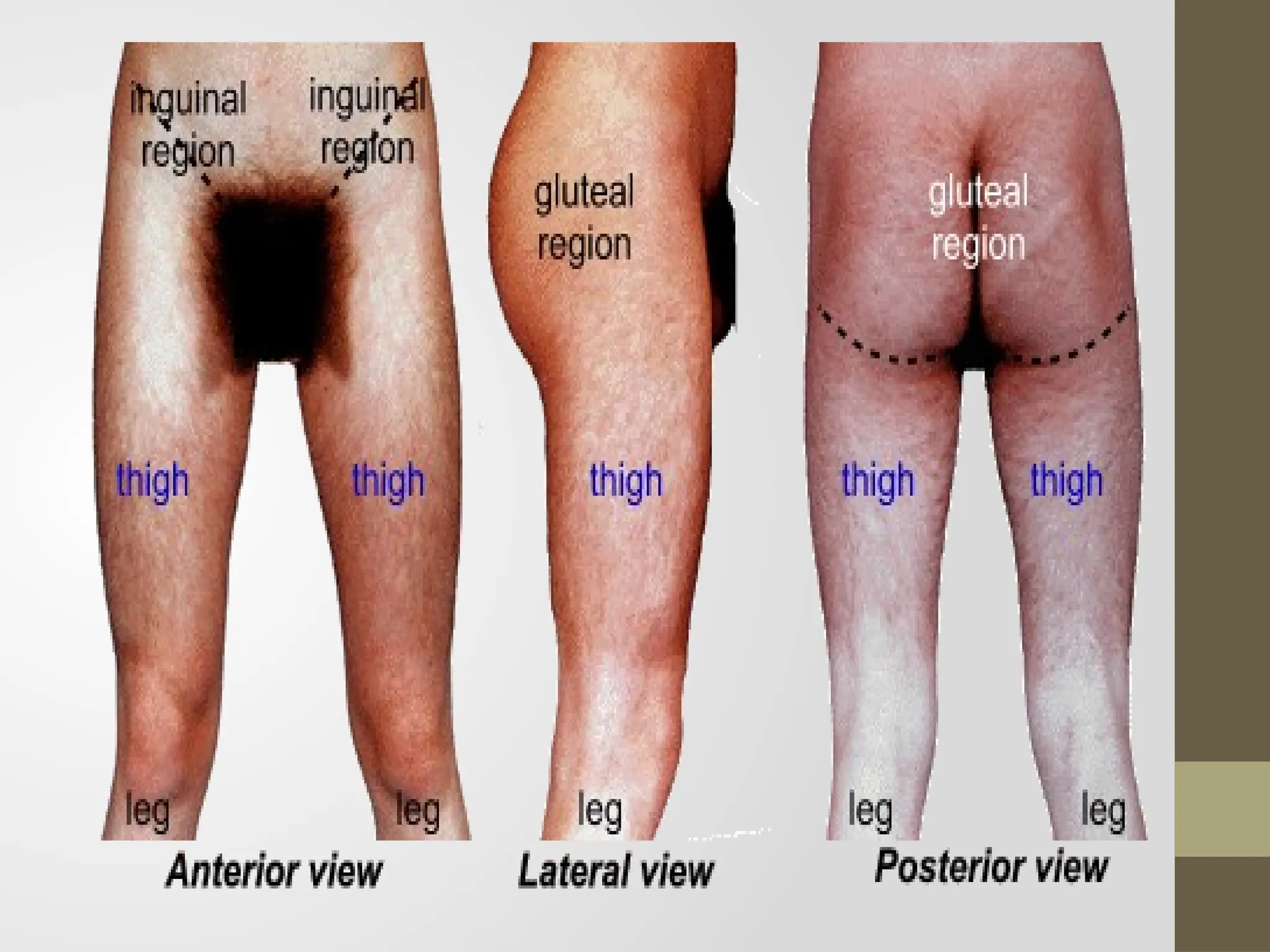
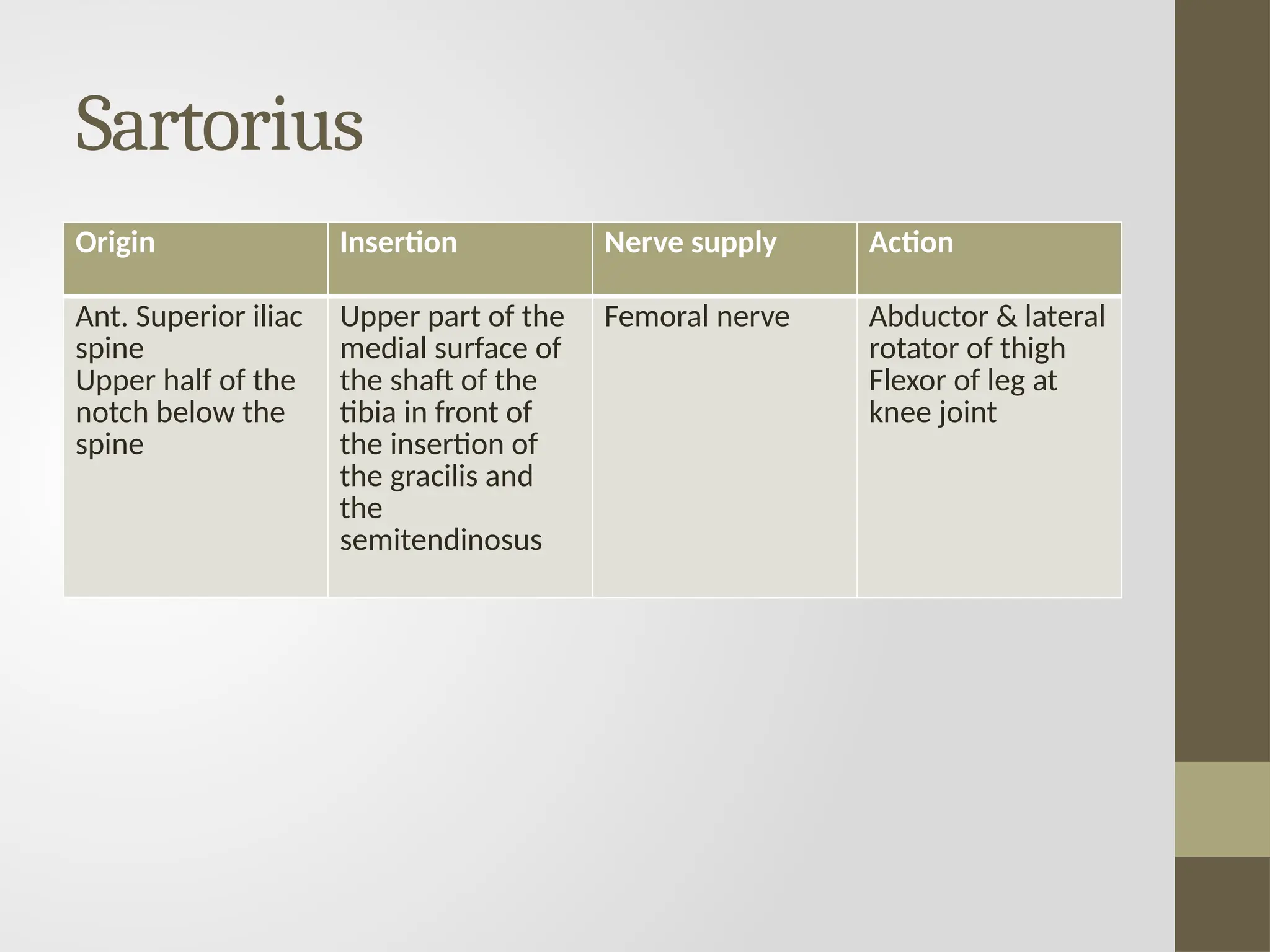
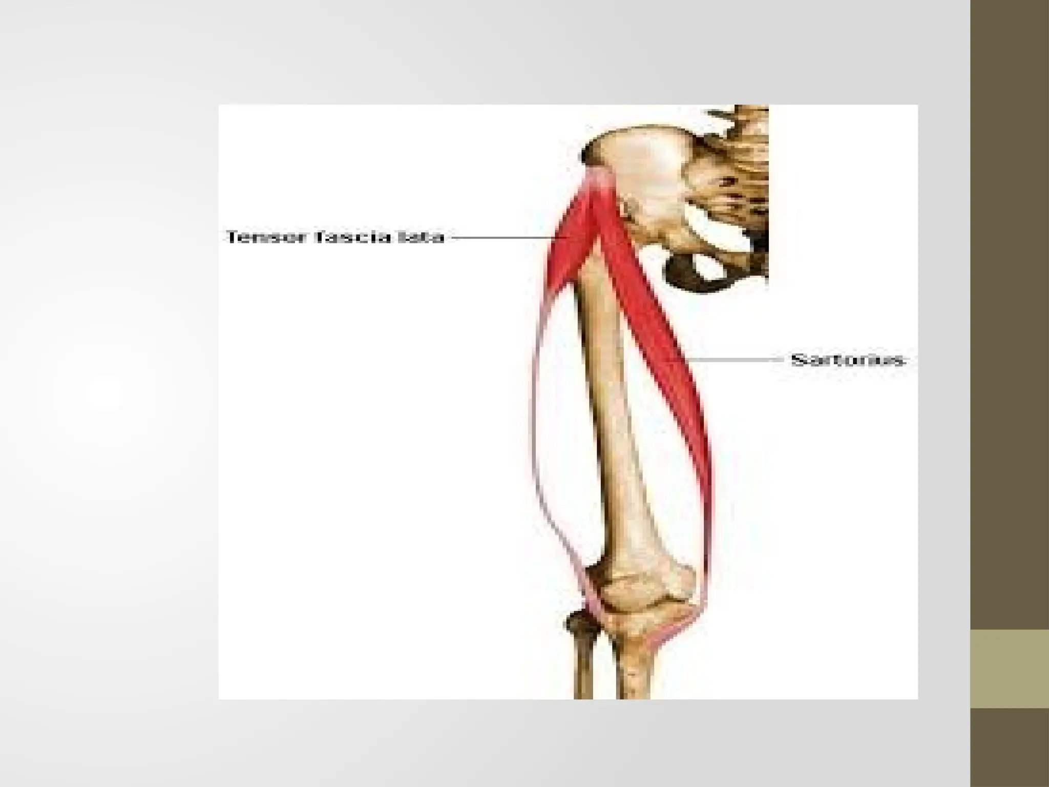
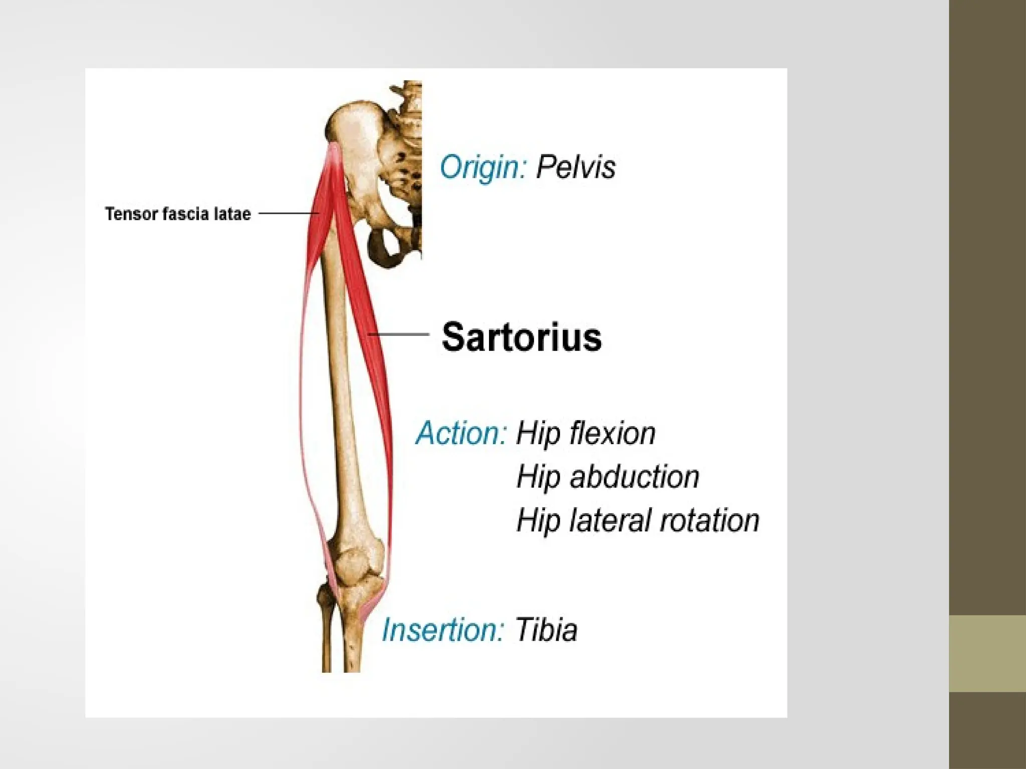
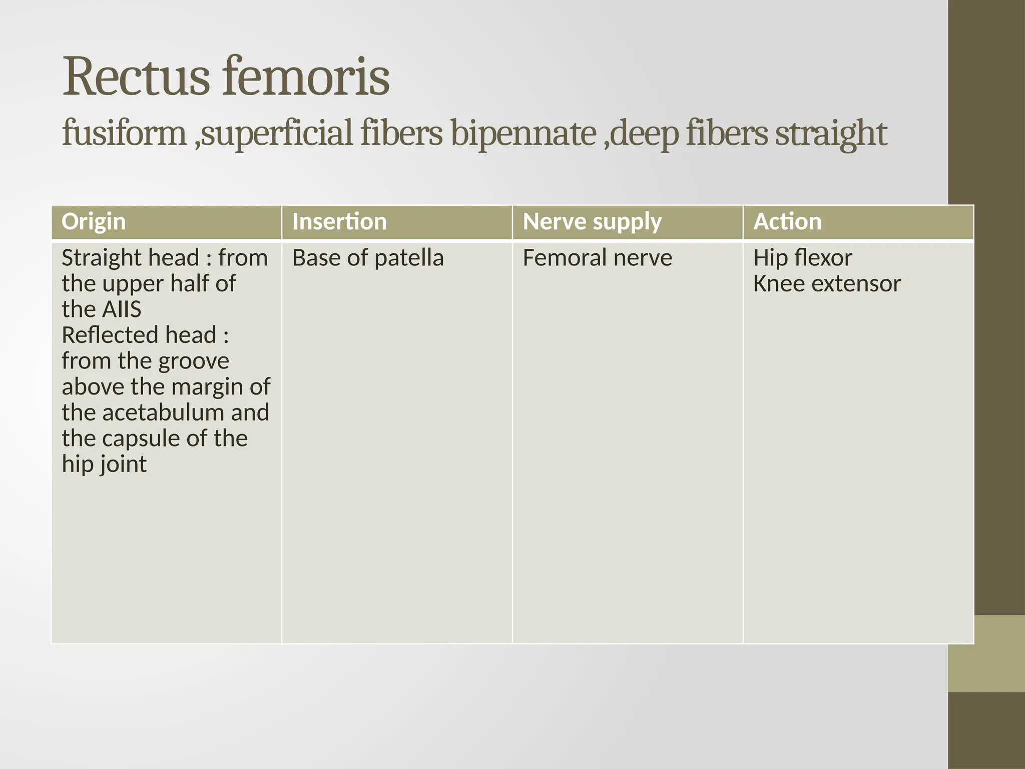
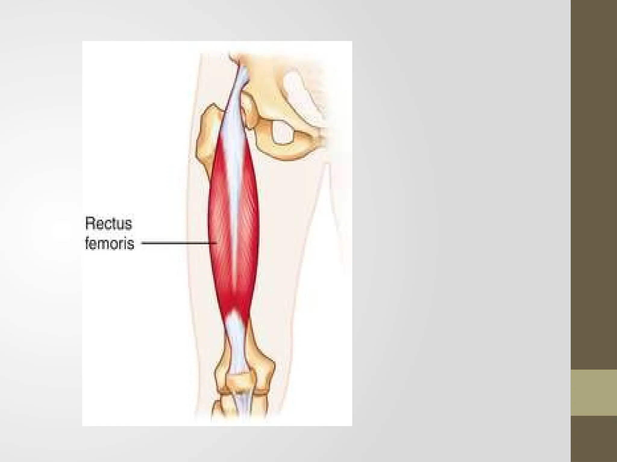
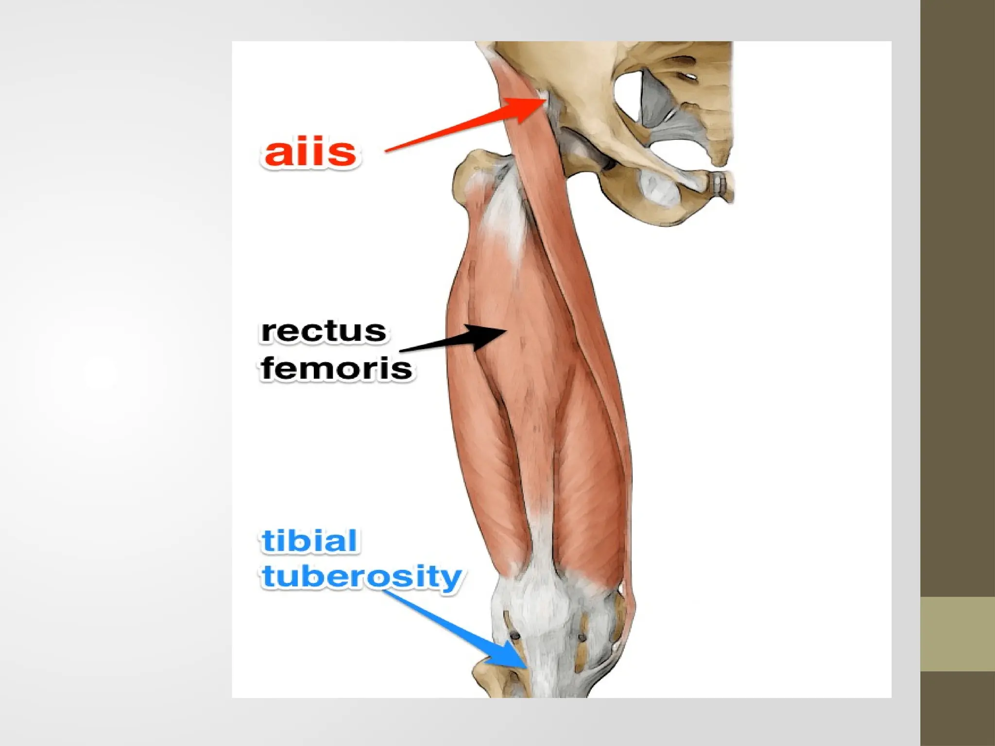
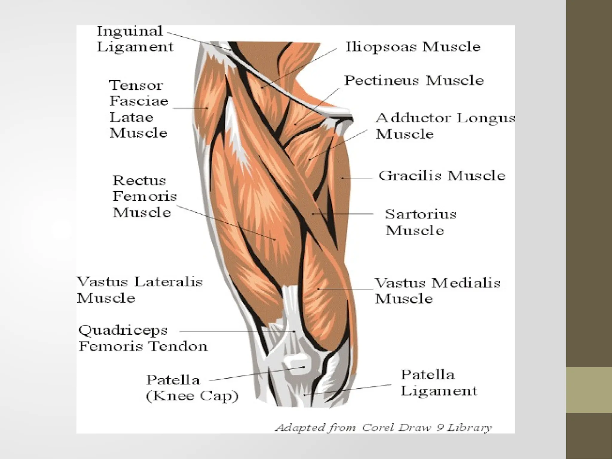
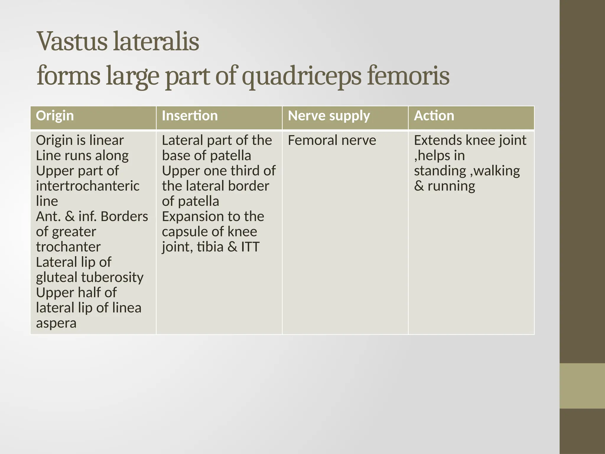
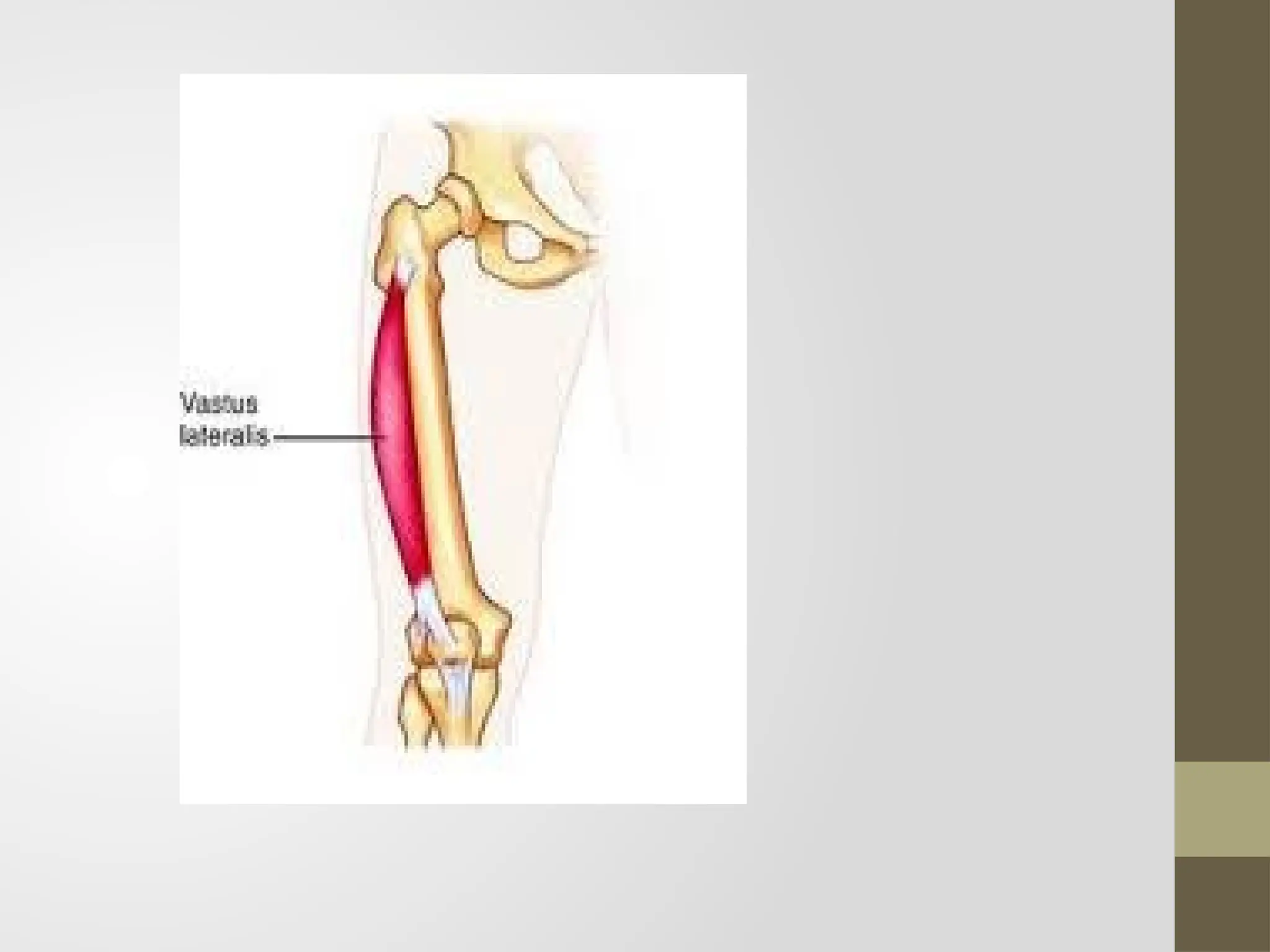
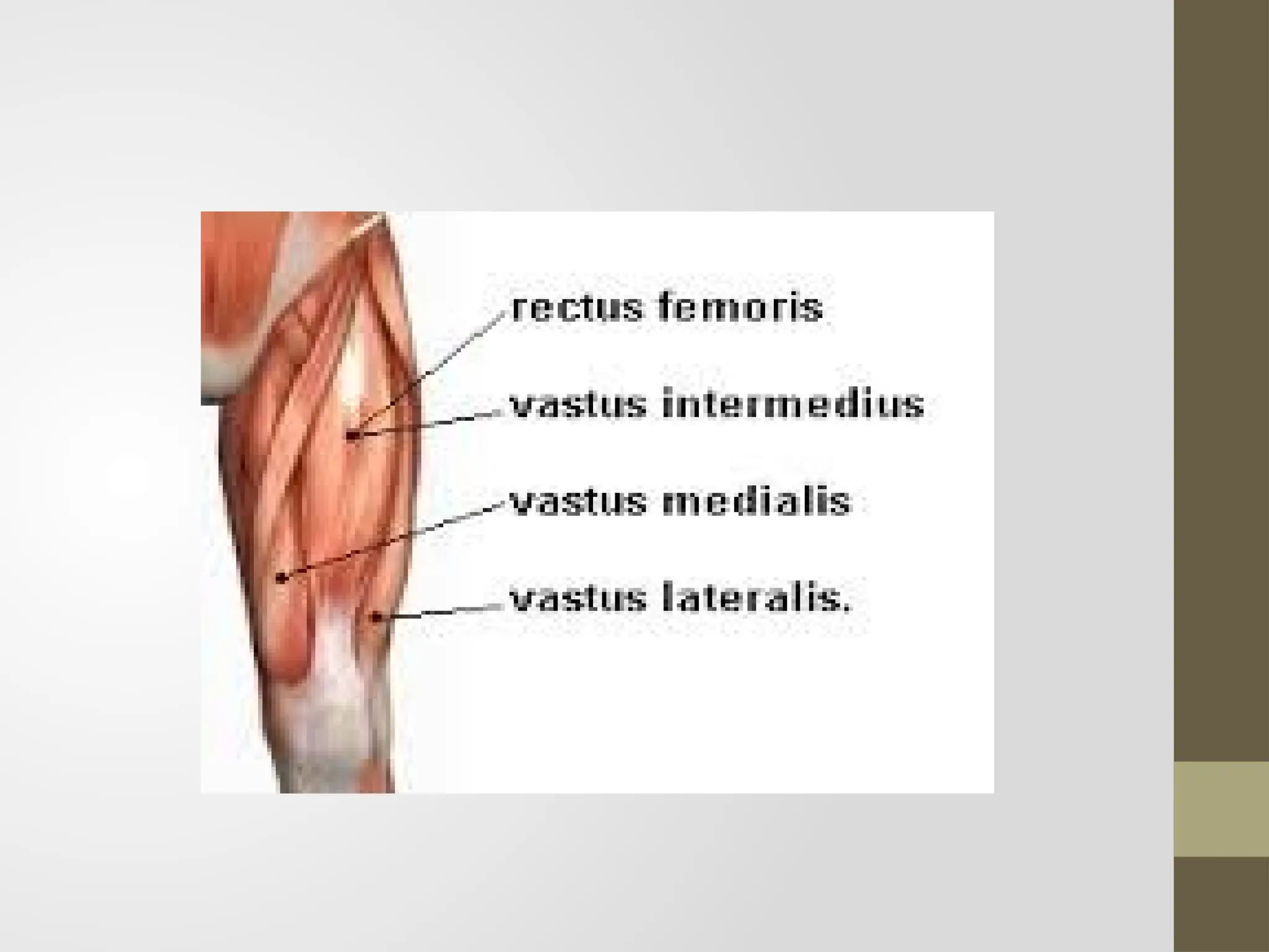
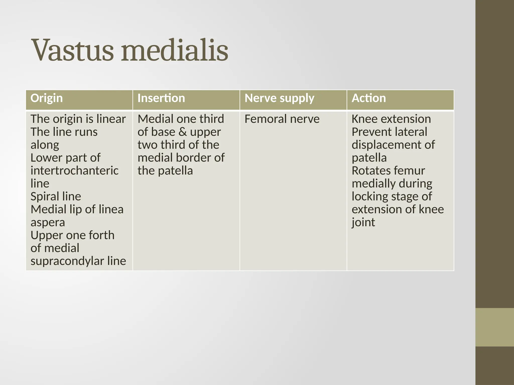
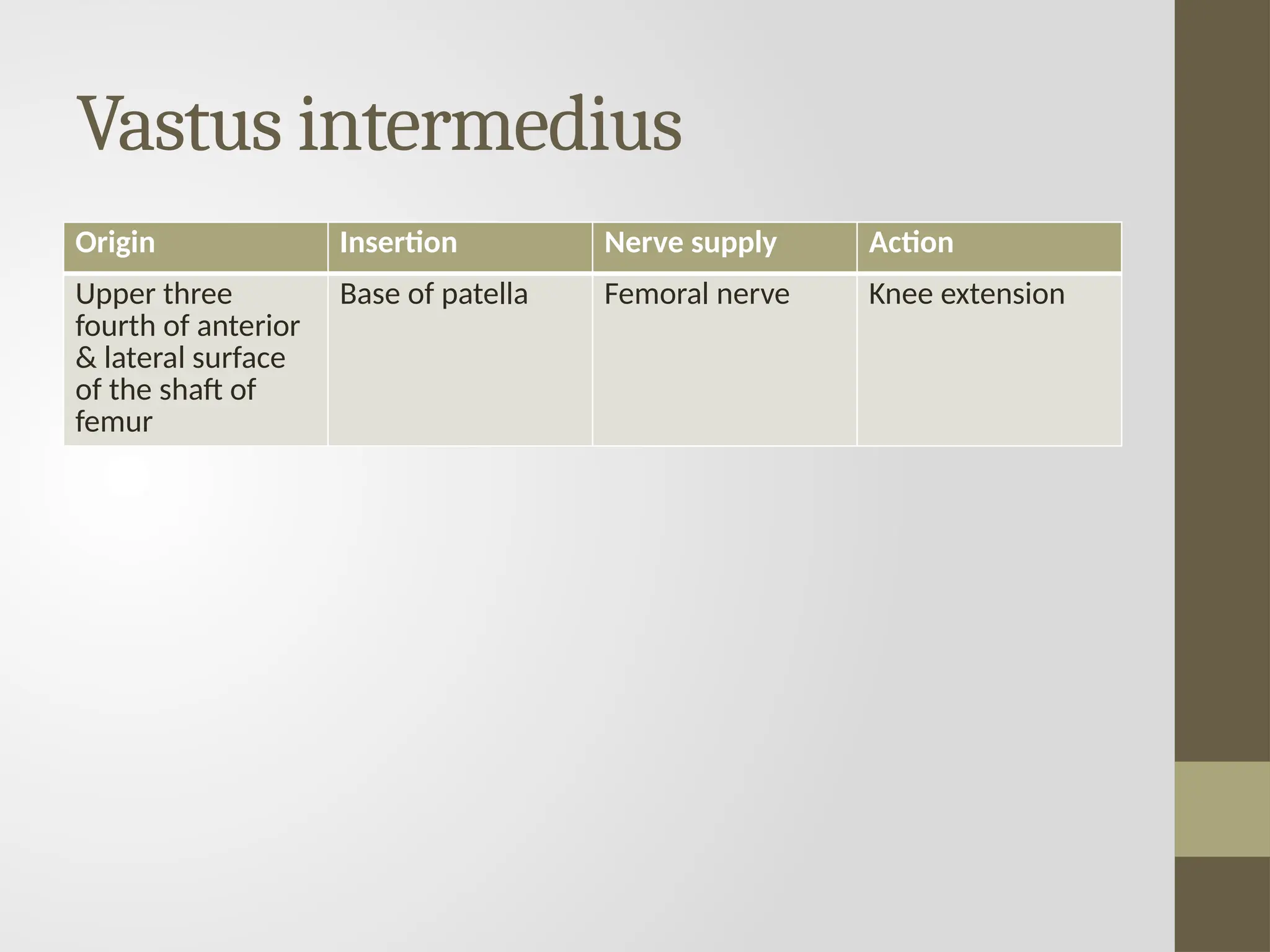
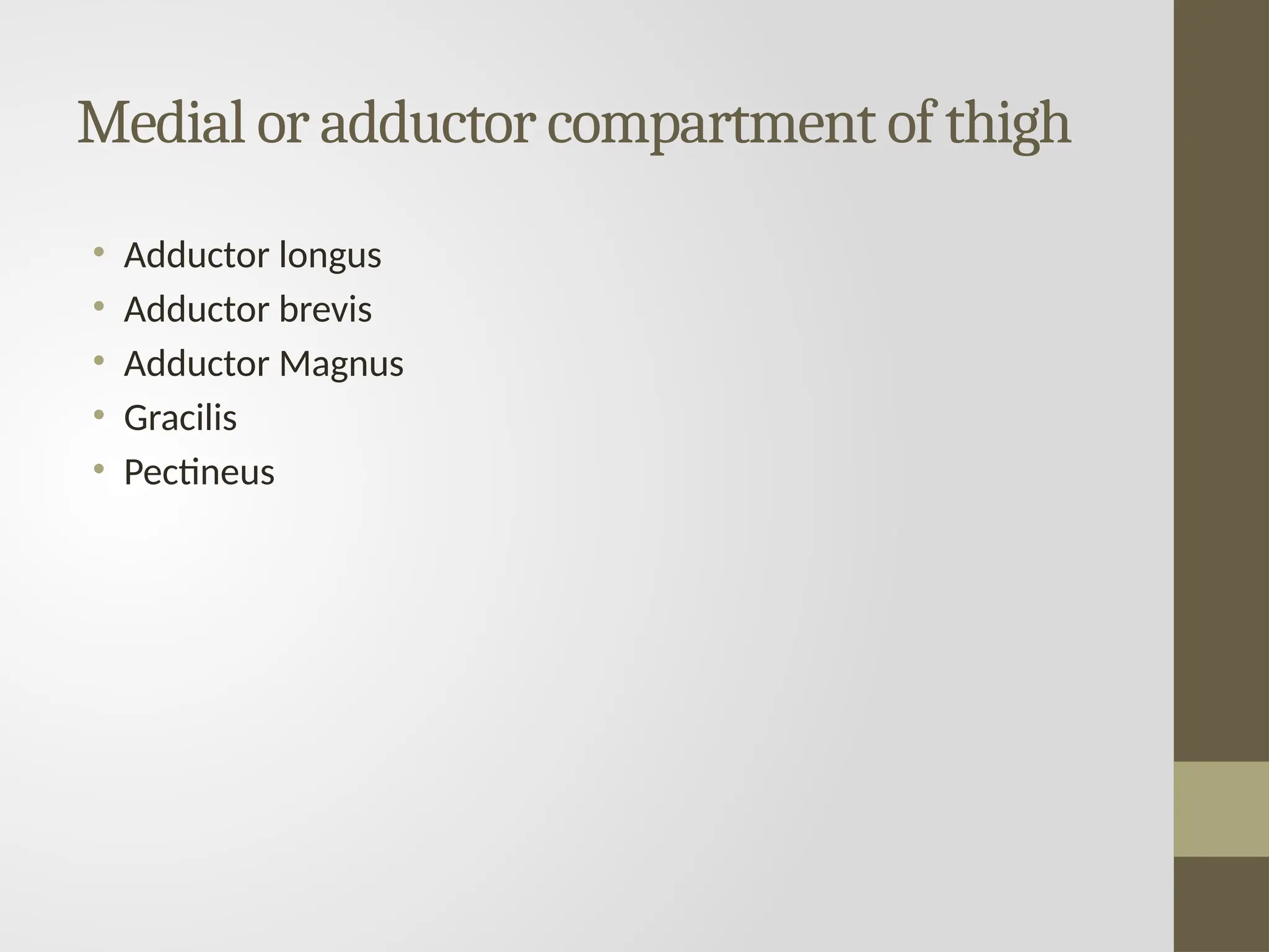
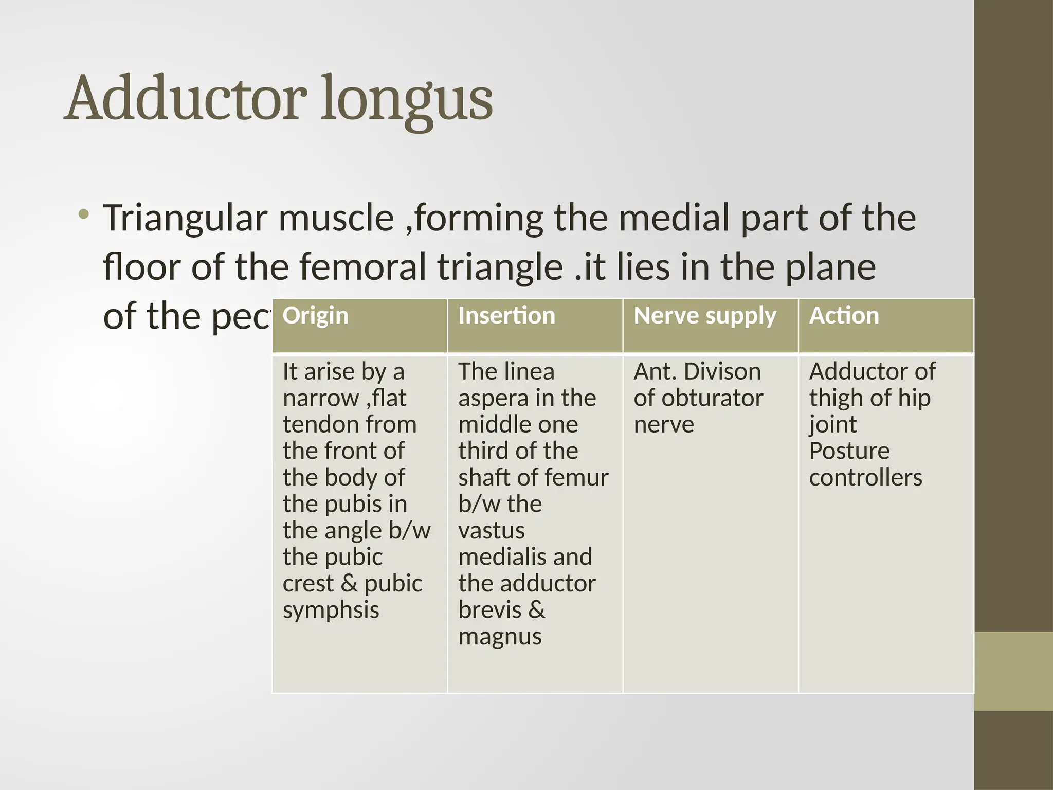
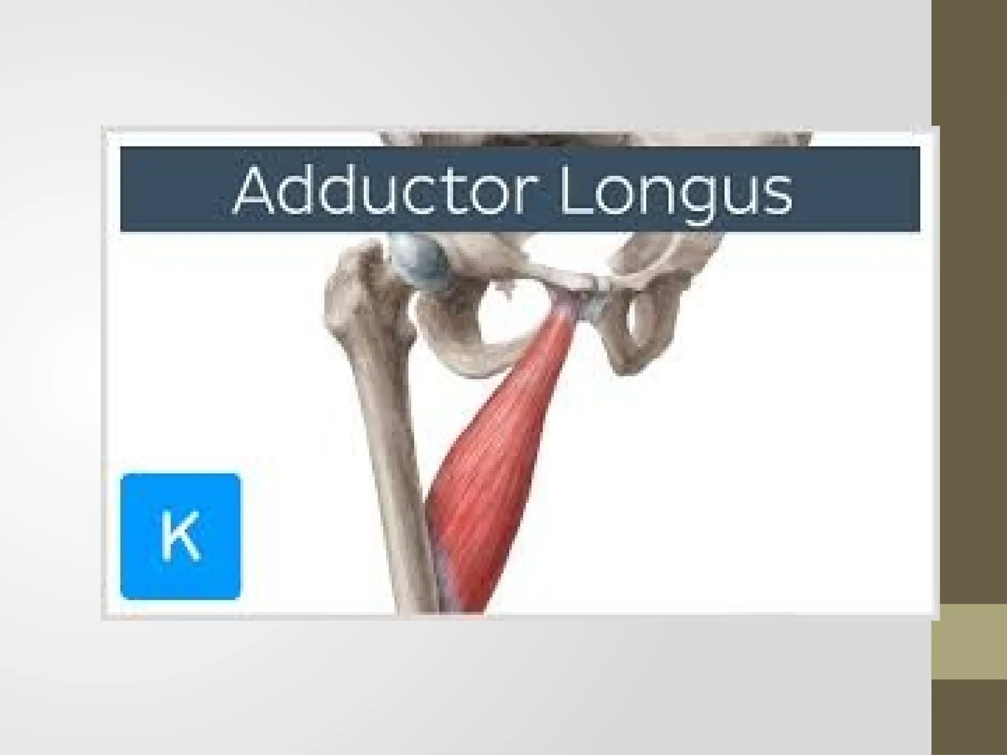
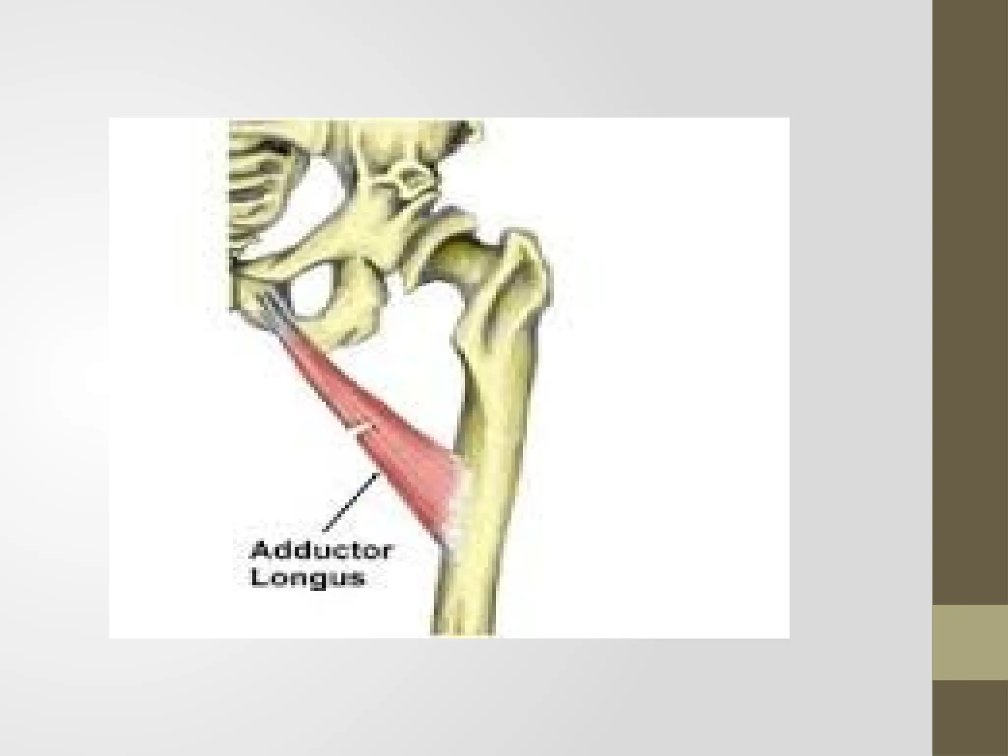
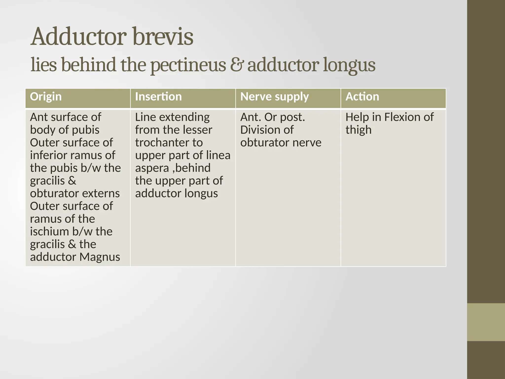
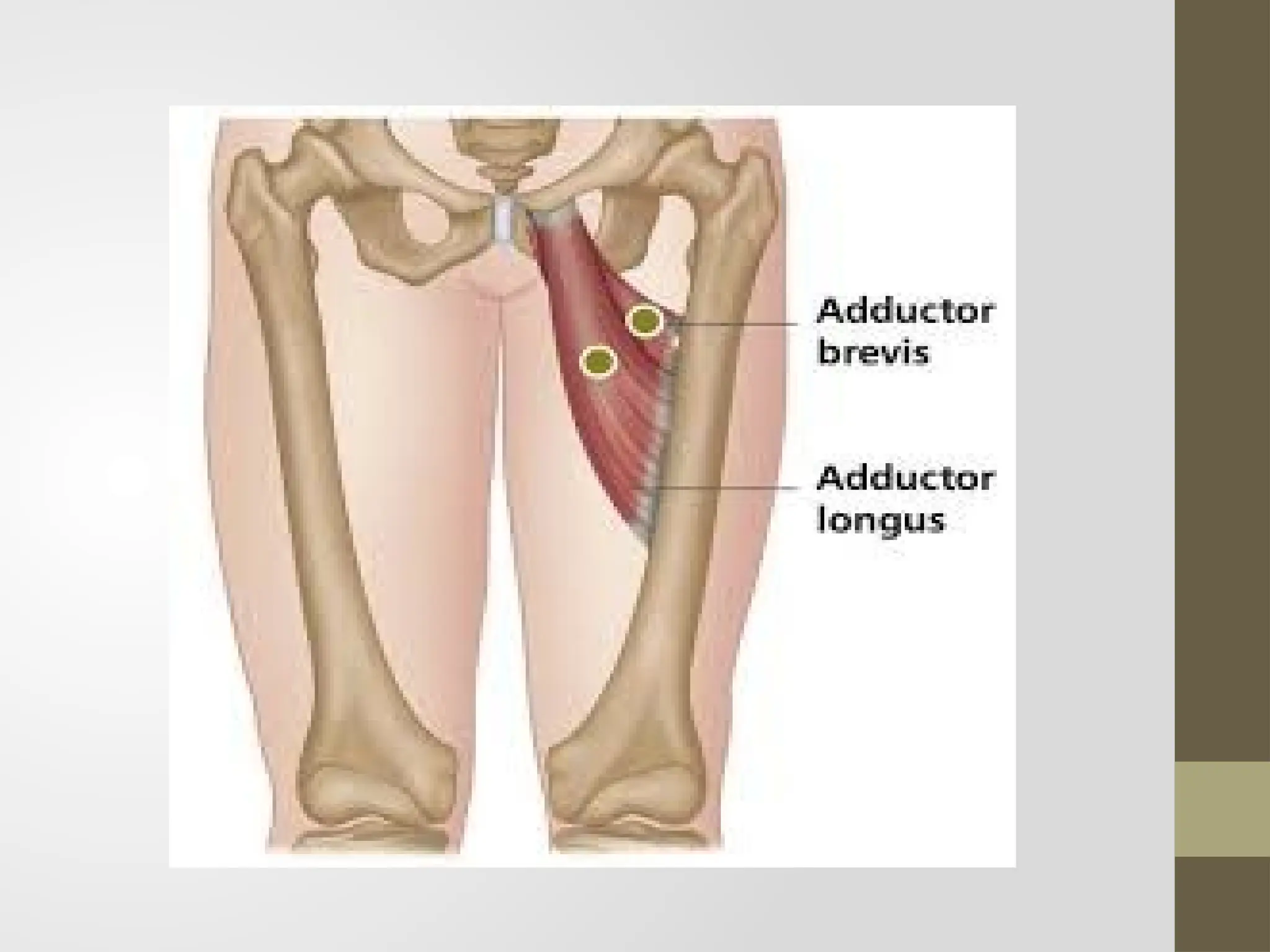
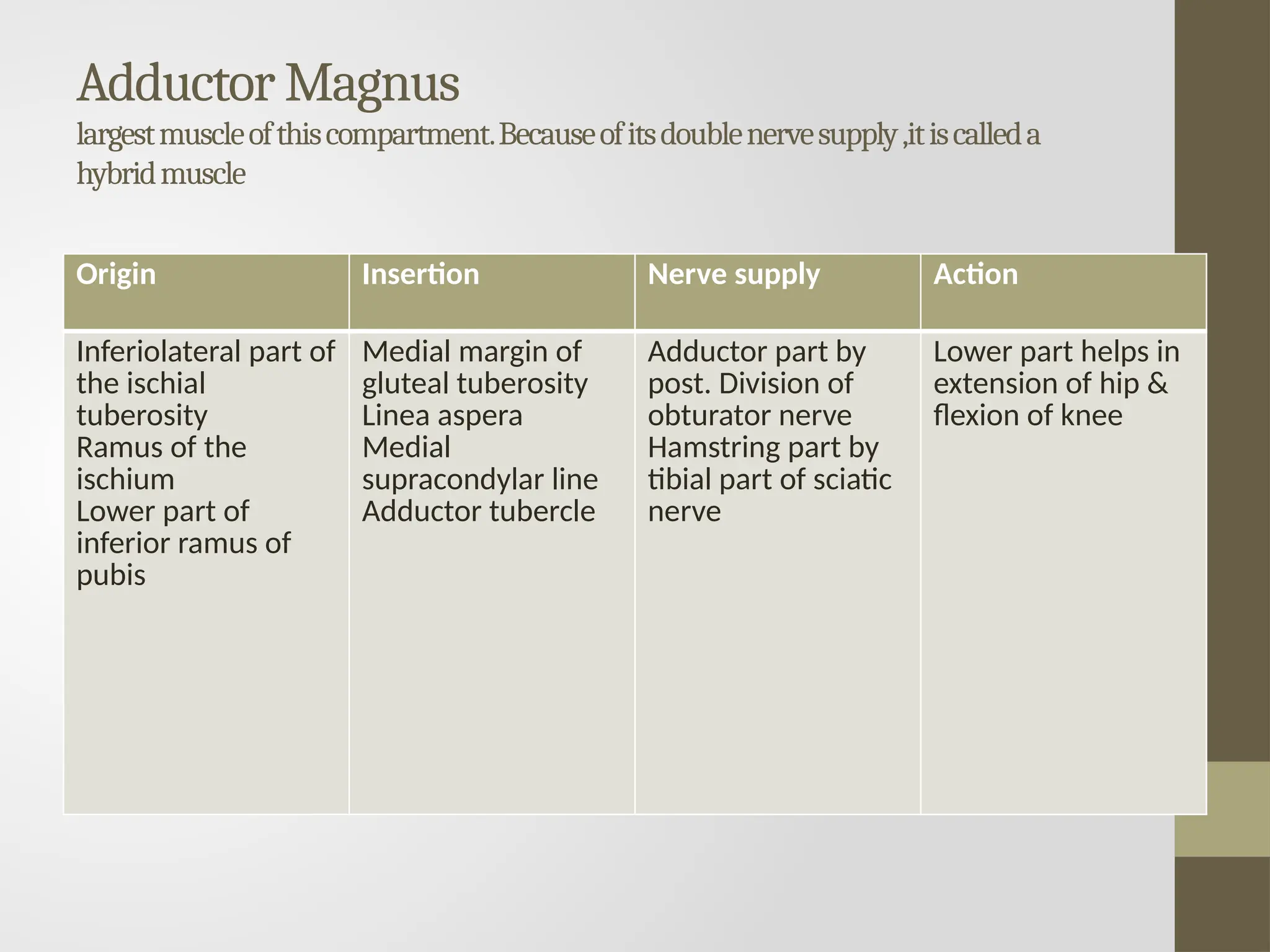
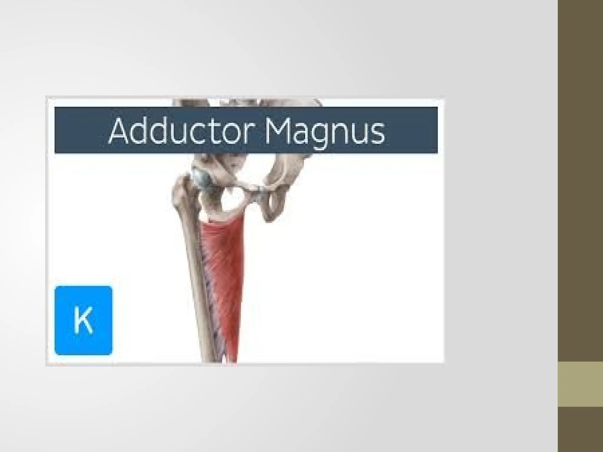
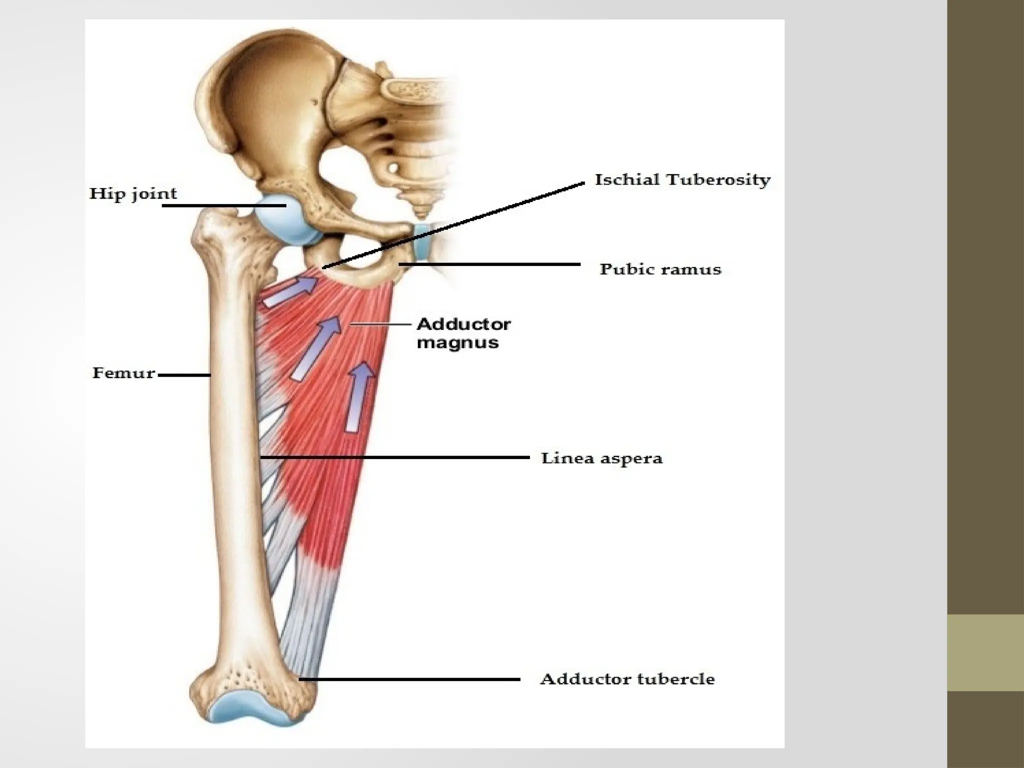
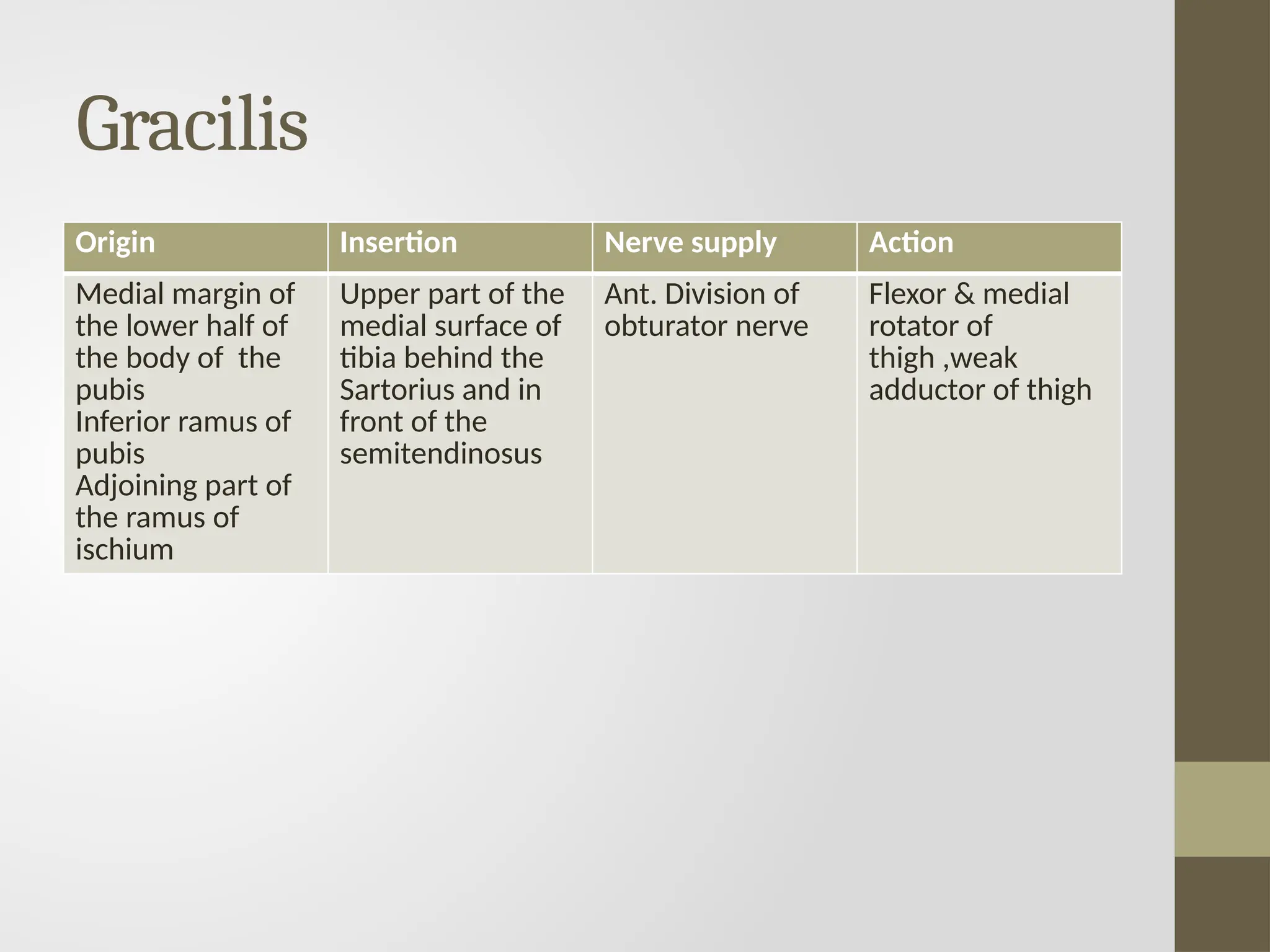
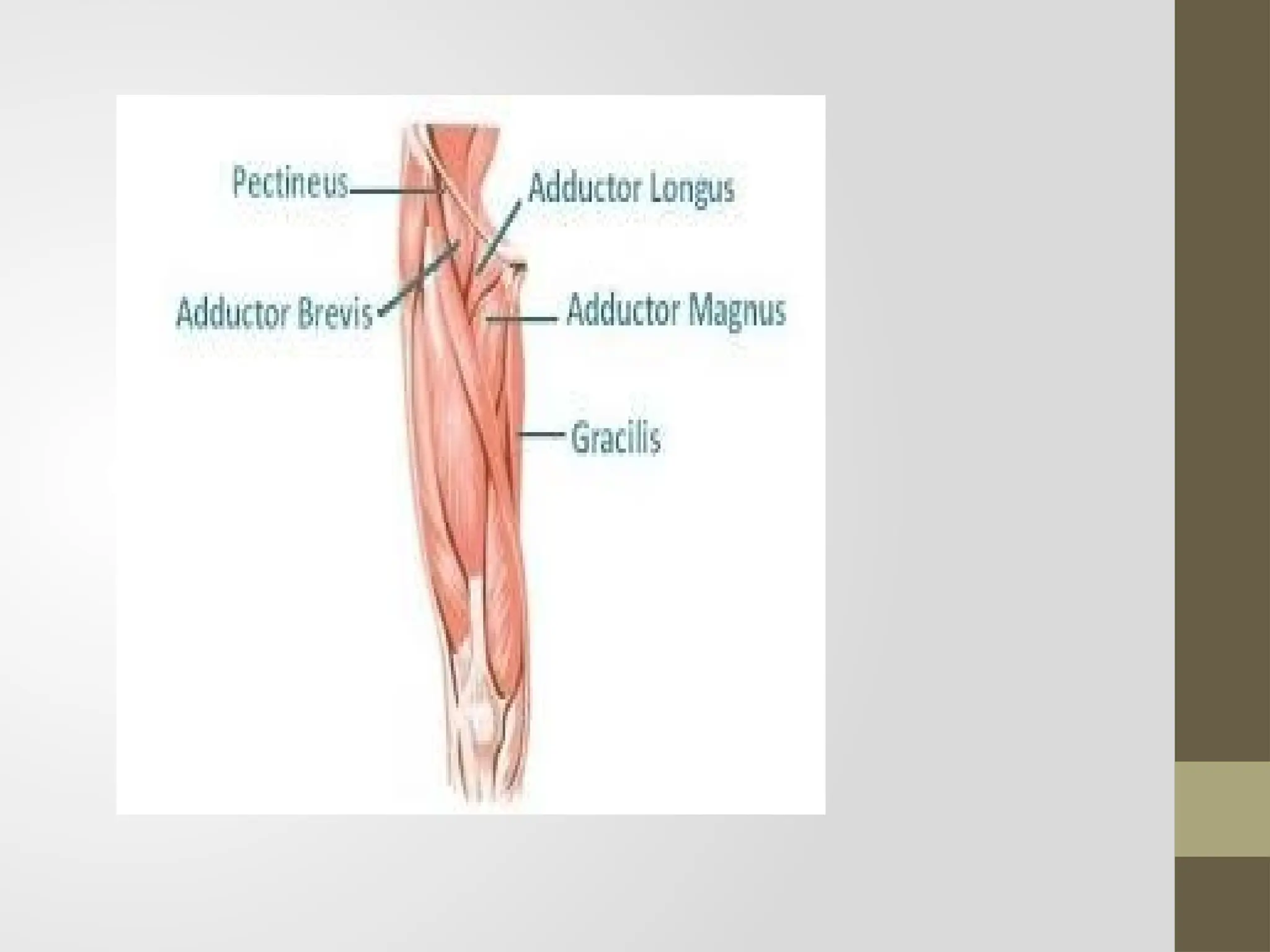
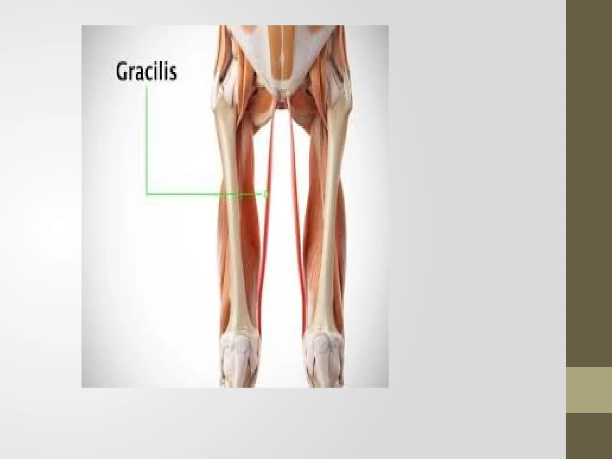
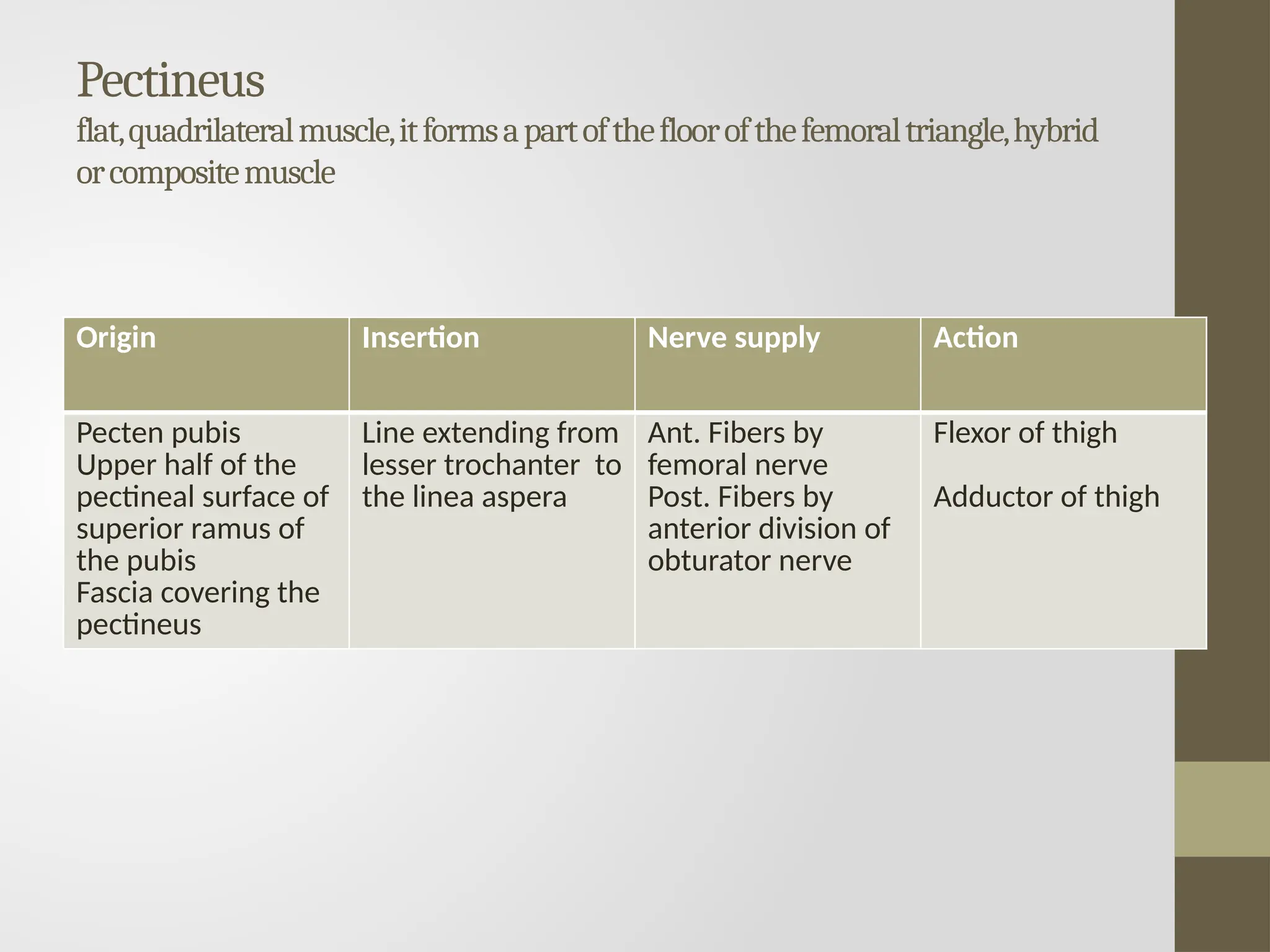
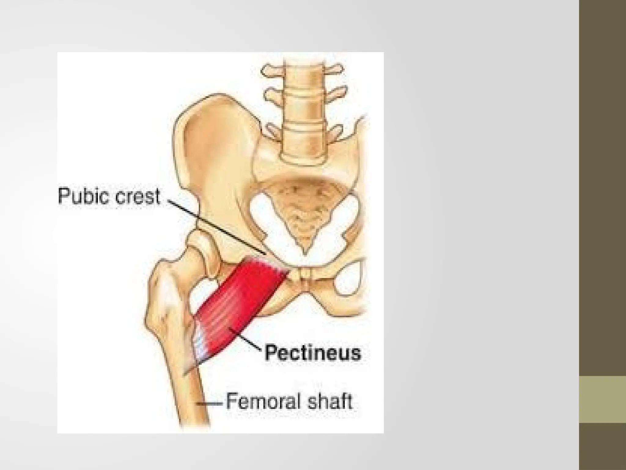
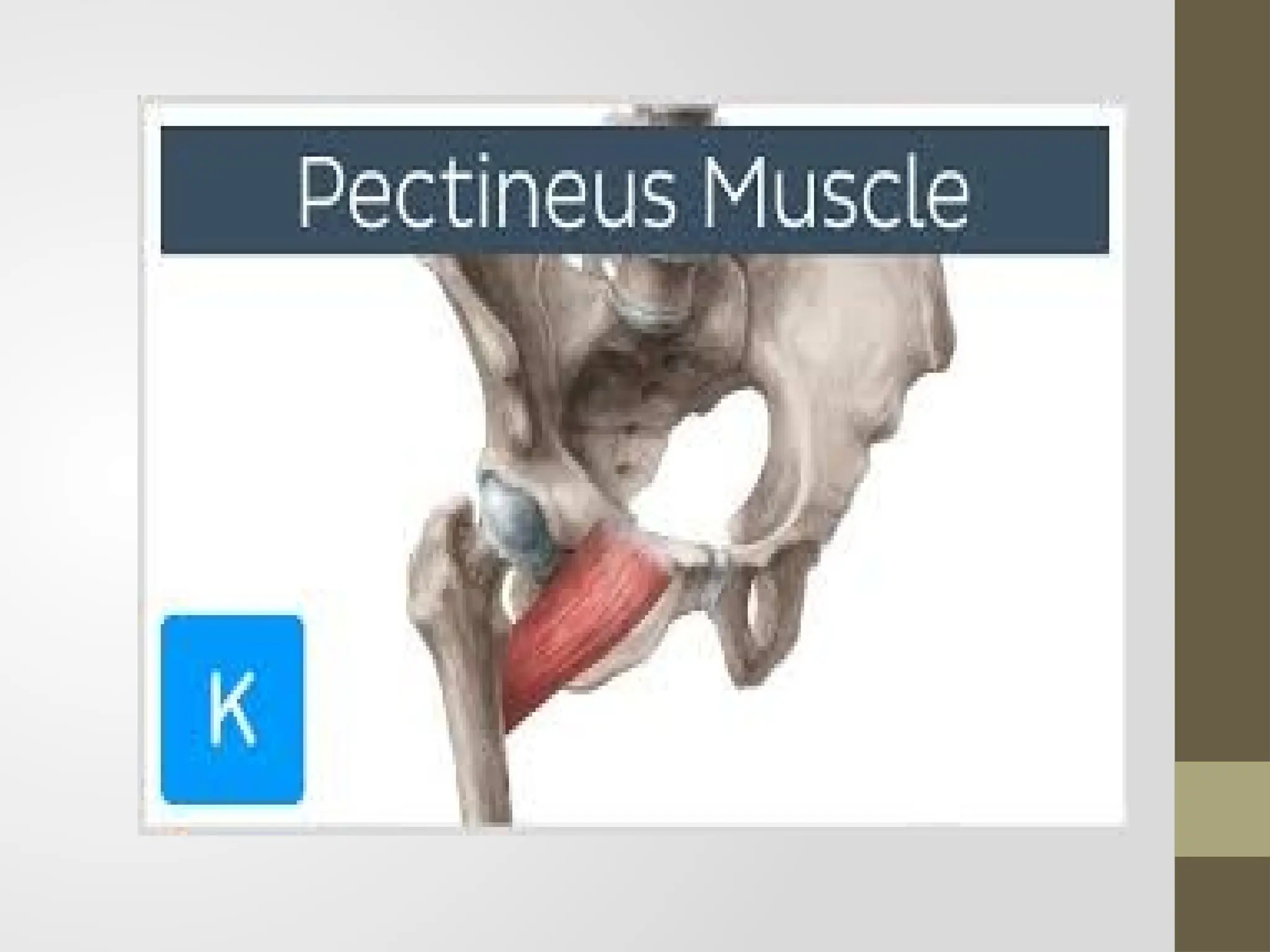
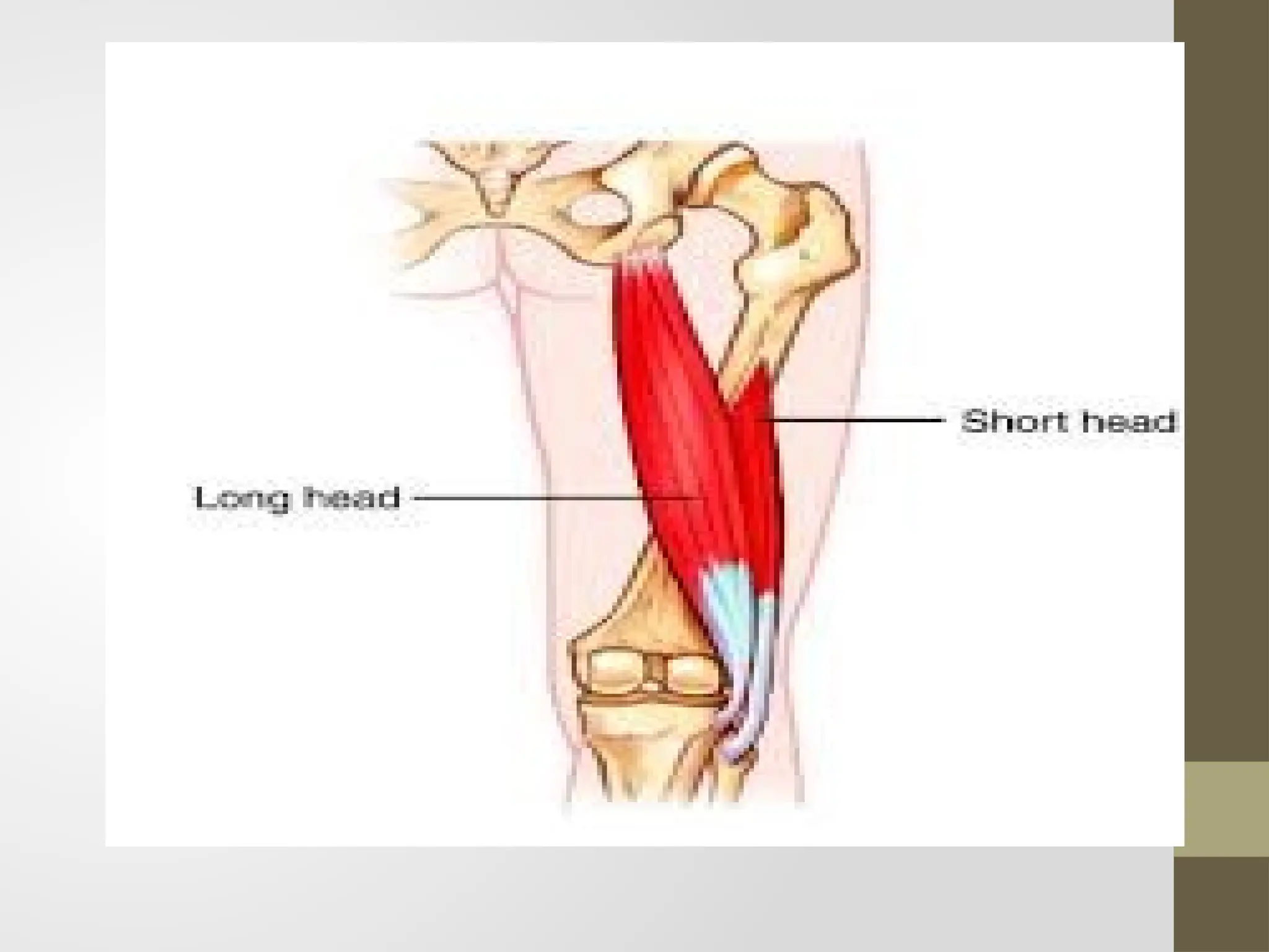
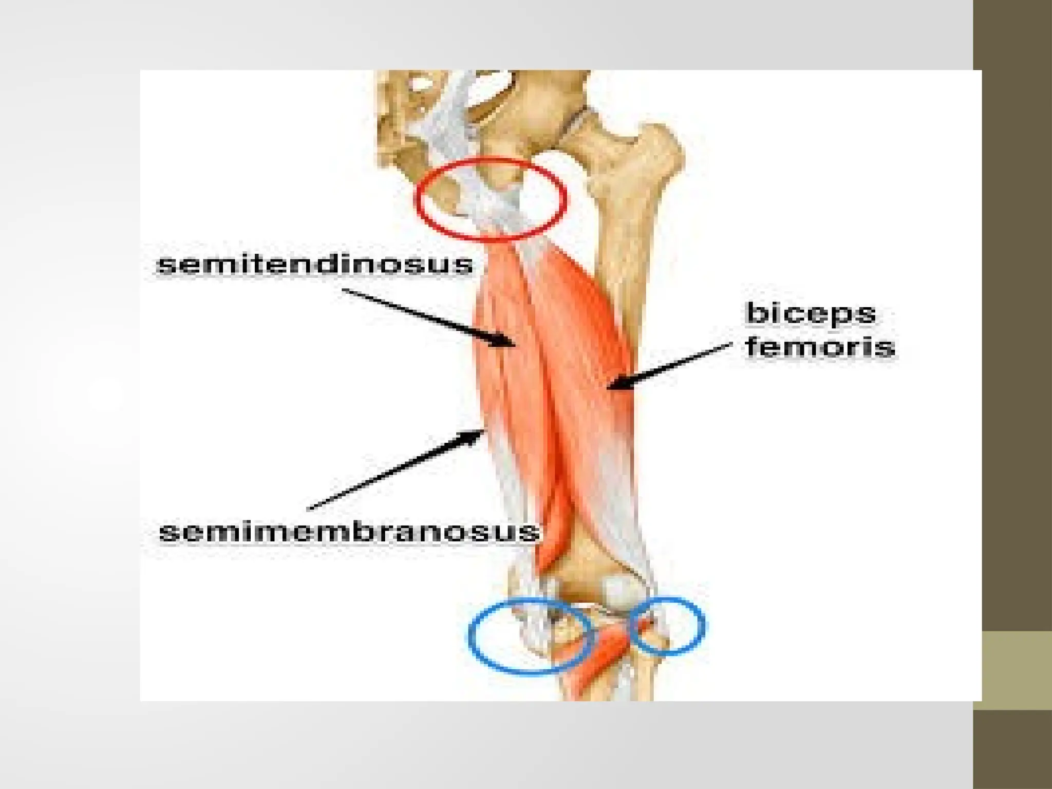
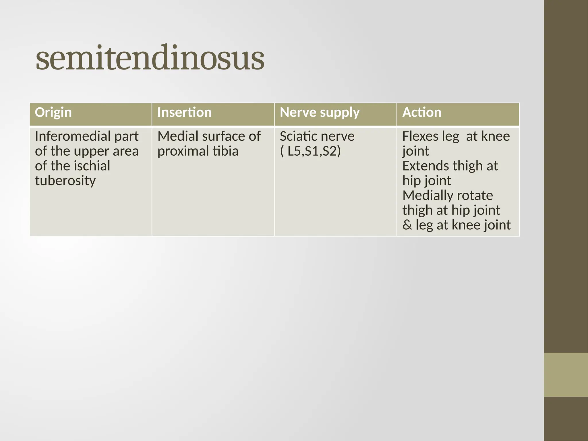
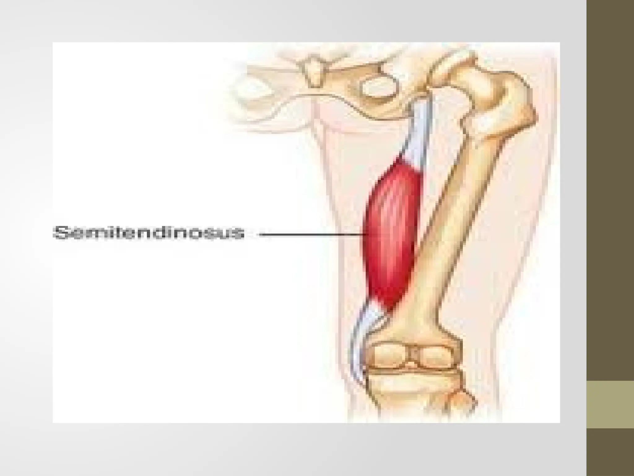
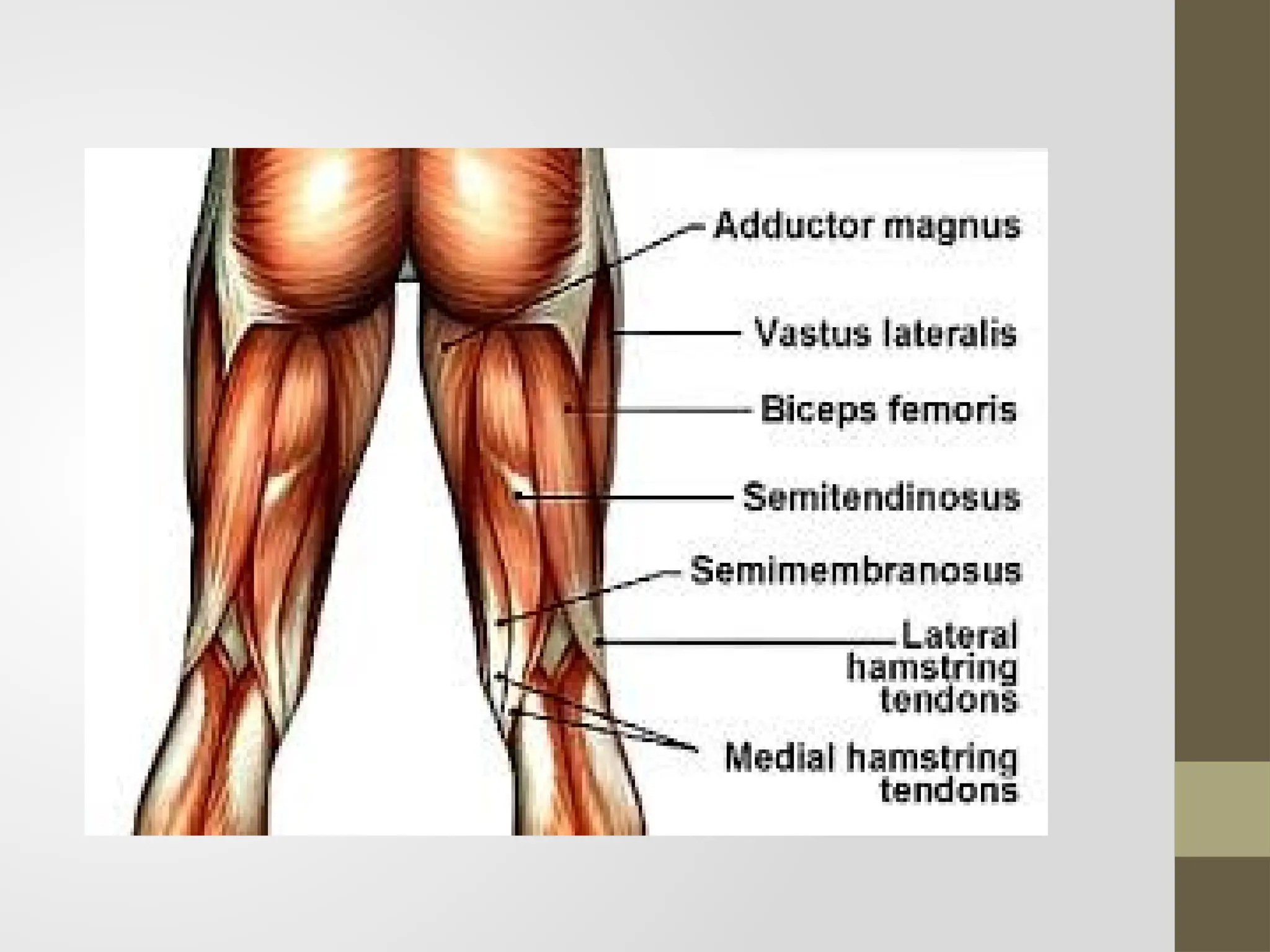
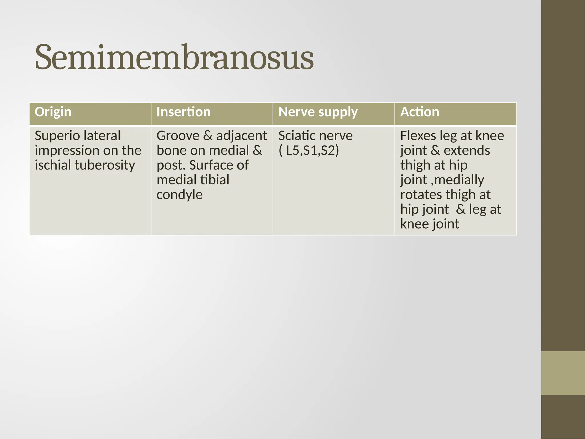
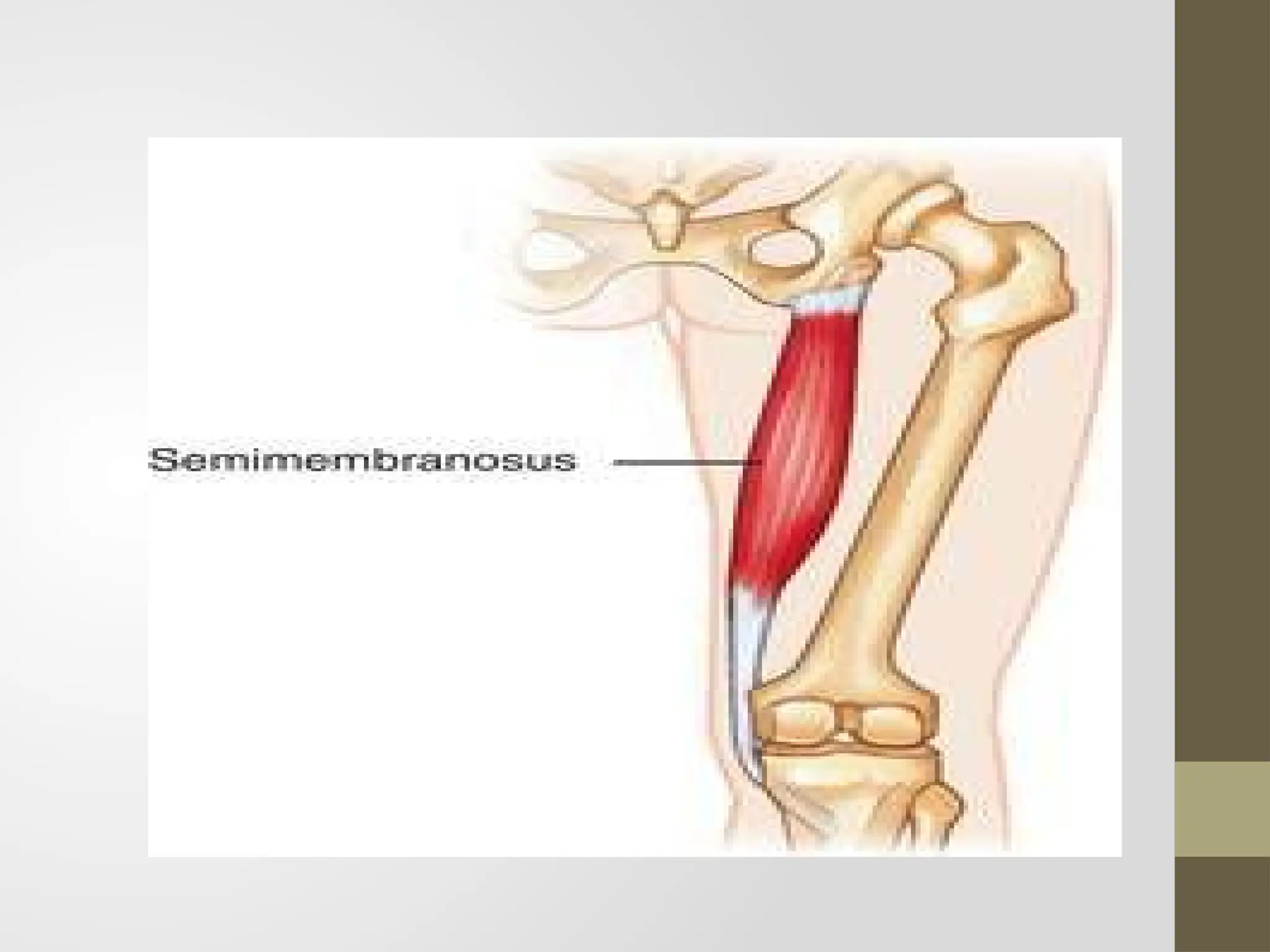
Muscles of Thigh
Presented by
Dr.Anam Maqbool(PT)
Lecturer KCHS
Thigh
Thigh is the region of the lower limb that is approximately
between the hip & knee joint
Anteriorly it is separated from the abdominal wall by the
inguinal ligament
Posteriorly it is separated from the gluteal region by the
gluteal fold superficially and by the inferior margin of the
gluteus Maximus and quadrates femoris on deeper planes
Compartments of thigh
The thigh is divided into three compartments by
intermuscular septa between the posterior aspect of
the femur and the fascia lata( the thick layer of deep
fascia that completely surrounds or invest the thigh)
Anterior compartment : contains muscle that
mainly extend the leg at the knee joint
Posterior compartment :contains muscle that
mainly extend the thigh at hip joint and flex the leg
at the knee joint
Medial compartment : contains muscle that mainly
adduct the thigh at the hip joint
Cont.…
The sciatic nerve innervates the muscles in the
posterior compartment of the thigh, the femoral
nerve innervates muscles in the anterior
compartment of the thigh & the obturator nerve
innervates most muscles in the medial compartment
of the thigh.
The major artery ,vein & lymphatic channels enter
the thigh anterior to pelvic bone and pass through
the femoral triangle inferior to inguinal ligament,
vessels & nerves passing between the thigh and leg
pass through the popliteal fossa posterior to knee
joint
Muscles of anterior or extensor
compartment of thigh
• Sartorius
• Quadriceps Femoris
 Rectus femoris
 Three Vasti
 Vastus medialis
 Vastus Intermedius
Vastus lateralis
Sartorius
Origin Insertion Nerve supply Action
Ant. Superior iliac
spine
Upper half of the
notch below the
spine
Upper part of the
medial surface of
the shaft of the
tibia in front of
the insertion of
the gracilis and
the
semitendinosus
Femoral nerve Abductor & lateral
rotator of thigh
Flexor of leg at
knee joint
Quadricepsfemoris
Rectus femoris
fusiform,superficialfibersbipennate,deepfibersstraight
Origin Insertion Nerve supply Action
Straight head : from
the upper half of
the AIIS
Reflected head :
from the groove
above the margin of
the acetabulum and
the capsule of the
hip joint
Base of patella Femoral nerve Hip flexor
Knee extensor
Vastus lateralis
forms large part of quadriceps femoris
Origin Insertion Nerve supply Action
Origin is linear
Line runs along
Upper part of
intertrochanteric
line
Ant. & inf. Borders
of greater
trochanter
Lateral lip of
gluteal tuberosity
Upper half of
lateral lip of linea
aspera
Lateral part of the
base of patella
Upper one third of
the lateral border
of patella
Expansion to the
capsule of knee
joint, tibia & ITT
Femoral nerve Extends knee joint
,helps in
standing ,walking
& running
Vastus medialis
Origin Insertion Nerve supply Action
The origin is linear
The line runs
along
Lower part of
intertrochanteric
line
Spiral line
Medial lip of linea
aspera
Upper one forth
of medial
supracondylar line
Medial one third
of base & upper
two third of the
medial border of
the patella
Femoral nerve Knee extension
Prevent lateral
displacement of
patella
Rotates femur
medially during
locking stage of
extension of knee
joint
Vastus intermedius
Origin Insertion Nerve supply Action
Upper three
fourth of anterior
& lateral surface
of the shaft of
femur
Base of patella Femoral nerve Knee extension
Medial or adductor compartment of thigh
• Adductor longus
• Adductor brevis
• Adductor Magnus
• Gracilis
• Pectineus
Adductor longus
• Triangular muscle ,forming the medial part of the
floor of the femoral triangle .it lies in the plane
of the pectineus
Origin Insertion Nerve supply Action
It arise by a
narrow ,flat
tendon from
the front of
the body of
the pubis in
the angle b/w
the pubic
crest & pubic
symphsis
The linea
aspera in the
middle one
third of the
shaft of femur
b/w the
vastus
medialis and
the adductor
brevis &
magnus
Ant. Divison
of obturator
nerve
Adductor of
thigh of hip
joint
Posture
controllers
Adductor brevis
lies behind the pectineus & adductor longus
Origin Insertion Nerve supply Action
Ant surface of
body of pubis
Outer surface of
inferior ramus of
the pubis b/w the
gracilis &
obturator externs
Outer surface of
ramus of the
ischium b/w the
gracilis & the
adductor Magnus
Line extending
from the lesser
trochanter to
upper part of linea
aspera ,behind
the upper part of
adductor longus
Ant. Or post.
Division of
obturator nerve
Help in Flexion of
thigh
Adductor Magnus
largestmuscleofthiscompartment.Becauseofitsdoublenervesupply,itiscalleda
hybridmuscle
Origin Insertion Nerve supply Action
Inferiolateral part of
the ischial
tuberosity
Ramus of the
ischium
Lower part of
inferior ramus of
pubis
Medial margin of
gluteal tuberosity
Linea aspera
Medial
supracondylar line
Adductor tubercle
Adductor part by
post. Division of
obturator nerve
Hamstring part by
tibial part of sciatic
nerve
Lower part helps in
extension of hip &
flexion of knee
Gracilis
Origin Insertion Nerve supply Action
Medial margin of
the lower half of
the body of the
pubis
Inferior ramus of
pubis
Adjoining part of
the ramus of
ischium
Upper part of the
medial surface of
tibia behind the
Sartorius and in
front of the
semitendinosus
Ant. Division of
obturator nerve
Flexor & medial
rotator of
thigh ,weak
adductor of thigh
Pectineus
flat,quadrilateralmuscle,itformsapartofthefloorofthefemoraltriangle,hybrid
orcompositemuscle
Origin Insertion Nerve supply Action
Pecten pubis
Upper half of the
pectineal surface of
superior ramus of
the pubis
Fascia covering the
pectineus
Line extending from
lesser trochanter to
the linea aspera
Ant. Fibers by
femoral nerve
Post. Fibers by
anterior division of
obturator nerve
Flexor of thigh
Adductor of thigh
Muscles of back of thigh
• Semitendinosus
• Semimembranosus
• Biceps femoris
Biceps femoris
Origin Insertion Nerve supply Action
Long head ;
inferomedial part
of the upper area
of ischial
tuberosity
Short head ;
lateral lip of linea
aspera
Head of fibula Sciatic nerve
( L5,S1,S2)
Flexes leg at knee
joints , extends &
laterally rotates
thigh at hip joint
& laterally rotate
leg at knee joint
semitendinosus
Origin Insertion Nerve supply Action
Inferomedial part
of the upper area
of the ischial
tuberosity
Medial surface of
proximal tibia
Sciatic nerve
( L5,S1,S2)
Flexes leg at knee
joint
Extends thigh at
hip joint
Medially rotate
thigh at hip joint
& leg at knee joint
Semimembranosus
Origin Insertion Nerve supply Action
Superio lateral
impression on the
ischial tuberosity
Groove & adjacent
bone on medial &
post. Surface of
medial tibial
condyle
Sciatic nerve
( L5,S1,S2)
Flexes leg at knee
joint & extends
thigh at hip
joint ,medially
rotates thigh at
hip joint & leg at
knee joint
ANYQUESTIONs?
Learningsour
ces
lectur
eslides
B
Dchau
rasi
a
’sHumanA
natom
y(volume2)
GRA
Y’SA
nat
om
y(3r
d
edit
ion)
i
nter
net
OurnexttopicwillbeMusclesofleg
THANKYOU

lec#05 Muscles of Thigh.pptx anatomy lecture

  • 1.
    Muscles of Thigh Presentedby Dr.Anam Maqbool(PT) Lecturer KCHS
  • 2.
    Thigh Thigh is theregion of the lower limb that is approximately between the hip & knee joint Anteriorly it is separated from the abdominal wall by the inguinal ligament Posteriorly it is separated from the gluteal region by the gluteal fold superficially and by the inferior margin of the gluteus Maximus and quadrates femoris on deeper planes
  • 5.
    Compartments of thigh Thethigh is divided into three compartments by intermuscular septa between the posterior aspect of the femur and the fascia lata( the thick layer of deep fascia that completely surrounds or invest the thigh) Anterior compartment : contains muscle that mainly extend the leg at the knee joint Posterior compartment :contains muscle that mainly extend the thigh at hip joint and flex the leg at the knee joint Medial compartment : contains muscle that mainly adduct the thigh at the hip joint
  • 6.
    Cont.… The sciatic nerveinnervates the muscles in the posterior compartment of the thigh, the femoral nerve innervates muscles in the anterior compartment of the thigh & the obturator nerve innervates most muscles in the medial compartment of the thigh. The major artery ,vein & lymphatic channels enter the thigh anterior to pelvic bone and pass through the femoral triangle inferior to inguinal ligament, vessels & nerves passing between the thigh and leg pass through the popliteal fossa posterior to knee joint
  • 7.
    Muscles of anterioror extensor compartment of thigh • Sartorius • Quadriceps Femoris  Rectus femoris  Three Vasti  Vastus medialis  Vastus Intermedius Vastus lateralis
  • 8.
    Sartorius Origin Insertion Nervesupply Action Ant. Superior iliac spine Upper half of the notch below the spine Upper part of the medial surface of the shaft of the tibia in front of the insertion of the gracilis and the semitendinosus Femoral nerve Abductor & lateral rotator of thigh Flexor of leg at knee joint
  • 11.
  • 12.
    Rectus femoris fusiform,superficialfibersbipennate,deepfibersstraight Origin InsertionNerve supply Action Straight head : from the upper half of the AIIS Reflected head : from the groove above the margin of the acetabulum and the capsule of the hip joint Base of patella Femoral nerve Hip flexor Knee extensor
  • 16.
    Vastus lateralis forms largepart of quadriceps femoris Origin Insertion Nerve supply Action Origin is linear Line runs along Upper part of intertrochanteric line Ant. & inf. Borders of greater trochanter Lateral lip of gluteal tuberosity Upper half of lateral lip of linea aspera Lateral part of the base of patella Upper one third of the lateral border of patella Expansion to the capsule of knee joint, tibia & ITT Femoral nerve Extends knee joint ,helps in standing ,walking & running
  • 19.
    Vastus medialis Origin InsertionNerve supply Action The origin is linear The line runs along Lower part of intertrochanteric line Spiral line Medial lip of linea aspera Upper one forth of medial supracondylar line Medial one third of base & upper two third of the medial border of the patella Femoral nerve Knee extension Prevent lateral displacement of patella Rotates femur medially during locking stage of extension of knee joint
  • 20.
    Vastus intermedius Origin InsertionNerve supply Action Upper three fourth of anterior & lateral surface of the shaft of femur Base of patella Femoral nerve Knee extension
  • 21.
    Medial or adductorcompartment of thigh • Adductor longus • Adductor brevis • Adductor Magnus • Gracilis • Pectineus
  • 22.
    Adductor longus • Triangularmuscle ,forming the medial part of the floor of the femoral triangle .it lies in the plane of the pectineus Origin Insertion Nerve supply Action It arise by a narrow ,flat tendon from the front of the body of the pubis in the angle b/w the pubic crest & pubic symphsis The linea aspera in the middle one third of the shaft of femur b/w the vastus medialis and the adductor brevis & magnus Ant. Divison of obturator nerve Adductor of thigh of hip joint Posture controllers
  • 25.
    Adductor brevis lies behindthe pectineus & adductor longus Origin Insertion Nerve supply Action Ant surface of body of pubis Outer surface of inferior ramus of the pubis b/w the gracilis & obturator externs Outer surface of ramus of the ischium b/w the gracilis & the adductor Magnus Line extending from the lesser trochanter to upper part of linea aspera ,behind the upper part of adductor longus Ant. Or post. Division of obturator nerve Help in Flexion of thigh
  • 27.
    Adductor Magnus largestmuscleofthiscompartment.Becauseofitsdoublenervesupply,itiscalleda hybridmuscle Origin InsertionNerve supply Action Inferiolateral part of the ischial tuberosity Ramus of the ischium Lower part of inferior ramus of pubis Medial margin of gluteal tuberosity Linea aspera Medial supracondylar line Adductor tubercle Adductor part by post. Division of obturator nerve Hamstring part by tibial part of sciatic nerve Lower part helps in extension of hip & flexion of knee
  • 30.
    Gracilis Origin Insertion Nervesupply Action Medial margin of the lower half of the body of the pubis Inferior ramus of pubis Adjoining part of the ramus of ischium Upper part of the medial surface of tibia behind the Sartorius and in front of the semitendinosus Ant. Division of obturator nerve Flexor & medial rotator of thigh ,weak adductor of thigh
  • 33.
    Pectineus flat,quadrilateralmuscle,itformsapartofthefloorofthefemoraltriangle,hybrid orcompositemuscle Origin Insertion Nervesupply Action Pecten pubis Upper half of the pectineal surface of superior ramus of the pubis Fascia covering the pectineus Line extending from lesser trochanter to the linea aspera Ant. Fibers by femoral nerve Post. Fibers by anterior division of obturator nerve Flexor of thigh Adductor of thigh
  • 36.
    Muscles of backof thigh • Semitendinosus • Semimembranosus • Biceps femoris
  • 37.
    Biceps femoris Origin InsertionNerve supply Action Long head ; inferomedial part of the upper area of ischial tuberosity Short head ; lateral lip of linea aspera Head of fibula Sciatic nerve ( L5,S1,S2) Flexes leg at knee joints , extends & laterally rotates thigh at hip joint & laterally rotate leg at knee joint
  • 40.
    semitendinosus Origin Insertion Nervesupply Action Inferomedial part of the upper area of the ischial tuberosity Medial surface of proximal tibia Sciatic nerve ( L5,S1,S2) Flexes leg at knee joint Extends thigh at hip joint Medially rotate thigh at hip joint & leg at knee joint
  • 43.
    Semimembranosus Origin Insertion Nervesupply Action Superio lateral impression on the ischial tuberosity Groove & adjacent bone on medial & post. Surface of medial tibial condyle Sciatic nerve ( L5,S1,S2) Flexes leg at knee joint & extends thigh at hip joint ,medially rotates thigh at hip joint & leg at knee joint
  • 45.
  • 46.
  • 47.
  • 48.