The gluteal region is located between the trunk and lower extremity. It includes the buttocks and hip region. The gluteal region contains important muscles like the gluteus maximus, medius, and minimus. It is innervated by the superior and inferior gluteal nerves. The piriformis muscle passes through the greater sciatic foramen. Other muscles in the region include the obturator internus, quadratus femoris, and hamstring muscles. Major blood vessels and nerves like the sciatic nerve also pass through the gluteal region.
Above power point wil give detailed explanation aboutthe cubital fossa.knowledge of this cubital fossa is clinically very important for all clinicians.
Above power point wil give detailed explanation aboutthe cubital fossa.knowledge of this cubital fossa is clinically very important for all clinicians.
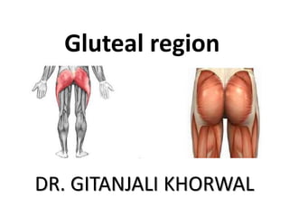
Gluteal region Power point presentation by Dr Monika sharma
PG Dept. of Anatomy,
Shri krishna Ayurvedic govt College, Kurukshetra
The gluteal region refers to the general region of the buttocks that is situated on the posterior aspect of the pelvic girdle. It is bounded anteriorly by the pelvic girdle, superiorly by the iliac crest and inferiorly by the gluteal folds.
The gluteal muscles (buttock muscles) are a muscle group consisting of the gluteus maximus (the largest and thereby strongest muscle in the body), gluteus medius, gluteus minimus and tensor fasciae latae muscles.
Strong gluteals are important for proper pelvic alignment, propulsion during walking and running, and even standing on one leg. Gluteals also help support the lower back during lifting, and help prevent knee injuries.
Prix Galien International 2024 Forum ProgramLevi Shapiro
June 20, 2024, Prix Galien International and Jerusalem Ethics Forum in ROME. Detailed agenda including panels:
- ADVANCES IN CARDIOLOGY: A NEW PARADIGM IS COMING
- WOMEN’S HEALTH: FERTILITY PRESERVATION
- WHAT’S NEW IN THE TREATMENT OF INFECTIOUS,
ONCOLOGICAL AND INFLAMMATORY SKIN DISEASES?
- ARTIFICIAL INTELLIGENCE AND ETHICS
- GENE THERAPY
- BEYOND BORDERS: GLOBAL INITIATIVES FOR DEMOCRATIZING LIFE SCIENCE TECHNOLOGIES AND PROMOTING ACCESS TO HEALTHCARE
- ETHICAL CHALLENGES IN LIFE SCIENCES
- Prix Galien International Awards Ceremony
The prostate is an exocrine gland of the male mammalian reproductive system
It is a walnut-sized gland that forms part of the male reproductive system and is located in front of the rectum and just below the urinary bladder
Function is to store and secrete a clear, slightly alkaline fluid that constitutes 10-30% of the volume of the seminal fluid that along with the spermatozoa, constitutes semen
A healthy human prostate measures (4cm-vertical, by 3cm-horizontal, 2cm ant-post ).
It surrounds the urethra just below the urinary bladder. It has anterior, median, posterior and two lateral lobes
It’s work is regulated by androgens which are responsible for male sex characteristics
Generalised disease of the prostate due to hormonal derangement which leads to non malignant enlargement of the gland (increase in the number of epithelial cells and stromal tissue)to cause compression of the urethra leading to symptoms (LUTS
New Directions in Targeted Therapeutic Approaches for Older Adults With Mantl...i3 Health
i3 Health is pleased to make the speaker slides from this activity available for use as a non-accredited self-study or teaching resource.
This slide deck presented by Dr. Kami Maddocks, Professor-Clinical in the Division of Hematology and
Associate Division Director for Ambulatory Operations
The Ohio State University Comprehensive Cancer Center, will provide insight into new directions in targeted therapeutic approaches for older adults with mantle cell lymphoma.
STATEMENT OF NEED
Mantle cell lymphoma (MCL) is a rare, aggressive B-cell non-Hodgkin lymphoma (NHL) accounting for 5% to 7% of all lymphomas. Its prognosis ranges from indolent disease that does not require treatment for years to very aggressive disease, which is associated with poor survival (Silkenstedt et al, 2021). Typically, MCL is diagnosed at advanced stage and in older patients who cannot tolerate intensive therapy (NCCN, 2022). Although recent advances have slightly increased remission rates, recurrence and relapse remain very common, leading to a median overall survival between 3 and 6 years (LLS, 2021). Though there are several effective options, progress is still needed towards establishing an accepted frontline approach for MCL (Castellino et al, 2022). Treatment selection and management of MCL are complicated by the heterogeneity of prognosis, advanced age and comorbidities of patients, and lack of an established standard approach for treatment, making it vital that clinicians be familiar with the latest research and advances in this area. In this activity chaired by Michael Wang, MD, Professor in the Department of Lymphoma & Myeloma at MD Anderson Cancer Center, expert faculty will discuss prognostic factors informing treatment, the promising results of recent trials in new therapeutic approaches, and the implications of treatment resistance in therapeutic selection for MCL.
Target Audience
Hematology/oncology fellows, attending faculty, and other health care professionals involved in the treatment of patients with mantle cell lymphoma (MCL).
Learning Objectives
1.) Identify clinical and biological prognostic factors that can guide treatment decision making for older adults with MCL
2.) Evaluate emerging data on targeted therapeutic approaches for treatment-naive and relapsed/refractory MCL and their applicability to older adults
3.) Assess mechanisms of resistance to targeted therapies for MCL and their implications for treatment selection
Title: Sense of Taste
Presenter: Dr. Faiza, Assistant Professor of Physiology
Qualifications:
MBBS (Best Graduate, AIMC Lahore)
FCPS Physiology
ICMT, CHPE, DHPE (STMU)
MPH (GC University, Faisalabad)
MBA (Virtual University of Pakistan)
Learning Objectives:
Describe the structure and function of taste buds.
Describe the relationship between the taste threshold and taste index of common substances.
Explain the chemical basis and signal transduction of taste perception for each type of primary taste sensation.
Recognize different abnormalities of taste perception and their causes.
Key Topics:
Significance of Taste Sensation:
Differentiation between pleasant and harmful food
Influence on behavior
Selection of food based on metabolic needs
Receptors of Taste:
Taste buds on the tongue
Influence of sense of smell, texture of food, and pain stimulation (e.g., by pepper)
Primary and Secondary Taste Sensations:
Primary taste sensations: Sweet, Sour, Salty, Bitter, Umami
Chemical basis and signal transduction mechanisms for each taste
Taste Threshold and Index:
Taste threshold values for Sweet (sucrose), Salty (NaCl), Sour (HCl), and Bitter (Quinine)
Taste index relationship: Inversely proportional to taste threshold
Taste Blindness:
Inability to taste certain substances, particularly thiourea compounds
Example: Phenylthiocarbamide
Structure and Function of Taste Buds:
Composition: Epithelial cells, Sustentacular/Supporting cells, Taste cells, Basal cells
Features: Taste pores, Taste hairs/microvilli, and Taste nerve fibers
Location of Taste Buds:
Found in papillae of the tongue (Fungiform, Circumvallate, Foliate)
Also present on the palate, tonsillar pillars, epiglottis, and proximal esophagus
Mechanism of Taste Stimulation:
Interaction of taste substances with receptors on microvilli
Signal transduction pathways for Umami, Sweet, Bitter, Sour, and Salty tastes
Taste Sensitivity and Adaptation:
Decrease in sensitivity with age
Rapid adaptation of taste sensation
Role of Saliva in Taste:
Dissolution of tastants to reach receptors
Washing away the stimulus
Taste Preferences and Aversions:
Mechanisms behind taste preference and aversion
Influence of receptors and neural pathways
Impact of Sensory Nerve Damage:
Degeneration of taste buds if the sensory nerve fiber is cut
Abnormalities of Taste Detection:
Conditions: Ageusia, Hypogeusia, Dysgeusia (parageusia)
Causes: Nerve damage, neurological disorders, infections, poor oral hygiene, adverse drug effects, deficiencies, aging, tobacco use, altered neurotransmitter levels
Neurotransmitters and Taste Threshold:
Effects of serotonin (5-HT) and norepinephrine (NE) on taste sensitivity
Supertasters:
25% of the population with heightened sensitivity to taste, especially bitterness
Increased number of fungiform papillae
Explore natural remedies for syphilis treatment in Singapore. Discover alternative therapies, herbal remedies, and lifestyle changes that may complement conventional treatments. Learn about holistic approaches to managing syphilis symptoms and supporting overall health.
Lung Cancer: Artificial Intelligence, Synergetics, Complex System Analysis, S...Oleg Kshivets
RESULTS: Overall life span (LS) was 2252.1±1742.5 days and cumulative 5-year survival (5YS) reached 73.2%, 10 years – 64.8%, 20 years – 42.5%. 513 LCP lived more than 5 years (LS=3124.6±1525.6 days), 148 LCP – more than 10 years (LS=5054.4±1504.1 days).199 LCP died because of LC (LS=562.7±374.5 days). 5YS of LCP after bi/lobectomies was significantly superior in comparison with LCP after pneumonectomies (78.1% vs.63.7%, P=0.00001 by log-rank test). AT significantly improved 5YS (66.3% vs. 34.8%) (P=0.00000 by log-rank test) only for LCP with N1-2. Cox modeling displayed that 5YS of LCP significantly depended on: phase transition (PT) early-invasive LC in terms of synergetics, PT N0—N12, cell ratio factors (ratio between cancer cells- CC and blood cells subpopulations), G1-3, histology, glucose, AT, blood cell circuit, prothrombin index, heparin tolerance, recalcification time (P=0.000-0.038). Neural networks, genetic algorithm selection and bootstrap simulation revealed relationships between 5YS and PT early-invasive LC (rank=1), PT N0—N12 (rank=2), thrombocytes/CC (3), erythrocytes/CC (4), eosinophils/CC (5), healthy cells/CC (6), lymphocytes/CC (7), segmented neutrophils/CC (8), stick neutrophils/CC (9), monocytes/CC (10); leucocytes/CC (11). Correct prediction of 5YS was 100% by neural networks computing (area under ROC curve=1.0; error=0.0).
CONCLUSIONS: 5YS of LCP after radical procedures significantly depended on: 1) PT early-invasive cancer; 2) PT N0--N12; 3) cell ratio factors; 4) blood cell circuit; 5) biochemical factors; 6) hemostasis system; 7) AT; 8) LC characteristics; 9) LC cell dynamics; 10) surgery type: lobectomy/pneumonectomy; 11) anthropometric data. Optimal diagnosis and treatment strategies for LC are: 1) screening and early detection of LC; 2) availability of experienced thoracic surgeons because of complexity of radical procedures; 3) aggressive en block surgery and adequate lymph node dissection for completeness; 4) precise prediction; 5) adjuvant chemoimmunoradiotherapy for LCP with unfavorable prognosis.
Couples presenting to the infertility clinic- Do they really have infertility...Sujoy Dasgupta
Dr Sujoy Dasgupta presented the study on "Couples presenting to the infertility clinic- Do they really have infertility? – The unexplored stories of non-consummation" in the 13th Congress of the Asia Pacific Initiative on Reproduction (ASPIRE 2024) at Manila on 24 May, 2024.
263778731218 Abortion Clinic /Pills In Harare ,sisternakatoto
263778731218 Abortion Clinic /Pills In Harare ,ABORTION WOMEN’S CLINIC +27730423979 IN women clinic we believe that every woman should be able to make choices in her pregnancy. Our job is to provide compassionate care, safety,affordable and confidential services. That’s why we have won the trust from all generations of women all over the world. we use non surgical method(Abortion pills) to terminate…Dr.LISA +27730423979women Clinic is committed to providing the highest quality of obstetrical and gynecological care to women of all ages. Our dedicated staff aim to treat each patient and her health concerns with compassion and respect.Our dedicated group ABORTION WOMEN’S CLINIC +27730423979 IN women clinic we believe that every woman should be able to make choices in her pregnancy. Our job is to provide compassionate care, safety,affordable and confidential services. That’s why we have won the trust from all generations of women all over the world. we use non surgical method(Abortion pills) to terminate…Dr.LISA +27730423979women Clinic is committed to providing the highest quality of obstetrical and gynecological care to women of all ages. Our dedicated staff aim to treat each patient and her health concerns with compassion and respect.Our dedicated group of receptionists, nurses, and physicians have worked together as a teamof receptionists, nurses, and physicians have worked together as a team wwww.lisywomensclinic.co.za/
Tom Selleck Health: A Comprehensive Look at the Iconic Actor’s Wellness Journeygreendigital
Tom Selleck, an enduring figure in Hollywood. has captivated audiences for decades with his rugged charm, iconic moustache. and memorable roles in television and film. From his breakout role as Thomas Magnum in Magnum P.I. to his current portrayal of Frank Reagan in Blue Bloods. Selleck's career has spanned over 50 years. But beyond his professional achievements. fans have often been curious about Tom Selleck Health. especially as he has aged in the public eye.
Follow us on: Pinterest
Introduction
Many have been interested in Tom Selleck health. not only because of his enduring presence on screen but also because of the challenges. and lifestyle choices he has faced and made over the years. This article delves into the various aspects of Tom Selleck health. exploring his fitness regimen, diet, mental health. and the challenges he has encountered as he ages. We'll look at how he maintains his well-being. the health issues he has faced, and his approach to ageing .
Early Life and Career
Childhood and Athletic Beginnings
Tom Selleck was born on January 29, 1945, in Detroit, Michigan, and grew up in Sherman Oaks, California. From an early age, he was involved in sports, particularly basketball. which played a significant role in his physical development. His athletic pursuits continued into college. where he attended the University of Southern California (USC) on a basketball scholarship. This early involvement in sports laid a strong foundation for his physical health and disciplined lifestyle.
Transition to Acting
Selleck's transition from an athlete to an actor came with its physical demands. His first significant role in "Magnum P.I." required him to perform various stunts and maintain a fit appearance. This role, which he played from 1980 to 1988. necessitated a rigorous fitness routine to meet the show's demands. setting the stage for his long-term commitment to health and wellness.
Fitness Regimen
Workout Routine
Tom Selleck health and fitness regimen has evolved. adapting to his changing roles and age. During his "Magnum, P.I." days. Selleck's workouts were intense and focused on building and maintaining muscle mass. His routine included weightlifting, cardiovascular exercises. and specific training for the stunts he performed on the show.
Selleck adjusted his fitness routine as he aged to suit his body's needs. Today, his workouts focus on maintaining flexibility, strength, and cardiovascular health. He incorporates low-impact exercises such as swimming, walking, and light weightlifting. This balanced approach helps him stay fit without putting undue strain on his joints and muscles.
Importance of Flexibility and Mobility
In recent years, Selleck has emphasized the importance of flexibility and mobility in his fitness regimen. Understanding the natural decline in muscle mass and joint flexibility with age. he includes stretching and yoga in his routine. These practices help prevent injuries, improve posture, and maintain mobilit
Ethanol (CH3CH2OH), or beverage alcohol, is a two-carbon alcohol
that is rapidly distributed in the body and brain. Ethanol alters many
neurochemical systems and has rewarding and addictive properties. It
is the oldest recreational drug and likely contributes to more morbidity,
mortality, and public health costs than all illicit drugs combined. The
5th edition of the Diagnostic and Statistical Manual of Mental Disorders
(DSM-5) integrates alcohol abuse and alcohol dependence into a single
disorder called alcohol use disorder (AUD), with mild, moderate,
and severe subclassifications (American Psychiatric Association, 2013).
In the DSM-5, all types of substance abuse and dependence have been
combined into a single substance use disorder (SUD) on a continuum
from mild to severe. A diagnosis of AUD requires that at least two of
the 11 DSM-5 behaviors be present within a 12-month period (mild
AUD: 2–3 criteria; moderate AUD: 4–5 criteria; severe AUD: 6–11 criteria).
The four main behavioral effects of AUD are impaired control over
drinking, negative social consequences, risky use, and altered physiological
effects (tolerance, withdrawal). This chapter presents an overview
of the prevalence and harmful consequences of AUD in the U.S.,
the systemic nature of the disease, neurocircuitry and stages of AUD,
comorbidities, fetal alcohol spectrum disorders, genetic risk factors, and
pharmacotherapies for AUD.
Title: Sense of Smell
Presenter: Dr. Faiza, Assistant Professor of Physiology
Qualifications:
MBBS (Best Graduate, AIMC Lahore)
FCPS Physiology
ICMT, CHPE, DHPE (STMU)
MPH (GC University, Faisalabad)
MBA (Virtual University of Pakistan)
Learning Objectives:
Describe the primary categories of smells and the concept of odor blindness.
Explain the structure and location of the olfactory membrane and mucosa, including the types and roles of cells involved in olfaction.
Describe the pathway and mechanisms of olfactory signal transmission from the olfactory receptors to the brain.
Illustrate the biochemical cascade triggered by odorant binding to olfactory receptors, including the role of G-proteins and second messengers in generating an action potential.
Identify different types of olfactory disorders such as anosmia, hyposmia, hyperosmia, and dysosmia, including their potential causes.
Key Topics:
Olfactory Genes:
3% of the human genome accounts for olfactory genes.
400 genes for odorant receptors.
Olfactory Membrane:
Located in the superior part of the nasal cavity.
Medially: Folds downward along the superior septum.
Laterally: Folds over the superior turbinate and upper surface of the middle turbinate.
Total surface area: 5-10 square centimeters.
Olfactory Mucosa:
Olfactory Cells: Bipolar nerve cells derived from the CNS (100 million), with 4-25 olfactory cilia per cell.
Sustentacular Cells: Produce mucus and maintain ionic and molecular environment.
Basal Cells: Replace worn-out olfactory cells with an average lifespan of 1-2 months.
Bowman’s Gland: Secretes mucus.
Stimulation of Olfactory Cells:
Odorant dissolves in mucus and attaches to receptors on olfactory cilia.
Involves a cascade effect through G-proteins and second messengers, leading to depolarization and action potential generation in the olfactory nerve.
Quality of a Good Odorant:
Small (3-20 Carbon atoms), volatile, water-soluble, and lipid-soluble.
Facilitated by odorant-binding proteins in mucus.
Membrane Potential and Action Potential:
Resting membrane potential: -55mV.
Action potential frequency in the olfactory nerve increases with odorant strength.
Adaptation Towards the Sense of Smell:
Rapid adaptation within the first second, with further slow adaptation.
Psychological adaptation greater than receptor adaptation, involving feedback inhibition from the central nervous system.
Primary Sensations of Smell:
Camphoraceous, Musky, Floral, Pepperminty, Ethereal, Pungent, Putrid.
Odor Detection Threshold:
Examples: Hydrogen sulfide (0.0005 ppm), Methyl-mercaptan (0.002 ppm).
Some toxic substances are odorless at lethal concentrations.
Characteristics of Smell:
Odor blindness for single substances due to lack of appropriate receptor protein.
Behavioral and emotional influences of smell.
Transmission of Olfactory Signals:
From olfactory cells to glomeruli in the olfactory bulb, involving lateral inhibition.
Primitive, less old, and new olfactory systems with different path
New Drug Discovery and Development .....NEHA GUPTA
The "New Drug Discovery and Development" process involves the identification, design, testing, and manufacturing of novel pharmaceutical compounds with the aim of introducing new and improved treatments for various medical conditions. This comprehensive endeavor encompasses various stages, including target identification, preclinical studies, clinical trials, regulatory approval, and post-market surveillance. It involves multidisciplinary collaboration among scientists, researchers, clinicians, regulatory experts, and pharmaceutical companies to bring innovative therapies to market and address unmet medical needs.
2. Gluteal region
• The transitional area between the trunk and
the lower extremity.
• The gluteal region includes the rounded,
posterior buttocks and the laterally placed hip
region.
5. Gluteal Aponeurosis
• This is attached to the
lateral border of the
iliac crest superiorly,
and
• splits anteriorly to
enclose tensor fasciae
latae and posteriorly
to enclose gluteus
maximus.
6.
7. Muscles of Gluteal region
Superficial Layer
• Gluteus maximus
• Tensor fasciae latae
8. Muscles of Gluteal region
Intermediate layer
• Gluteus medius
• Piriformis
• Superior gemellus.
• Tendon of obturator
internus.
• Inferior gemellus
• Quadratus femoris
• Upper part of
Adductor magnus
• And Hamstrings
9. Muscles of Gluteal region
Deep layer
• Gluteus minimus
• Reflected head of
rectus femoris
• Tendinous insertion
of obturator
externus
10. Gluteus Maximus
Origins: posterior end of the iliac crest,
posterior surface of the sacrum, coccyx and
sacrotuberous ligament.
Insertions: ilio-tibial tract( 3/4)and gluteal
tuberosity.(1/4 )
Innervation: inferior gluteal nerve - [ Ventral
rami of L5, S1,2] - emerges below the
piriformis muscle to penetrate the deep
surface of the gluteus maximus with
accompanying vessels.
11. Actions
• Extensor at hip joint during
running and climbing upstairs.
• Chief antigravity muscle in the
standing up from a seated position.
• Strong lateral rotation of the thigh.
Its upper fibres are active in
powerful abduction of the thigh.
• It is a tensor of the fascia lata, and
through the iliotibial tract it
stabilizes the femur on the tibia
when the extensor muscles of the
knee are relaxed.
12. Tensor Fascia Lata
Small muscle close to the anterior
border of the gluteus medius, at the dorsal
surface of the ASIS.
Origin: outer lip of iliac crest from ASIS to
tubercle of iliac crest.
Insertion: ilio-tibial tract.
Innervation - superior gluteal nerve.
Action - helps in flexion and abduction of the
thigh. Maintains extension of knee joint.
13. Structures under cover of gluteus maximus
• Bones
• Ligaments
• Bursae
Trochanteric
Gluteofemoral
Ischial
• Muscles
• Blood vessels and
• Nerves
• Arterial Anastomosis
Trochanteric
cruciate
16. GLUTEUS MEDIUS
Covered partially by Gluteus maximus
Origins: dorsal surface of the ilium
between the anterior and posterior
gluteal lines and from the gluteal
aponeurosis.
Insertion: lateral surface of the greater trochanter on an
oblique ridge.
17. GLUTEUS MINIMUS
Covered completely by Gluteus
medius.
Origins: gluteal surface of the ilium
between the anterior and inferior
gluteal lines upto margin of greater
sciatic notch.
Insertion: lateral part of anterior surface of the greater
trochanter.
18. • Innervation of Gluteus medius and minimus:
superior gluteal nerve [L4, 5, S1] – that emerges
above the piriformis muscle, with accompanying
vessels, to penetrate the deep surface of the
muscle.
• Actions
Abduction of the thigh and medial rotation.
Preventing the unsupported side of pelvis from
sagging downward during locomotion.
Lurching Gait
20. • Trendelenburgs sign is positive in
paralysis of gluteus medius & minimus,
congenital dislocation of hip joint,
fracture of the neck of femur
21. Piriformis
Origin: antero-lateral
surface and border of the
sacrum.
Insertion: the fibers are emerge laterally through the
greater sciatic foramen as a narrow tendon attached to
the posterior inturned upper border of the greater
trochanter.
Innervation - “nerve to the piriformis” [S1, 2.]
Action - lateral rotator and abductor of the thigh.
23. Obturator internus
• Origin : inner surface
of obturator membrane and
Adjoining ischio-pubic ramus.
• Insertion: Tendon makes a right
angle bend at lesser sciatic foramen
to insert to the medial surface of
greater trochanter above and in
front of the trochanteric fossa
24. Obturator internus
• It is accompanied by Superior and
Inferior Gemelli and insert at
superior and inferior margin of
the insertion of obturator
internus.
• Superior Gemellus from ischial
spine.
• Inferior Gemellus from lower
margin of lesser sciatic notch.
25. Obturator internus
• Nerve supply :
Nerve to obturator internus also
supplies Sup. Gemellus (L5,S1,S2)
Inf. Gemellus is supplied by nerve
to Quadratus femoris (L4,L5,S1)
• Action:
Lateral rotation at Hip joint
26. Quadratus Femoris
• Origin:
Linear origin from external surface
of ischial tuberosity.
• Insertion:
Quadrate tubercle near middle of
intertrochanteric crest.
• Innervation: nerve to Quadratus
femoris (L4,L5,S1)
• Action: Lateral rotation of hip
32. Superior gluteal nerve
Ventral rami of
L4, L5, S1
Inferior gluteal nerve
Ventral rami of
L5, S1, S2
Pudendal nerve
Ventral rami of
S2, S3, S4
33. Posterior femoral cutaneous nerve
D (S1, S2)
V (S2, S3)
It descends on the back of
the thigh, and in the popliteal
fossa it pierces the deep fascia
and supplies the skin on the back
of the thigh and the upper part
of the leg
Branches:
a) Gluteal
b) Perineal
c) Perforating
34. • Nerve to obturator
internus
Ventral rami of
V (L5, S1, S2)
• Nerve to quadrator
femoris
Ventral rami of
V (L4, L5, S1)
37. • Superior gluteal artery divides into
Superficial branch
Deep branch- upper branch and lower branch
SPINOUS ANASTOMOSIS
1. Upper branch of Superior
gluteal artery
2. Superficial and deep
circumflex iliac arteries
3. Ascending branch of lateral
circumflex femoral artery
4. Iliac branch of ilio-lumbar
artery.
38. • Superior gluteal artery
Branch of posterior division of Internal Iliac artery.
Branches:-
a) Muscular branches
b) Anastomotic branches
c) Arteria nervi ischiadici
• Internal pudendal artery
Branch of anterior
division of Internal Iliac artery
39.
40. CRUCIATE ANASTOMOSIS
•Descending branch of inferior
gluteal artery
•Ascending branch of 1st
perforating artery
•Medially- transverse branch of
medial circumflex femoral
artery
•Laterally- transverse branch of
lateral circumflex femoral artery
43. Trochanteric anastomosis
Descending branch of superior gluteal artery
ascending branches of medial & lateral
circumflex femoral artery
Branch from inferior gluteal artery
situated near the trochanteric fossa of the
femur & supplies the head of femur and
retinacular fibers of neck
45. Hamstring Muscles
Common name applied to the muscles in the
Posterior compartment.
They have a common origin from the ischial
tuberosity and crosses knee joint to insert on tibia
or fibula.
They are innervated by the tibial component of
sciatic nerve.
They also have a common primary function of
flexing the leg, but they also help to extend and
adduct the thigh.
Their blood supply comes principally from the
perforating branches of the deep femoral artery.
46. adductor magnus
long head of biceps femoris
semitendinosus
semimembranosus short head of biceps femoris
popliteal vessels in the
popliteal fossa
sartorius
gracilis
47. Origin - Common from Ischial tuberosity.
Insertion - One of the leg bones.
Nerve supply-Tibial part of sciatic nerve.
Common action Extensors of hip joint.
Flexors of knee joint.
48. True hamstrings-
• Semimembranosus
• Semitendinosus
Modified hamstrings
•Long head of biceps femoris-
•Sacrotuberous ligament morphologically
degenerated part.
•Ischial head of adductor magnus-
Tibial collateral ligament represents the
morphological degenerated part of adductor magnus
49. Biceps Femoris - most lateral muscled with
a “long head” from the ischial tuberosity,
and a “short head” from the middle of the
linea aspera and the lateral supracondylar
ridge.
*The two heads unite to form a common
tendon, which deviates lateral to its
insertion into the apex of the head of the
fibula where it is joined by an extension of
the iliotibial tract.
•The short head receives a branch from
the common peroneal nerve; it also helps
in the lateral rotation of the leg.
50. Semitendinosus - usually fusiform tapering
distally into a long cylindrical tendon at the
popliteal region to be inserted to the upper
medial surface of the tibia, adjacent to the
attachments of the sartorius and gracilis.
Semimembranosus - usually has a fleshy belly
that form a thick flattened tendon that inserts at
the back of the medial condyle of the tibia, the
tendon contributes to the formation of the
“oblique popliteal ligament” of the knee joint,
which reinforces the posterior capsule of the
joint.