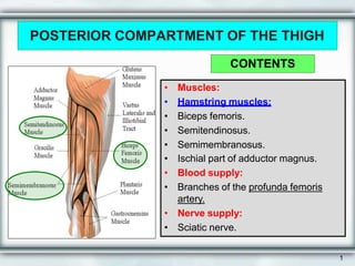
1. POSTERIOR COMPARTMENT OF THE THIGH
• Muscles:
• Hamstring muscles:
• Biceps femoris.
• Semitendinosus.
• Semimembranosus.
• Ischial part of adductor magnus.
• Blood supply:
• Branches of the profunda femoris
artery.
• Nerve supply:
• Sciatic nerve.
CONTENTS
1
2. Biceps Femoris : • Origin:
– The long head from the ischial
tuberosity.
– The short head from the linea
aspera .
• Insertion:
• Mainly into the head of the fibula.
Nerve supply:
• The long head is supplied by the tibial
part of sciatic;
• The short head is supplied by the
common peroneal part of the sciatic.
Action :
• Flexion of knee.
• Lateral rotation of flexed leg.
• Long head: extends hip.
Lateral
rotation
2
Medial
rotation
3. SEMITENDINOSUS
• Origin:
• Ischial tuberosity.
• Insertion:
• Upper part of the medial
surface of the shaft of the
tibia (SGS).
Nerve supply:
• Tibial portion of the sciatic.
Action:
• Flexes and medially rotates
the leg at the knee joint;
• Extends the thigh at the hip
joint.
3
4. SEMIMEMBRANOSUS
• Origin:
• Ischial tuberosity.
• Insertion:
• Posterior surface of the medial
condyle of the tibia.
• It forms the oblique popliteal
ligament, which reinforces the
capsule on the back of the
knee joint.
Nerve supply:
• Tibial portion of the sciatic
nerve.
Action:
• Flexes and medially rotates
the leg at the knee joint;
• Extends the thigh at the hip.
4
5. ADDUCTOR MAGNUS (HAMSTRING PART)
• Origin:
• Ischial ramus and ischial
tuberosity
• Insertion:
• Adductor tubercle of the
medial condyle of the
femur.
• Nerve supply:
• The tibial portion of the
sciatic.
• Action:
• Extends the thigh at the
hip joint.
5
6. BLOOD SUPPLY
• The four perforating branches
of the profunda femoris artery
(deep artery of thigh) provide a
rich blood supply to this
compartment.
• The profunda femoris vein
drains the greater part of the
blood from the compartment.
6
7. NERVE SUPPLY
• Sciatic Nerve
• The sciatic nerve, is a
branch of the sacral
plexus (L4 and 5; S1, 2,
and 3), leaves the gluteal
region as it descends in
the midline of the thigh.
• It lies on the posterior
aspect of the adductor
magnus.
• In the lower third of the
thigh it ends by dividing
into tibial and common
peroneal nerves.
7
8. • Diamond shaped space
• Behind knee joint
• Formed between muscles
• Posterior compartment of thigh
and leg
Popleteal fossa
9. Location
• Diamond shaped space
• Behind knee joint
• Formed between muscles
• Posterior compartment of
thigh and leg
Outline of right
popliteal fossa
10. Boundaries
• Superolaterally: biceps femoris
• Superomedially: semitendinosus,
semimembranosus
supplemented: gracilis, sartorius,
adductor magnus
• Inferolaterally: lateral head of
gastrocnemius,Plantaris
• Inferomedially: medial head of
gastrocnemius
11. Floor
Above downwards
• Popliteal surface of
femur
• Capsule of knee
joint
• Oblique popliteal
ligament
• Popliteal fascia
• Popliteal muscle
13. Superficial Fascia Over Roof
• Short saphenous vein
• Cutaneous nerve
-Posterior cutaneous nerve of
thigh
-Posterior division of medial
cutaneous Nerve of thigh
-Peroneal (sural)
communicating nerve
14. Content
Popliteal vessels and its branch
Tibial nerve and its branches
Common peroneal nerve and
its branches
Genicular branch of
obturator nerve Popliteal
lymph nodes
15. Relations In Fossa
• Upper part (medial
to lateral) Artery,
vein, nerve
• Middle part (superficial
to deep) Nerve, vein,
artery
• Lower part (medial
to lateral) Nerve,
vein, artery
16. Popliteal vessels
• Popliteal artery
DeepestBranches:
muscularcutaneous
genicular
• Popliteal vein
Continues as femoral vein
Tributaries: small
saphenousvein