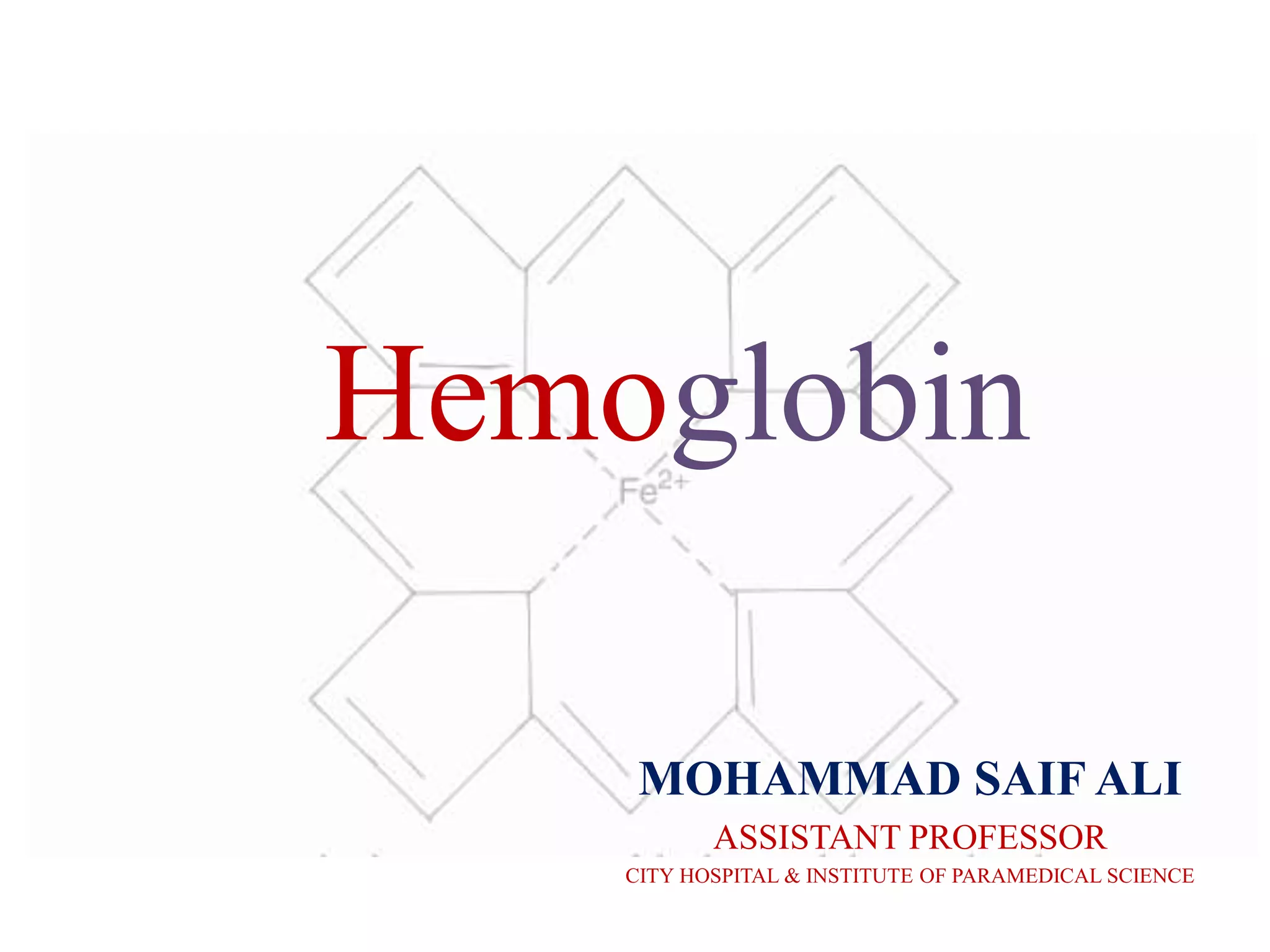
Title: Hemoglobin: The Oxygen-Carrying Marvel of Our Blood
Introduction:
Hemoglobin, a remarkable protein present in our red blood cells, plays a crucial role in maintaining our body's equilibrium. Its extraordinary ability to transport oxygen efficiently throughout the circulatory system ensures our survival and well-being. In this presentation, we will explore the fascinating world of hemoglobin, understanding its structure, function, and importance in sustaining human life.
Slide 1: What is Hemoglobin?
Hemoglobin, often represented as Hb, is a complex protein found in red blood cells (erythrocytes).
It is composed of four subunits, each carrying a heme group that binds to an oxygen molecule.
Slide 2: Structure of Hemoglobin
Display the 3D structure of hemoglobin, emphasizing the four globular subunits and heme groups.
Highlight the iron atom in each heme group, which is essential for oxygen binding.
Slide 3: Oxygen Binding
Describe how oxygen binds to the iron in the heme group, forming oxyhemoglobin.
Explain the cooperative binding nature of hemoglobin, where one oxygen molecule facilitates the binding of others.
Slide 4: Oxygen Release
Illustrate how hemoglobin releases oxygen in tissues with lower oxygen concentrations.
Discuss factors that influence oxygen release, such as pH and carbon dioxide levels.
Slide 5: Hemoglobin Variants
Introduce different hemoglobin variants, such as HbA, HbF, and HbS.
Briefly explain their unique properties and significance in various medical conditions.
Slide 6: Hemoglobin and Carbon Dioxide Transport
Explore how hemoglobin also aids in the transportation of carbon dioxide from tissues to the lungs.
Describe the formation of carbaminohemoglobin and its role in CO2 transport.
Slide 7: Hemoglobin Disorders
Highlight common hemoglobin disorders like anemia, sickle cell disease, and thalassemia.
Discuss their impact on overall health and the challenges faced by affected individuals.
Slide 8: Diagnostic and Clinical Applications
Showcase the clinical importance of hemoglobin analysis in diagnosing and monitoring various blood-related disorders.
Mention hemoglobin electrophoresis and other diagnostic techniques used in the assessment.
Slide 9: Regulation and Homeostasis
Explain how the body maintains a balance of hemoglobin levels to ensure optimal oxygen-carrying capacity.
Discuss the role of erythropoietin and iron metabolism in hemoglobin regulation.
Slide 10: Conclusion
Summarize the vital role of hemoglobin in transporting oxygen and maintaining physiological balance.
Emphasize the significance of understanding hemoglobin in clinical contexts and the advancement of medical treatments.
2. CHARACTERISTICS OF HEMOGLOBIN
• Hemoglobin is a conjugated protein synthesized
inside the immature erythrocyte in the red bone
marrow,
• It consists of two components
1) Heme (iron + protoporphyrin)
2) 2) Globin (amino acid chains).
It gives red color to blood.
3. Cont…
• In 1862, Felix Seyler identified the respiratory
protein hemoglobin,
• He discovered the characteristic color
spectrum of hemoglobin and proved that this
was the true coloring matter of the blood.
4.
5. Chemical Composition of Hemoglobin
• Normal adult hemoglobin (hemoglobin A) consists of four
heme groups and four polypeptide chains with a total of
574 amino acids.
• The polypeptide chains are organized into two alpha chains
and two beta chains.
• Each of the chains has an attached heme group
• Normal adult hemoglobin has 141 amino acids in each of
the alpha chains and 146 amino acids in each of the beta
chains.
• The specific sequence of these amino acids is known and is
important in the identification of abnormal hemoglobins
involving substitutions of specific amino acids.
6. Role of 2,3-BPG
• function of the hemoglobin molecule is the
transport of oxygen to the tissues.
• oxygen affinity of the Hb molecule is regulated by the
concentration of phosphates, particularly 2,3-
diphosphoglycerate also called 2,3-
biphosphoglycerate (2,3-DPG or 2,3-BPG).
• 2,3-DPG combines with the beta chains of
deoxyhemoglobin and diminishes the molecule's
affinity for oxygen.
7. • When the individual heme groups unload oxygen in
the tissues, the beta chains are pulled apart. This
permits the entrance of 2,3-DPG and the
establishment of salt bridges between the individual
chain
8. Hemoglobin Synthesis
• The heme part of hemoglobin is synthesized in a
series of steps in the mitochondria and the cytosol of
immature red blood cells,
• while the globin protein parts are synthesized by
ribosomes in the cytosol.
• Production of hemoglobin continues in the cell
throughout its early development from the
proerythroblast to the reticulocyte in the bone
marrow.
11. Globin Structure and Synthesis
• Both the structure and the production of
globin in the hemoglobin molecule are under
genetic control .
• The specific sequences of amino acids are
governed by the triplet code of DNA bases,
which are genetically inherited.
1. Alpha Globin Locus
2. Beta Globin Locus
12. Alpha Globin Locus
• Each chromosome 16 has two alpha globin
genes that are aligned one after the other on
the chromosome.
• For practical purposes, the two alpha globin
genes are identical.
13. Cont…
• Each cell has two chromosomes 16; a total of four
alpha globin genes exist in each cell.
• Each of the four genes produces about one
quarter of the alpha globin chains needed for
hemoglobin synthesis.
• The mechanism of this coordination is unknown
• The transiently expressed embryonic genes that
substitute for alpha very early in development
designated zeta, are also in the alpha globin
locus.
14.
15. Beta Globin Locus
• The genes in the beta globin locus are arranged
sequentially.
• The sequence of the genes is epsilon, gamma, delta,
and beta
• There are two copies of the gamma gene on each
chromosome 11.
16. Cont…
• The others are present in single copies.
• Each cell has two beta globin genes, one on each of
the two chromosomes 11 in the cell.
• These two beta globin genes express their globin
protein in a quantity that precisely matches that of
the four alpha globin genes.
• The polypeptide chains of globin are produced as are
other body proteins, on the ribosomes.
17. Cont…
• The alpha polypeptide chain unites with one
of three other chains to form a dimer and
ultimately a tetramer. In normal adult
hemoglobin (hemoglobin A), these chains are
two alpha and two beta chains.
18. Assembly of a Hemoglobin Molecule
• A hemoglobin molecule is assembled in sequential
steps
• These steps are as follows:
1. The α- and β -globin polypeptides are translated from
appropriate mRNAs
2. Once heme containing iron binds to each of the four
polypeptide chains, the protein folds into a native 3-D
structure.
3. The α and β units bind to each other by electrostatic
bonding.
4. Two dimers combine to form the functional
hemoglobin molecule, an α2 β2 tetramer
19. ONTOGENY OF HEMOGLOBIN
• In normal human development, several types
of hemoglobin are produced.
• These hemoglobin types are hemoglobin A
and a sub-fraction A1, hemoglobin A2, fetal
hemoglobin, and embryonic hemoglobin.
• Each of these hemoglobin types has a
distinctive composition of polypeptide chains.
20.
21. Embryonic hemoglobins
• Formed by immature erythrocytes in the yolk sac.
• These hemoglobins include Gower I, Gower II,
and Portland types.
• They are found in the human embryo and persist
until approximately 12 weeks of gestation.
• In these hemoglobin, the zeta chain is analogous
to the alpha chain of fetal and adult hemoglobin
and may combine with epsilon or gamma chains
to form various embryonic hemoglobin types.
22. Fetal hemoglobin (HbF)
• Fetal hemoglobin (hemoglobin F) is the predominant
hemoglobin variety in the fetus and the newborn.
• This hemoglobin type has two alpha and two gamma
chains.
• Gradually, hemoglobin F is replaced in the circulating
erythrocytes until the normal adult level of
hemoglobin F (less than 2%) is attained.
23. Hemoglobin A
• The shift in hemoglobin type from fetal to adult
hemoglobin reflects transcription of the β-
globulin chain.
• Adult hemoglobin is predominantly of the A
variety (95% to 97%)
• Hemoglobin A is composed of two alpha and two
beta polypeptide chains.
• Hemoglobin A2 is composed of two alpha and
two delta chains.
• the concentration of hemoglobin A2 in a normal
adult averages 2.5% of the total hemoglobin
24. Glycosylated Hemoglobin (HbA1c)
• The formation of glycosylated hemoglobin is a
slow, irreversible process that depends on the
concentration of glucose in the body.
• Concentration of hemoglobin A1 is 3% to 6% in
normal persons and 6% to 12% in both insulin-
dependent and non-insulin-dependent
diabetics.
25. Variant Forms Of Normal Hemoglobin
• Carboxyhemoglobin, sulfhemoglobin, and
methemoglobin are known as variant forms of
normal hemoglobin or dyshemoglobins.
• Unlike abnormal hemoglobin with permanent
structural rearrangements of the hemoglobin
molecule,
• These variants are typified by differing from normal
hemoglobin only by the molecule that replaces
oxygen.
• These dysfunctional hemoglobin are unable to
transport oxygen
26. Carboxyhemoglobin
• Hemoglobin has the capacity to combine with
carbon monoxide in the same proportion as
with oxygen, but the affinity of the
hemoglobin molecule for carbon monoxide is
210 times greater.
• Carbonmonoxide is highly toxic in
unventilated spaces.
• Carbon monoxide poisoning is the most
common type of accidental poisoning.
27. Sulfhemoglobin
• This variant of hemoglobin contains sulfur.
• In vitro and in the presence of oxygen,
hemoglobin reacts with hydrogen sulfide to
form a greenish derivative called
sulfhemoglobin
• Concentrations of sulfhemoglobin in vivo are
normally less than 1%
• Elevated concentrations result in cyanosis but
are usually other wise asymptomatic.
28. Methemoglobin
• Methemoglobin is a variant of hemoglobin, with iron
in the ferric state, that is incapable of combining with
oxygen.
• It can result from a metabolic defect .
• Normally, 2% methemoglobin is formed each day.
• At this concentration, the abnormal hemoglobin is
not harmful
• Because the reduced ability of the erythrocytes to
carry oxygen is insignificant.
• Cyanosis develops if methemoglobin levels
exceed10%; hypoxia develops if levels exceed 60%.
29. Possible Queations
• What do you understand by hemoglobin?
• Discuss hemoglobin synthesis? Explain
abnormal forms of hemoglobin?
• Explain the role of 2,3-BPG?