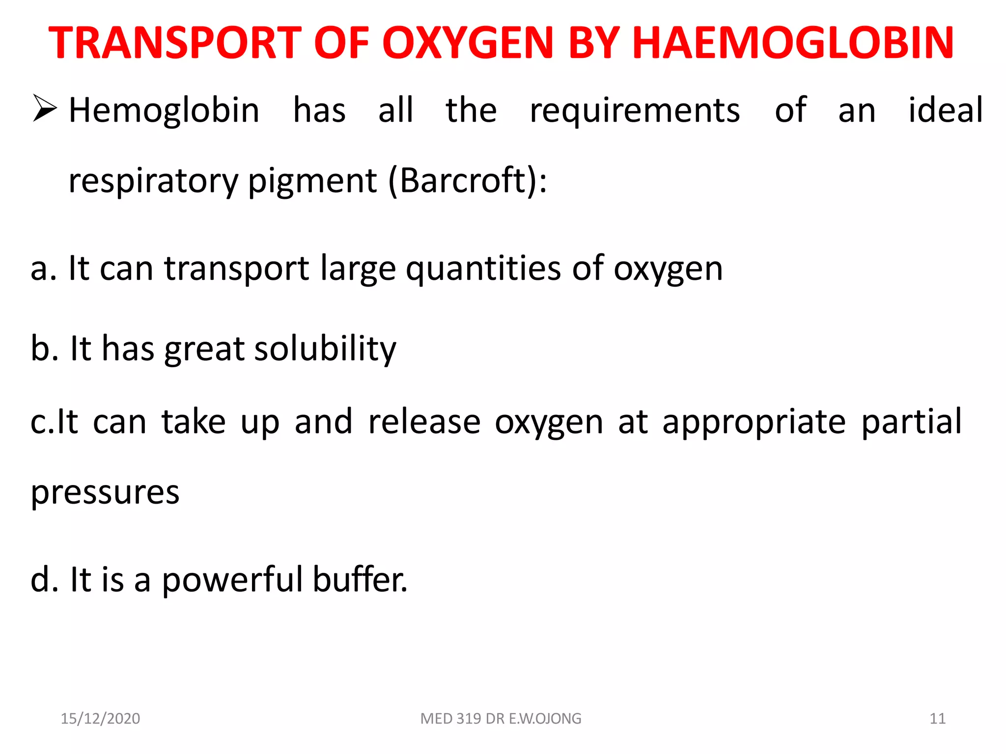
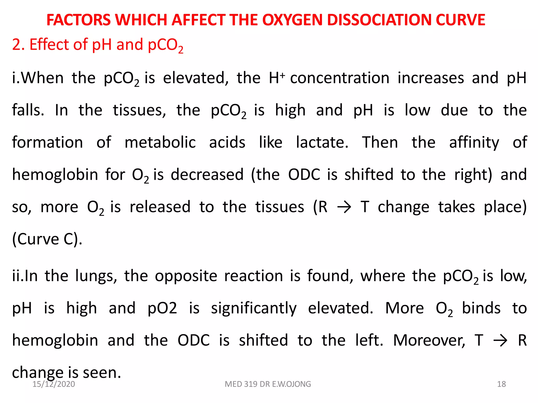
This document discusses the structure and function of haemoglobin and the transport of gases in the blood. It provides a history of discoveries about haemoglobin dating back to the 17th century. Key points covered include the tetrameric structure of haemoglobin, with each subunit binding one heme group and iron atom. Haemoglobin is able to efficiently transport oxygen and carbon dioxide via changes in its quaternary structure and binding of effectors like hydrogen ions, carbon dioxide and 2,3-BPG. The sigmoidal oxygen dissociation curve illustrates haemoglobin's ability to load and unload oxygen in the lungs and tissues respectively. Factors like pH, temperature and organic phosphates influence the curve.
Hemoglobin is the iron-containing protein in red blood cells that carries oxygen from the lungs to tissues and carbon dioxide from tissues back to the lungs. It consists of four polypeptide chains and a heme group, which gives blood its red color. Sickle cell anemia is a genetic blood disorder caused by a mutation in the hemoglobin gene, where glutamic acid is replaced by valine in the beta chain. This causes hemoglobin S to polymerize under low oxygen conditions, distorting red blood cells into a sickle shape and blocking blood vessels.
The document provides information about the digestive system, including both the gastrointestinal tract and accessory digestive organs. It discusses the organs that make up the GI tract, including the mouth, esophagus, stomach, and small and large intestines. It also covers the accessory organs like the liver, gallbladder and pancreas. The document describes the processes of ingestion, digestion, absorption, and defecation. It provides histological details of the layers of the alimentary tract and details of individual digestive organs and their functions.
Proteins are composed of amino acids that link together via peptide bonds. There are 20 naturally occurring amino acids that vary in properties like polarity, charge, and ability to form secondary structures. The sequence and interactions of amino acids give proteins their unique 3D structures and functions. Denaturation disrupts non-covalent bonds within proteins, altering their shapes and eliminating biological activity.
Primary structure of protein
Secondary structure of protein
Tertiary structure of protein
Quaternary structure of protein
Methods to determine protein structure
Conclusion
References
METHODS TO DETERMINE PROTEIN STRUCTURE
Each protein has a unique sequence of amino acids.
The amino acids are held together in a protein by
covalent peptide bonds or linkages.
A peptide bond are formed when amino group of an
amino acid combines with the carboxyl group of another.
The conformation of polypeptide chain by twisting or folding is referred to as secondary structure.
Two types of secondary structures α-helix and β-sheet are mainly identified.
α-Helical structure was proposed by Pauling and Corey in 1951.
It occurs when the sequence of amino acids are linked by hydrogen bonds.
Each turn of α-helix contains 3.6 amino acids.
β-pleated sheets are composed of two or more segments of fully extended peptide chains.
β-Sheets may be arranged either in parallel or anti-parallel direction.
Many globular proteins contain combinations of α-helix and β-pleated sheet secondary structure, these patterns are called supersecondary structures also called motifs.
The three dimensional arrangement of protein structure is referred to as tertiary structure.
It is a compact structure with hydrophobic side chains held interior while the hydrophilic groups are on the surface.
This type of arrangement provide stability of the molecule.
Besides the H-bongs, disulfide bonds, ionic interactions, hydrophobic interactions also contribute to the tertiary structure.
This document provides an overview of lymph nodes, including their anatomy, components, and function. Lymph nodes are oval shaped structures distributed along lymphatic vessels that filter lymph and help trap foreign substances. They have an outer cortex containing lymphoid follicles and T cells, and an inner medulla containing sinuses that drain lymph. Lymph nodes play an important role in the immune system by filtering lymph and providing an environment for immune cells like lymphocytes and antigen-presenting cells to interact with antigens.
The document provides an introduction to biochemistry by Dr. V.P. Acharya. It discusses the pioneers of Indian and Western medicine such as Charaka, Sushruta, and Hippocrates. It then summarizes the evolution of biochemistry from the 19th century to modern times. The rest of the document describes the molecular and functional organization of the cell, including the structure and functions of organelles like the nucleus, endoplasmic reticulum, Golgi apparatus, lysosomes, peroxisomes, mitochondria, and cytoskeleton.
This document summarizes polysaccharides and glycans. It discusses homopolysaccharides including fructosan, galactosan, and glucosans such as starch and glycogen. Starch is made of amylose and amylopectin and forms helical structures with iodine. Cellulose is composed of beta-glucose units linked by beta-1,4 bonds, forming long straight chains strengthened by hydrogen bonds. Glycosaminoglycans discussed include hyaluronic acid, chondroitin sulfate, keratin sulfate, dermatan sulfate, and heparan sulfate. Proteoglycans are composed of core proteins with covalently linked glycosaminoglycan side chains. They
This document provides an introduction to biochemistry, including definitions and key concepts. It discusses biochemistry as the study of the structure and function of biomolecules in living organisms. The document outlines that living things are composed of common elements like carbon, hydrogen, oxygen, nitrogen and complex macromolecules including carbohydrates, proteins, nucleic acids and lipids. It also describes important cellular structures like the cell membrane, nucleus, mitochondria and chloroplasts. Finally, the document provides overviews of the four major macromolecule classes and how pH and buffers are important concepts in biochemistry.
MCQs on General Biochemistry - DentosphereRaman Dhungel
This is only a portion of the MCQs. For viewing the complete list of MCQs, please visit: bit.ly/dentistrymcqs
Multiple Choice Questions in General Biochemistry, Practice these MCQs for scoring better in Competitive Entrance Examinations like AIIMS, NEET MDS, AIPG, BPKIHS,KU and TU Entrance Exams.
This document provides information on porphyrins, their chemistry, metabolism, biosynthesis, and degradation. It discusses how porphyrins bind metallic ions like iron to form metalloporphyrins. The biosynthesis of porphyrins occurs in multiple steps in both the mitochondria and cytosol of liver and red blood cells. Porphyrins are degraded in the spleen, liver, and bone marrow. The document also covers the regulation, clinical features, applications, and conclusions regarding porphyrins and heme synthesis and metabolism.
This document discusses the chemistry of carbohydrates. It states that carbohydrates are synthesized in plants through photosynthesis and are a major source of energy in our diets. Carbohydrates can be classified as monosaccharides, disaccharides, or polysaccharides depending on their size. Important monosaccharides include glucose, fructose, and ribose. Glucose is the primary sugar transported in blood and used for energy by tissues. Carbohydrates exist in solution both as open-chain and cyclic ring forms.
This document provides information about carbohydrates. It begins by defining carbohydrates and describing their main biological functions. It then discusses the three main classes of carbohydrates: monosaccharides, disaccharides, and polysaccharides. For each class, key examples are provided and their structures and properties are explained. The document also covers topics like stereochemistry of carbohydrates, glycosaminoglycans, and important monosaccharides and polysaccharides like starch, cellulose, and glycogen. In summary, it serves as a comprehensive overview of carbohydrate structure, classification, and functions in biological systems.
1. Carbohydrates can be classified as monosaccharides, disaccharides, oligosaccharides, or polysaccharides depending on the number of sugar molecules present.
2. Monosaccharides exist as both open-chain and ring forms, with the ring forms being more stable. The rings can be pyranoses or furanoses depending on whether they have 6 or 5 members.
3. Monosaccharides also exist as optical isomers called enantiomers that are non-superimposable mirror images of each other. Their naming depends on their relation to D-glyceraldehyde.
1) Organisms require chemical energy stored in high-energy compounds for processes like muscle contraction and active transport.
2) High-energy compounds include ATP, phosphoenolpyruvate, and acetyl-CoA, which contain high-energy bonds like phosphoanhydride and thioester bonds.
3) ATP is the most common energy currency in cells. It stores and transports chemical energy through its high-energy phosphoanhydride bonds, which are hydrolyzed to fuel energetic reactions.
The HMP shunt, also known as the pentose phosphate pathway or phosphogluconate pathway, is an alternative pathway to glycolysis and the TCA cycle for glucose oxidation. It is more anabolic in nature and concerned with biosynthesis of NADPH and pentoses. Approximately 10% of glucose enters this pathway daily, with the liver and RBCs metabolizing around 30% of glucose through this pathway. The HMP shunt occurs in the cytosol and generates NADPH and pentoses like ribose-5-phosphate, which are important for lipid, steroid, and nucleic acid synthesis. No ATP is directly utilized or produced in the HMP shunt.
Myoglobin is a protein found in muscle tissue that binds oxygen. It was the first protein whose three-dimensional structure was determined using X-ray crystallography in the 1950s-60s. Myoglobin facilitates oxygen transport within muscles through reversible binding of oxygen to an iron-containing heme group. It stores oxygen to help meet rapid energy demands in muscle cells and prevents accumulation of toxic nitric oxide.
Hormones are chemical messengers that are produced by endocrine glands and circulated in the bloodstream to regulate physiological functions in target cells. There are several types of hormones based on their site of production and target cells, including endocrine hormones, autocrine hormones, and paracrine hormones. The major endocrine glands that produce hormones are the hypothalamus, pituitary gland, thyroid gland, adrenal glands, and gonads. Hormones can also be classified based on their chemical structure as peptides, steroids, amino acid derivatives, or other types. Additionally, hormones are classified based on their mechanism of action as either intracellular receptors or surface receptors, with the latter utilizing second messenger systems to carry
This document provides an overview of carbohydrate metabolism. It begins with an introduction to nutrition and carbohydrates, discussing the classification and functions of carbohydrates. It then describes the major metabolic pathways involved in carbohydrate metabolism, including glycolysis, the citric acid cycle, gluconeogenesis, and others. For each pathway, it provides details on the reactions, enzymes involved, energy production, and some clinical aspects. It also discusses the role of hormones in carbohydrate metabolism and dental aspects. The document concludes with a summary and references section.
- Carbohydrates consist of carbon, hydrogen, and oxygen in a ratio of 1:2:1. They include sugars, starches, and fiber.
- Sugars can be simple monosaccharides like glucose or combined to form more complex disaccharides and polysaccharides.
- Carbohydrates exist as both linear chains and ring structures. The ring form is favored in water.
- Carbohydrates serve as the primary energy source for the body but excess consumption can contribute to obesity, diabetes, and metabolic syndrome.
This document discusses enzyme inhibition. It defines inhibitors as substances that bind to enzymes and interfere with their activity, preventing the formation of enzyme-substrate complexes or their breakdown into products. There are two main types of inhibitors - reversible inhibitors, which bind non-covalently and can dissociate, and irreversible inhibitors, which bind covalently and permanently inactivate the enzyme. Reversible inhibition is further divided into competitive, uncompetitive and non-competitive inhibition based on whether the inhibitor binds the enzyme, enzyme-substrate complex, or both. Irreversible inhibitors permanently alter the enzyme's active site groups essential for activity. Examples of different types of inhibitors are provided.
This document provides information about carbohydrates. It discusses that carbohydrates are the most abundant organic molecules in nature and an important source of energy for cells. Carbohydrates can also act as structural components and be involved in cell membranes, surface antigens, and extracellular substances. The document further describes different types of carbohydrates including monosaccharides, disaccharides, and polysaccharides. It provides examples and characteristics of important carbohydrates such as glucose, fructose, sucrose, lactose, and glycogen. Reaction and derivatives of monosaccharides are also summarized.
1) Biological oxidation involves the conversion of energy from foods like carbohydrates and lipids into ATP through electron transport chain and oxidative phosphorylation in mitochondria.
2) The electron transport chain involves a series of protein complexes that transfer electrons from electron donors like NADH to final acceptor oxygen, creating a proton gradient that drives ATP synthesis.
3) Through the chemiosmotic hypothesis, the potential energy of the proton gradient is used by ATP synthase to phosphorylate ADP into ATP, coupling electron transport to oxidative phosphorylation.
The blood buffer system maintains blood pH between 6.8-7.4 by using bicarbonate, carbonic acid, and other buffers. When acids enter the bloodstream, bicarbonate buffers help prevent acidosis by neutralizing them. Similarly, when bases enter, bicarbonate helps prevent alkalosis. Abnormal pH outside this range can denature enzymes and cells, stopping bodily functions and potentially causing death. Acidosis results from excess acid and alkalosis from excess base, requiring treatment of the underlying cause to restore pH balance.
Lipids are a group of naturally occurring molecules that include fats, waxes, sterols, and fat-soluble vitamins. They serve important functions like storing energy, signaling, and as structural components of cell membranes. The document defines lipids and discusses their chemistry, classifications, structures, and biological importance. Key points covered include that lipids are insoluble in water but soluble in organic solvents, and include triglycerides, fatty acids, and other compounds.
Proteoglycans are complex macromolecules consisting of a core protein with one or more glycosaminoglycan chains attached. They are found mainly in connective tissues and help modulate cellular development processes. Glycoproteins contain oligosaccharide chains covalently bonded to amino acids on their polypeptide side chains. They are found in cellular membranes and function in cellular recognition. Some examples of glycoproteins discussed are mucins, transferrins, fibrinogen, follicle-stimulating hormone, and erythropoietin.
Hemoglobin and myoglobin are two important proteins involved in the transport...tekalignpawulose09
1. Hemoglobin:
• Hemoglobin is found in red blood cells and is responsible for carrying oxygen from the lungs to the tissues and organs of the body.
• It consists of four protein subunits, each containing a heme group with an iron atom that binds to oxygen.
• Hemoglobin also helps in the transport of carbon dioxide from tissues back to the lungs for exhalation.
• The function of hemoglobin is vital for the body's oxygen transport system and overall metabolism.
2. Myoglobin:
• Myoglobin is a protein found in muscle tissues and serves as an oxygen reservoir for muscle cells.
• It contains a single heme group that binds to oxygen, similar to hemoglobin.
• Myoglobin stores oxygen in muscle cells and releases it when needed, helping in the supply of oxygen during muscle activity.
• This protein helps muscles sustain aerobic metabolism and endurance during physical activities.
This document summarizes key information about heme chemistry and hemoglobin. It discusses the structure and function of hemoglobin, including its role in oxygen transport and delivery to tissues. Hemoglobin is a protein composed of globin and heme groups that allows for the reversible binding and transport of oxygen in the blood. Factors like pH, carbon dioxide levels, and cooperativity between heme groups influence the oxygen binding affinity of hemoglobin.
HEMOGLOBIN - STRUCTURE IN RELATION TO FUNCTIONMuunda Mudenda
Hemoglobin is a protein in red blood cells that transports oxygen and carbon dioxide throughout the body. There are three main types of hemoglobin in humans: hemoglobin A, hemoglobin F, and hemoglobin A2. Hemoglobin has a structure of four subunits, each containing a globular protein chain and a heme group with iron. The structure allows hemoglobin to reversibly bind oxygen in the lungs and release it in tissues through a conformational change between tense and relaxed states. Diseases can arise from structural variants that decrease stability or from disorders in globin chain synthesis.
The document discusses the molecular basis of hemoglobin and hemoglobinopathies. It describes the structures and functions of myoglobin and hemoglobin, comparing their oxygen binding properties. Hemoglobin has a sigmoidal oxygen binding curve due to its allosteric properties arising from quaternary structure. Effectors like CO2, H+, chloride ions, and 2,3-BPG regulate hemoglobin's affinity for oxygen. The molecular basis of sickle cell anemia is explained, with the point mutation causing hemoglobin tetramers to aggregate and deform red blood cells, clogging capillaries. ELISA principles and importance for serodiagnosis of infectious diseases is briefly outlined.
Oxygen is essential for aerobic respiration in humans. It undergoes a "cascade" of decreasing partial pressure from the atmosphere into the mitochondria of cells. Key steps include uptake in the lungs (PaO2 of 100 mmHg), transport in blood bound to hemoglobin and dissolved in plasma, delivery to tissues, and cellular uptake and use. Hemoglobin's oxygen-binding curve allows for efficient oxygen loading in the lungs and unloading in tissues. Factors like pH, CO2, and 2,3-DPG regulate the curve to facilitate oxygen transport.
[Brief]Structure and functions of hemoglobin and myglobin (Bio-Inorganic chem...Anim60
This ppt is made from the bio-inorganic point of view for those who are having difficulty in finding the correct type and quality of information. This ppt has all the important points which one needs to know about this topic.
Red blood cells (erythrocytes) transport oxygen and carbon dioxide throughout the body. Hemoglobin in red blood cells binds oxygen in the lungs and releases it in tissues. Factors like pH, carbon dioxide levels, and 2,3-BPG affect hemoglobin's affinity for oxygen. Sickle cell anemia and thalassemias are genetic disorders affecting hemoglobin and red blood cells. Bilirubin is produced from hemoglobin breakdown; jaundice occurs when bilirubin levels are elevated due to issues with production, conjugation, or excretion of bilirubin.
The document discusses the history and structure of hemoglobin. It describes key discoveries such as the identification of red blood cells in 1665, the isolation of hemoglobin in 1862, and the determination of hemoglobin's role in oxygen transport in 1904. The document then provides details on the structure of hemoglobin, including that it is composed of heme and globin. Hemoglobin contains four heme groups, each containing an iron ion, and it exists as an alpha-2 beta-2 tetramer in its main form HbA. The document also reviews factors that affect hemoglobin's oxygen binding such as pH, temperature, and 2,3-BPG levels.
This document summarizes the key functions and processes related to blood, red blood cells, hemoglobin, oxygen transport and carbon dioxide transport. It discusses how hemoglobin binds and transports oxygen and carbon dioxide in the body, and how pH, carbon dioxide levels and 2,3-BPG affect hemoglobin's affinity for oxygen. It also summarizes sickle cell anemia and thalassemias.
The document provides an overview of hemoglobin structure and function including:
- Hemoglobin transports oxygen from the lungs to tissues and carbon dioxide from tissues to the lungs.
- It is composed of heme and globin proteins. Heme contains iron that reversibly binds oxygen.
- Factors like pH, carbon dioxide levels, and 2,3-BPG affect oxygen binding affinity and allow delivery of oxygen to tissues.
- Abnormal hemoglobins cannot transport oxygen due to structural defects.
Hemoglobin is a protein in red blood cells that transports oxygen from the lungs to tissues and carbon dioxide from tissues back to the lungs. It is composed of four polypeptide chains and a heme group which binds oxygen. Hemoglobin has a cooperative binding of oxygen that allows it to efficiently deliver oxygen to tissues via an oxygen dissociation curve. Factors like pH, carbon dioxide levels, and 2,3-bisphosphoglycerate regulate hemoglobin's affinity for oxygen and facilitate unloading of oxygen in tissues.
The oxyhemoglobin dissociation curve shows the relationship between oxygen concentration and hemoglobin saturation in the blood. It demonstrates how hemoglobin binds to oxygen in the lungs when partial pressure of oxygen is high, and releases oxygen into tissues where partial pressure is low. Several factors can shift the curve left or right, changing hemoglobin's affinity for oxygen and impacting how much oxygen is unloaded to tissues. These include pH, carbon dioxide levels, 2,3-DPG, temperature, and certain conditions like methemoglobinemia.
The Bohr effect describes how hemoglobin's affinity for oxygen decreases as blood acidity increases, such as when carbon dioxide levels rise. This allows hemoglobin to release more oxygen to tissues. Specifically, increased hydrogen ions or carbon dioxide causes hemoglobin to take up fewer oxygen molecules in the tissues and release more oxygen. This facilitates oxygen delivery to tissues with higher oxygen needs. The effect depends on cooperative interactions between heme groups in hemoglobin that increase its sensitivity to changes in acidity.
Oxygen is carried in the blood bound to hemoglobin (98.5%) and dissolved in plasma (1.5%). Hemoglobin binds up to four oxygen molecules in a reversible reaction. The oxygen-hemoglobin dissociation curve relates hemoglobin saturation to oxygen partial pressure and has a sigmoid shape due to hemoglobin's quaternary structure. Carbon dioxide is transported as bicarbonate (70%), carbamino compounds (23%), and dissolved in plasma (7%). The chloride shift exchanges bicarbonate and chloride between red blood cells and plasma to facilitate carbon dioxide transport.
Hemoglobin is an oxygen-binding protein in red blood cells. It is composed of four polypeptide subunits - two alpha chains and two beta chains - as well as a heme group containing iron. The heme group gives hemoglobin its red color and allows it to carry oxygen from the lungs to tissues and carbon dioxide from tissues back to the lungs. Mutations in hemoglobin genes can lead to hemoglobinopathies like thalassemias, sickle cell anemia, and hemoglobin M disease. These disorders disrupt hemoglobin's ability to carry oxygen and can cause anemia.
Transport of o2 and co2 between respiratory surfaceAbuzar Tabusam
This document summarizes oxygen transport in blood and the factors that influence oxygen binding to hemoglobin. It discusses how hemoglobin transports oxygen by reversibly binding to it, and how oxygen dissociation curves describe this relationship. The curves are sigmoid for vertebrate hemoglobin and hyperbolic for other respiratory pigments. Factors like pH, temperature, CO2 levels, and organic phosphates influence the oxygen affinity by inducing conformational changes in hemoglobin. Together, these mechanisms facilitate oxygen loading in the lungs and unloading in tissues to meet metabolic demand.
Seminario 2 Respiratory function of hemoglobinMijail JN
Hemoglobin actively regulates oxygen transport through allosteric effects. Binding of oxygen causes a conformational change that increases hemoglobin's affinity for additional oxygen, promoting cooperative binding in a sigmoid relationship. Hemoglobin's affinity is reduced by protons, carbon dioxide, and 2,3-BPG, maximizing oxygen unloading in tissues via the Bohr and Haldane effects. This optimized coupling of oxygen and carbon dioxide transport is vital for respiration.
This document discusses oxygen and carbon dioxide transport in the blood. It notes that up to four oxygen molecules can bind to each hemoglobin molecule. The oxygen dissociation curve is sigmoid shaped, with hemoglobin binding oxygen rapidly up to a partial pressure of 60mmHg. Carbon dioxide is also reversibly transported in blood, primarily as bicarbonate ions formed from the reaction of carbon dioxide and water catalyzed by carbonic anhydrase in red blood cells. The partial pressures of oxygen and carbon dioxide differ between arterial, venous, interstitial, and intracellular fluids in a manner that facilitates uptake of oxygen by tissues and removal of carbon dioxide.
The structure of a protein determines its function. For enzymes, the binding site or active site allows substrates to bind via lock-and-key or induced fit mechanisms. Myoglobin and hemoglobin both contain heme groups to bind oxygen, but differ in structure - myoglobin is spherical while hemoglobin is a tetramer. This allows hemoglobin to exhibit cooperative binding and transport oxygen more efficiently than myoglobin via conformational changes.
Similar to Haemoglobin and Gas Transport.pptx (20)
This document discusses the mechanism and regulation of hydrochloric acid secretion in the stomach. It contains the following key points:
1) Hydrochloric acid is secreted by parietal cells in the stomach through an energy-requiring process that transports protons against a concentration gradient. This involves the K+-ATPase pump and generation of hydrogen ions from carbonic acid.
2) Gastrin and histamine are the main stimulators of acid secretion. Gastrin secretion is inhibited by acidic pH in a feedback loop.
3) Tests of gastric function include analysis of resting gastric contents, pentagastrin testing to measure maximal acid output, and insulin-induced hypoglycemia testing.
The document discusses peptic ulcer disease, specifically focusing on the role of Helicobacter pylori infection as the main cause of gastric and duodenal ulcers. It details how H. pylori infection increases the risk of gastric cancer and peptic ulcers. The document then discusses various diagnostic tests for H. pylori infection and peptic ulcer disease, including biopsy, urea breath tests, and stool antigen tests. It also discusses the recommended treatment of peptic ulcer disease when patients are infected with H. pylori.
This document discusses peptic ulcer disease and its main cause, Helicobacter pylori bacteria. It notes that H. pylori infection is present in about half of the world's population and increases the risk of gastric cancer and peptic ulcers. At least 95% of duodenal ulcer patients are infected with H. pylori. The document discusses various tests used to diagnose H. pylori infection, including urea breath tests and biopsy tests. Eradication of H. pylori through treatment is recommended for patients with peptic ulcers who test positive for the infection.
Spectrophotometry and colorimetry are analytical techniques that use light to determine properties of substances. Spectrophotometry measures how much light is absorbed by a solution and can be used to determine concentration. It works by passing light through a sample and measuring the absorption at specific wavelengths. Many factors can affect the measurements, including concentration, path length, and calibration. Spectrophotometry has wide applications in fields like chemistry, medicine, food science and more.
This document discusses the principles and applications of nephelometry and turbidimetry. Both methods measure the scattering of light by particles in solution, but differ in how the scattered light is measured. Nephelometry measures scattered light at an angle, usually 90 degrees, to the incident light beam. Turbidimetry measures light transmitted through the solution in the direction of the incident beam. Factors that affect scattering include particle concentration, size, shape, wavelength of light, and refractive indices of particles and solvent. Applications include determining concentrations of substances like proteins, sulfate, and ammonia in biochemical and environmental analysis.
The document discusses hemoglobin synthesis, which consists of two parts: globin synthesis and heme synthesis. Globin is synthesized through transcription and translation of genes on chromosomes 16 and 11. Heme is synthesized through the Shemin cycle, where glycine and succinyl-CoA are condensed to form delta-aminolevulinic acid, which undergoes further modifications to ultimately form protoporphyrin IX. Protoporphyrin then binds iron to form heme. Heme synthesis is regulated by feedback inhibition of delta-aminolevulinic acid synthase, and both heme and globin are required for the formation of functional hemoglobin.
This document discusses pilot studies and pretesting of surveys. It defines a pilot study as a small-scale trial of the research process to assess feasibility, while pretesting specifically refers to testing survey questions to ensure they are clear and understood by respondents. The key objectives of pilot studies and pretesting are to validate research tools and methods, identify any issues, and improve the quality and rigor of the full research project. Sample sizes of 10-30% of the planned study are typically used for pilot testing.
This document provides an overview of proteins and amino acids. It discusses the 20 standard amino acids that make up proteins, how they are joined by peptide bonds, and how amino acids are classified. It also outlines several important roles of proteins in biological processes, including enzymatic catalysis, transport, mechanical support, and growth regulation.
This document discusses carbohydrates, including their classification, roles, and functions. Carbohydrates can be classified as monosaccharides, disaccharides, oligosaccharides, or polysaccharides based on the number of sugar units present. Examples of monosaccharides are glucose and galactose. Important polysaccharides include glycogen, starch, and cellulose. Carbohydrates serve important structural and metabolic roles, such as providing energy, storing energy as glycogen, detoxifying the body, and being components of genetic material and cellular structures.
TEST BANK For An Introduction to Brain and Behavior, 7th Edition by Bryan Kol...rightmanforbloodline
TEST BANK For An Introduction to Brain and Behavior, 7th Edition by Bryan Kolb, Ian Q. Whishaw, Verified Chapters 1 - 16, Complete Newest Versio
TEST BANK For An Introduction to Brain and Behavior, 7th Edition by Bryan Kolb, Ian Q. Whishaw, Verified Chapters 1 - 16, Complete Newest Version
Here is the updated list of Top Best Ayurvedic medicine for Gas and Indigestion and those are Gas-O-Go Syp for Dyspepsia | Lavizyme Syrup for Acidity | Yumzyme Hepatoprotective Capsules etc
share - Lions, tigers, AI and health misinformation, oh my!.pptxTina Purnat
• Pitfalls and pivots needed to use AI effectively in public health
• Evidence-based strategies to address health misinformation effectively
• Building trust with communities online and offline
• Equipping health professionals to address questions, concerns and health misinformation
• Assessing risk and mitigating harm from adverse health narratives in communities, health workforce and health system
Basavarajeeyam is a Sreshta Sangraha grantha (Compiled book ), written by Neelkanta kotturu Basavaraja Virachita. It contains 25 Prakaranas, First 24 Chapters related to Rogas& 25th to Rasadravyas.
Clinic ^%[+27633867063*Abortion Pills For Sale In Tembisa Central19various
Clinic ^%[+27633867063*Abortion Pills For Sale In Tembisa Central Clinic ^%[+27633867063*Abortion Pills For Sale In Tembisa CentralClinic ^%[+27633867063*Abortion Pills For Sale In Tembisa CentralClinic ^%[+27633867063*Abortion Pills For Sale In Tembisa CentralClinic ^%[+27633867063*Abortion Pills For Sale In Tembisa Central
Does Over-Masturbation Contribute to Chronic Prostatitis.pptxwalterHu5
In some case, your chronic prostatitis may be related to over-masturbation. Generally, natural medicine Diuretic and Anti-inflammatory Pill can help mee get a cure.
Promoting Wellbeing - Applied Social Psychology - Psychology SuperNotesPsychoTech Services
A proprietary approach developed by bringing together the best of learning theories from Psychology, design principles from the world of visualization, and pedagogical methods from over a decade of training experience, that enables you to: Learn better, faster!
3. 15/12/2020 MED 319 DR E.W.OJONG 3
HISTORY OF HAEMOGLOBIN
Marcello Malpighi described the RBCs in 1665.
Felix Hoppe Seyler in 1862 isolated pure hemoglobin.
Christian Bohr in 1904 discovered that hemoglobin is the
transporter of oxygen.
In 1912 Kuster established the structure of hemoglobin.
Hans Fischer synthesized heme in laboratory in 1920
(Nobel prize, 1930).
Perutz (Nobel prize, 1962) studied the three dimensional
structure of hemoglobin.
5. 15/12/2020 MED 319 DR E.W.OJONG 5
STRUCTURE OF HAEMOGLOBIN
i. Normal level of Hemoglobin (Hb) in blood in males is
14-16 g/dl and in females, 13-15 g/ dl.
Hb is globular in shape.
The adult Hb (HbA) has 2 alpha chains and 2 beta chains.
Molecular weight of HbA is 67,000 Daltons (66,684 to be
exact).
Hb F (fetal Hb) is made up of 2 alpha and 2 gamma
chains.
6. 15/12/2020 MED 319 DR E.W.OJONG 6
STRUCTURE OF HAEMOGLOBIN
Hb A2 has 2 alpha and 2 delta chains.
Normal adult blood contains 97% HbA, about 2% HbA 2
and about 1% HbF.
Alpha chain gene is on chromosome 16 while the beta,
gamma and delta chains are on chromosome 11.
Each alpha chain has 141 amino acids. The beta, gamma
and delta chains have 146 amino acids.
7. 15/12/2020 MED 319 DR E.W.OJONG 7
STRUCTURE OF HAEMOGLOBIN
There are 36 histidine residues in Hb molecule; these are
important in buffering action.
The 58th residue in alpha chain is called distal histidine,
because it is far away from the iron atom.
The 87th residue in alpha chain is called proximal
histidine, because it lies near to the iron atom
The alpha and beta subunits are connected by relatively weak non-
covalent bonds like van der Waals forces, hydrogen bonds and
electrostatic forces.
8. ATTACHMENT OF HEME WITH GLOBIN
There are 4 heme residues per Hb molecule, one for each
subunit in Hb.
The 4 heme groups account for about 4% of the whole
mass of Hb. The heme is located in a hydrophobic cleft of
globin chain.
The iron atom of heme occupies the central position of
the porphyrin ring.
The reduced state is called ferrous (Fe++) and the
1o
5/1
x
2/i2d
020
ized state is ferric (
M
F
ED
e
31
+
9 D
+
R+
E.W
).O
.JONG 9
9. 15/12/2020 MED 319 DR E.W.OJONG 9
ATTACHMENT OF HEME WITH GLOBIN
The ferrous iron has 6 valencies and ferric has 5
valencies.
In hemoglobin, iron remains in the ferrous state.
Iron carries oxygen: The iron is linked to the pyrrole
nitrogen by 4 coordinate valiancy bonds and a fifth one to
the imidazole nitrogen of the proximal histidine
In oxy-Hb, the 6th valency of iron binds the O2
10. 15/12/2020 MED 319 DR E.W.OJONG 10
ATTACHMENT OF HEME WITH GLOBIN
The oxygen atom directly binds to Fe, and forms a
hydrogen bond with an imidazole nitrogen of the distal
histidine.
In deoxy-Hb, a water molecule is present between the
iron and distal histidine.
As the porphyrin molecule is in resonance, central iron
atom is linked by coordinate bond.
The distal histidine lies on the side of the heme ring
11. 15/12/2020 MED 319 DR E.W.OJONG 11
TRANSPORT OF OXYGEN BY HAEMOGLOBIN
Hemoglobin has all the requirements of an ideal
respiratory pigment (Barcroft):
a. It can transport large quantities of oxygen
b. It has great solubility
c.It can take up and release oxygen at appropriate partial
pressures
d. It is a powerful buffer.
12. 15/12/2020 MED 319 DR E.W.OJONG 12
OXYGEN DISSOCIATION CURVE
The ability of hemoglobin to load and unload oxygen at
physiological pO2 (partial pressure of oxygen) is shown by the
oxygen dissociation curve (ODC) .
Hemoglobin is oxygenated and not oxidized.
At the oxygen tension in the pulmonary alveoli, the Hb is 97%
saturated with oxygen. Normal blood with 15 gm/dl of Hb can carry
1.34 x 15 = 20 ml of O2 /dl of blood.
In the tissue capillaries, where the pO2 is only 40 mm of Hg,
theoretically, Hb saturation is 75%. Thus under NTP conditions,
blood can release only 22% .
14. OXYGEN DISSOCIATION CURVE
But actually in tissue capillaries, where pO2 is 40 mm of
Hg, the Hb is about 60% saturated.
So physiologically, 40% of oxygen is released .
The pO2 in inspired air is 158 mm Hg; in alveolar air 100
mm Hg; in the blood in lungs, pO2 is 90 mm Hg; and in
capillary bed, it is 40 mm Hg. In tissues, oxygen is liberated
from hemoglobin. In lung capillaries, oxygen is taken up by
the hemoglobin. The following factors will affect the
o
15/1
xy
2/
g
20e
20n dissociation curv
Me
ED:319 DR E.W
.OJONG
15
16. FACTORS WHICH AFFECT THE OXYGEN DISSOCIATION CURVE
1. Heme-heme Interaction and Cooperativity
i.The sigmoid shape of the oxygen dissociation curve (ODC) is due to the allosteric
effect, or cooperativity. The equilibrium of Hb with oxygen is expressed by the Hill
equation (AV Hill, Nobel prize, 1922).
ii.The binding of oxygen to one heme residue increases the affinity of remaining
heme residues for oxygen (homotropic interaction). This is called positive
cooperativity (CURVE B).
15/12/2020 MED 319 DR E.W.OJONG 16
17. FACTORS WHICH AFFECT THE OXYGEN DISSOCIATION CURVE
1. Heme-heme Interaction and Cooperativity
iii. Thus each successive addition of O2, increases the affinity of Hb to oxygen
synergistically.
iv.Similarly, binding of 2,3-BPG at a site other than the oxygen binding site, lowers
the affinity for oxygen (heterotropic interaction).
v. The quaternary structure of oxy-Hb is described as R (relaxed) form; and that of
deoxyHb is T (tight) form.
vi.When oxygenation occurs the salt bonds are broken successively. Thus, on
oxygenation, the hemoglobin molecule can form two similar dimers.
15/12/2020 MED 319 DR E.W.OJONG 17
18. 15/12/2020 MED 319 DR E.W.OJONG 18
FACTORS WHICH AFFECT THE OXYGEN DISSOCIATION CURVE
2. Effect of pH and pCO2
i.When the pCO2 is elevated, the H+ concentration increases and pH
falls. In the tissues, the pCO2 is high and pH is low due to the
formation of metabolic acids like lactate. Then the affinity of
hemoglobin for O2 is decreased (the ODC is shifted to the right) and
so, more O2 is released to the tissues (R → T change takes place)
(Curve C).
ii.In the lungs, the opposite reaction is found, where the pCO2 is low,
pH is high and pO2 is significantly elevated. More O2 binds to
hemoglobin and the ODC is shifted to the left. Moreover, T → R
change is seen.
19. 15/12/2020 MED 319 DR E.W.OJONG 19
FACTORS WHICH AFFECT THE OXYGEN DISSOCIATION CURVE
3. The Bohr Effect
i.The influence of pH and pCO2 to facilitate oxygenation of Hb in the
lungs and deoxygenation at the tissues is known as the Bohr effect
(1904).
ii. Binding of CO2 forces the release of O2.
iii.When the pCO2 is high, CO2 diffuses into the red blood cells. The
carbonic anhydrase in the red cells favors the formation of carbonic
acid (H2CO3).
iv.When carbonic acid ionizes, the intracellular pH falls. The affinity of
Hb for O2 is decreased and O2 is unloaded to the tissues.
20. FACTORS WHICH AFFECT THE OXYGEN DISSOCIATION CURVE
2. The Bohr Effect
15/12/2020 MED 319 DR E.W.OJONG 20
21. 15/12/2020 MED 319 DR E.W.OJONG 21
FACTORS WHICH AFFECT THE OXYGEN DISSOCIATION CURVE
4. The Chloride Shift
i. When CO2 is taken up, the HCO3
¯ concentration within the cell
increases. This would diffuse out into the plasma. Simultaneously,
chloride ions from the plasma would enter in the cell to establish
electrical neutrality. This is called chloride shift or Hamburger
effect. Thus on venous side, RBCs are slightly bulged due to the
higher chloride ion concentration.
Ii When the blood reaches the lungs, the reverse reaction takes place.
The deoxyhemoglobin liberates protons. These would combine with
HCO3
– to form H2CO3 which is dissociated to CO2 and H2O by the
carbonic anhydrase. The CO 2 is expelled. As HCO – binds H+, more
3
HCO3
– from plasma enters the cell and Cl– gets out (reversal of chloride
shift).
22. FACTORS WHICH AFFECT THE OXYGEN DISSOCIATION CURVE
4. The Chloride Shift
a
15/12/2020 MED 319 DR E.W.OJONG 22
23. FACTORS WHICH AFFECT THE OXYGEN DISSOCIATION CURVE
4. The Chloride Shift
15/12/2020 MED 319 DR E.W.OJONG 23
24. 15/12/2020 MED 319 DR E.W.OJONG 24
FACTORS WHICH AFFECT THE OXYGEN DISSOCIATION CURVE
5. Effect of Temperature
The term p50 means, the pO2 at which Hb is half saturated
(50%) with O2. The p50 of normal Hb at 37oC is 26 mm Hg.
Elevation of temperature from 20 to 37oC causes 88%
increase in p50. Metabolic demand is low when there is
relative hypothermia. Shift in ODC to left at low temperature
results in release of less O2 to the tissues. On the other
hand, under febrile conditions, the increased needs of O2
are met by a shift in ODC to right (Curve D).
25. 16/12/2020 MED 319 DR E.W.OJONG 25
FACTORS WHICH AFFECT THE OXYGEN DISSOCIATION CURVE
6. Effect of 2,3-BPG
i. Normally the 2,3-bisphosphoglycerate level is 15 + 1.5 mg /g Hb.
The 2,3-BPG concentration is higher in young children compared
to the elderly.
ii. The 2,3-BPG is produced from 1,3-BPG, an intermediate of glycolytic
pathway .
iii.The 2,3-BPG, preferentially binds to deoxy-Hb and stabilizes the T
conformation. When the T form reverts to the R conformation, the
2,3- BPG is ejected. During oxygenation, BPG is released.
iv. The high oxygen affinity of fetal blood (HbF) is due to the inability of
gamma chains to bind 2,3-BPG.
26. FACTORS WHICH AFFECT THE OXYGEN DISSOCIATION CURVE
6. Effect of 2,3-BPG
16/12/2020 MED 319 DR E.W.OJONG 26
27. 16/12/2020 MED 319 DR E.W.OJONG 27
FACTORS WHICH AFFECT THE OXYGEN DISSOCIATION CURVE
Adaptation to High Altitude
1. Increase in the number of RBCs
2. Increase in concentration of Hb inside RBCs
3. Increase in BPG.
28. 16/12/2020 MED 319 DR E.W.OJONG 28
TRANSPORT OF CARBON DIOXIDE
At rest, about 200 ml of CO2 is produced per minute in tissues. The CO2
is carried by the following 3 ways.
1: Dissolved Form
About 10% of CO2 is transported as dissolved form.
– +
CO2 + H2O → H2CO3 → HCO3 + H
The hydrogen ions thus generated, are buffered by
the buffer systems of plasma.
2: Isohydric Transport of Carbon Dioxide
i.Isohydric transport constitutes about 75% of CO2. It means that there
is minimum change in pH during the transport. The H+ ions are
buffered by the deoxy-Hb and this is called the Haldane effect.
ii.In tissues: Inside tissues, pCO2 is high and carbonic acid is formed. It
ionizes to H+ and HCO3
– inside the RBCs.
29. TRANSPORT OF CARBON DIOXIDE
The H+ ions are buffered by deoxy-Hb and the HCO3
– diffuses out into
the plasma. In order to maintain ionic equilibrium, chloride ions are
taken into RBC. Thus the CO2 is transported from tissues to lungs, as
plasma bicarbonate, without significant lowering of pH. The H+ are
bound by N-terminal NH2 groups and also by the imidazole groups of
histidine residues.
16/12/2020 MED 319 DR E.W.OJONG 29
30. TRANSPORT OF CARBON DIOXIDE
iii. Oxy-Hb is More Negatively Charged Than Deoxy-Hb:
The iso-electric point of oxyhemoglobin is 6.6, while
that of deoxy-Hb is 6.8. Thus oxy- Hb is more
negatively charged than deoxy Hb. The reaction in
tissues may be written as
16/12/2020 MED 319 DR E.W.OJONG 30
31. TRANSPORT OF CARBON DIOXIDE
Therefore some cation is required to remove the extra
negative charge of Oxy-Hb. So H+ are trapped. Hemoglobin
binds 1 proton for every 2 oxygen molecules released. Or, 1
millimol of deoxy Hb can take up 0.6 mEq of H+, produced
from 0.6 mmol of carbonic acid.
iv. In the lungs: In lung capillaries, where the pO2 is high,
oxygenation of hemoglobin occurs. When 4 molecules of O2
are bound and one molecule of hemoglobin is fully
oxygenated, hydrogen ions are released.
16/12/2020 MED 319 DR E.W.OJONG 31
32. TRANSPORT OF CARBON DIOXIDE
v.The protons released in the RBC combine with HCO3– forming
H2CO3 which would dissociate to CO2, that is expelled through
pulmonary capillaries.
vi. As the HCO3
– level inside the erythrocyte falls, more and more
HCO – gets into the RBC, and chloride diffuses out (reversal of chloride
3
shift).
16/12/2020 MED 319 DR E.W.OJONG 32
33. 16/12/2020 MED 319 DR E.W.OJONG 33
TRANSPORT OF CARBON DIOXIDE
3: Carriage as Carbamino-Hemoglobin
The rest 15% of CO2 is carried as carbaminohemoglobin,
without much change in pH. A fraction of CO2 that enters
into the red cell is bound to Hb as a carbamino complex.
R–NH2 + CO2 --------- R–NH–COOH
The N-terminal amino group (valine) of each globin chain
forms carbamino complex with carbon dioxide. Deoxy-
hemoglobin binds CO2 in this manner more readily than oxy-
hemoglobin.
34. 16/12/2020 MED 319 DR E.W.OJONG 34
FOETAL HAEMOGLOBIN
1. HbF has 2 alpha chains and 2 gamma chains. Gamma
chain has 146 amino acids.
2. The differences in physicochemical properties
compared with HbA are:
a. Increased solubility of deoxy HbF
b. Slower electrophoretic mobility for HbF
c. Increased resistance of HbF to alkali denaturation
d. HbF has decreased interaction with 2,3-BPG.
when
35. 16/12/2020 MED 319 DR E.W.OJONG 35
FOETAL HAEMOGLOBIN
3.. The ODC of fetus and newborn are shifted to left. This
increase in O2 affinity is physiologically advantageous in
facilitating transplacental oxygen transport. The major
reason is the diminished binding of 2,3-BPG to HbF. When
pO2 is 20 mm Hg, the HbF is 50% saturated.
4.The synthesis of HbF starts by 7th week of gestation; it
becomes the predominant Hb by 28th week. At birth, 80%
of Hb is HbF. During the first 6 months of life, it decreases to
about 5% of total. There is a rapid postnatal rise in 2,3-BPG
content of RBC. However, HbF level may remain elevated in
children with anemia and beta thalassemia, as a
compensatory measure.
36. 16/12/2020 MED 319 DR E.W.OJONG 36
FOETAL HAEMOGLOBIN
In humans and other mammals, the
developing embryo and fetus express
different forms of hemoglobin than does the
mother.
The oxygen affinities of fetal hemoglobin are
considerably greater than that of maternal
hemoglobin.
This phenomenon fits with the fact that fetal
hemoglobin must be oxygenated in the
placenta, where the pO2 is lower than it is in
the lungs.
37. 16/12/2020 MED 319 DR E.W.OJONG 37
FOETAL HAEMOGLOBIN
The higher affinity (lower P 50) of fetal
hemoglobin is due to its lower affinity for
BPG. Because BPG binding and O2 binding
interfere with each other, reduced affinity
for the former means increased affinity for
the latter.
Fetal hemoglobin is replaced by the mature
form in human infants by about six months
of age.