Immunoglobulin
: Structure
BY: PROF. SHIVRAJ POPAT JADHAV,
DIVINE COLLEGE OF PHARMACY, SATANA
Definition
Immunoglobulin (Ig)
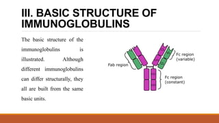
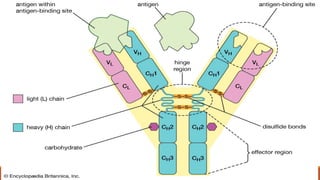
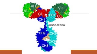
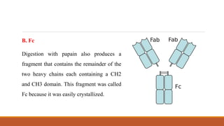
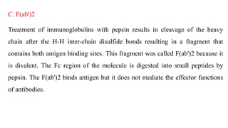
Immunoglobulins are glycoprotein molecules that are produced by plasma
cells in response to an immunogen and which function as antibodies.
The immunoglobulins derive their name from the finding that they migrate
with globular proteins when antibody-containing serum is placed in an
electrical field.
GENERAL FUNCTIONS OF
IMMUNOGLOBULINS
A. Antigen binding
Immunoglobulins bind specifically to one or a few closely related antigens. Each
immunoglobulin actually binds to a specific antigenic determinant. Antigen
binding by antibodies is the primary function of antibodies and can result in
protection of the host. The valency of antibody refers to the number of antigenic
determinants that an individual antibody molecule can bind. The valency of all
antibodies is at least two and in some instances more.
B. Effector Functions
Frequently the binding of an antibody to an antigen has no direct biological
effect. Rather, the significant biological effects are a consequence of secondary
"effector functions" of antibodies. The immunoglobulins mediate a variety of
these effector functions. Usually the ability to carry out a particular effector
function requires that the antibody bind to its antigen. Not every immunoglobulin
will mediate all effector functions. Such effector functions include:
1. Fixation of complement - This results in lysis of cells and release of biologically
active molecules.
2. Binding to various cell types - Phagocytic cells, lymphocytes, platelets, mast cells,
and basophils have receptors that bind immunoglobulins. This binding can activate the
cells to perform some function. Some immunoglobulins also bind to receptors on
placental trophoblasts, which results in transfer of the immunoglobulin across the
placenta. As a result, the transferred maternal antibodies provide immunity to the fetus
and newborn
III. BASIC STRUCTURE OF
IMMUNOGLOBULINS
The basic structure of the
immunoglobulins is
illustrated. Although
different immunoglobulins
can differ structurally, they
all are built from the same
basic units.
A. Heavy and Light Chains
All immunoglobulins have a four chain structure as their basic unit. They are composed of
two identical light chains (23kD) and two identical heavy chains (50-70kD).
B. Disulfide bonds
1. Inter-chain disulfide bonds - The heavy and light chains and the two heavy chains are
held together by inter-chain disulfide bonds and by non-covalent interactions
The number of inter-chain disulfide bonds varies among different immunoglobulin
molecules.
2. Intra-chain disulfide binds - Within each of the polypeptide chains there are
also intra-chain disulfide bonds.
C. Variable (V) and Constant (C) Regions
When the amino acid sequences of many different heavy chains and light chains were
compared, it became clear that both the heavy and light chain could be divided into two
regions based on variability in the amino acid sequences. These are the:
1. Light Chain - VL (110 amino acids) and CL (110 amino acids)
2. Heavy Chain - VH (110 amino acids) and CH (330-440 amino acids)
D. Hinge Region
This is the region at which the arms of the antibody molecule forms a Y. It is called the hinge
region because there is some flexibility in the molecule at this point.
E. Domains
Three dimensional images of the immunoglobulin molecule show that it is not straight as
depicted in figure. Rather, it is folded into globular regions each of which contains an intra-
chain disulfide bond. These regions are called domains.
◦ 1. Light Chain Domains - VL and CL
◦ 2. Heavy Chain Domains - VH, CH1 - CH3 (or CH4)
F. Oligosaccharides
Carbohydrates are attached to the CH2 domain in most immunoglobulins. However, in some
cases carbohydrates may also be attached at other locations.
IV. STRUCTURE OF THE
VARIABLE REGION
A. Hypervariable (HVR) or complementarity determining regions (CDR)
Comparisons of the amino acid sequences of the variable regions of immunoglobulins show
that most of the variability resides in three regions called the hypervariable regions or the
complementarity determining regions. Antibodies with different specificities (i.e. different
combining sites) have different complementarity determining regions while antibodies of the
exact same specificity have identical complementarity determining regions (i.e. CDR is the
antibody combining site). Complementarity determining regions are found in both the H and
the L chains.
B. Framework regions
The regions between the complementarity determining regions in the variable region are
called the framework regions. Based on similarities and differences in the framework
regions the immunoglobulin heavy and light chain variable regions can be divided into
groups and subgroups. These represent the products of different variable region genes.
V. IMMUNOGLOBULIN FRAGMENTS:
STRUCTURE/FUNCTION
RELATIONSHIPS
Immunoglobulin fragments produced by proteolytic digestion have proven very useful in elucidating
structure/function relationships in immunoglobulins.
A. Fab
Digestion with papain breaks the immunoglobulin molecule in the hinge region before the H-H inter-chain
disulfide bond. This results in the formation of two identical fragments that contain the light chain and the
VH and CH1 domains of the heavy chain.
Antigen binding - These fragments were called the Fab fragments because they contained the antigen
binding sites of the antibody. Each Fab fragment is monovalent whereas the original molecule was divalent.
The combining site of the antibody is created by both VH and VL. An antibody is able to bind a particular
antigenic determinant because it has a particular combination of VH and VL. Different combinations of a
VH and VL result in antibodies that can bind a different antigenic determinants.
B. Fc
Digestion with papain also produces a
fragment that contains the remainder of the
two heavy chains each containing a CH2
and CH3 domain. This fragment was called
Fc because it was easily crystallized.
C. F(ab')2
Treatment of immunoglobulins with pepsin results in cleavage of the heavy
chain after the H-H inter-chain disulfide bonds resulting in a fragment that
contains both antigen binding sites. This fragment was called F(ab')2 because it
is divalent. The Fc region of the molecule is digested into small peptides by
pepsin. The F(ab')2 binds antigen but it does not mediate the effector functions
of antibodies.
Immunoglobulin

Immunoglobulin

  • 1.
    Immunoglobulin : Structure BY: PROF.SHIVRAJ POPAT JADHAV, DIVINE COLLEGE OF PHARMACY, SATANA
  • 2.
    Definition Immunoglobulin (Ig) Immunoglobulins areglycoprotein molecules that are produced by plasma cells in response to an immunogen and which function as antibodies. The immunoglobulins derive their name from the finding that they migrate with globular proteins when antibody-containing serum is placed in an electrical field.
  • 3.
    GENERAL FUNCTIONS OF IMMUNOGLOBULINS A.Antigen binding Immunoglobulins bind specifically to one or a few closely related antigens. Each immunoglobulin actually binds to a specific antigenic determinant. Antigen binding by antibodies is the primary function of antibodies and can result in protection of the host. The valency of antibody refers to the number of antigenic determinants that an individual antibody molecule can bind. The valency of all antibodies is at least two and in some instances more.
  • 4.
    B. Effector Functions Frequentlythe binding of an antibody to an antigen has no direct biological effect. Rather, the significant biological effects are a consequence of secondary "effector functions" of antibodies. The immunoglobulins mediate a variety of these effector functions. Usually the ability to carry out a particular effector function requires that the antibody bind to its antigen. Not every immunoglobulin will mediate all effector functions. Such effector functions include:
  • 5.
    1. Fixation ofcomplement - This results in lysis of cells and release of biologically active molecules. 2. Binding to various cell types - Phagocytic cells, lymphocytes, platelets, mast cells, and basophils have receptors that bind immunoglobulins. This binding can activate the cells to perform some function. Some immunoglobulins also bind to receptors on placental trophoblasts, which results in transfer of the immunoglobulin across the placenta. As a result, the transferred maternal antibodies provide immunity to the fetus and newborn
  • 6.
    III. BASIC STRUCTUREOF IMMUNOGLOBULINS The basic structure of the immunoglobulins is illustrated. Although different immunoglobulins can differ structurally, they all are built from the same basic units.
  • 8.
    A. Heavy andLight Chains All immunoglobulins have a four chain structure as their basic unit. They are composed of two identical light chains (23kD) and two identical heavy chains (50-70kD). B. Disulfide bonds 1. Inter-chain disulfide bonds - The heavy and light chains and the two heavy chains are held together by inter-chain disulfide bonds and by non-covalent interactions The number of inter-chain disulfide bonds varies among different immunoglobulin molecules. 2. Intra-chain disulfide binds - Within each of the polypeptide chains there are also intra-chain disulfide bonds.
  • 9.
    C. Variable (V)and Constant (C) Regions When the amino acid sequences of many different heavy chains and light chains were compared, it became clear that both the heavy and light chain could be divided into two regions based on variability in the amino acid sequences. These are the: 1. Light Chain - VL (110 amino acids) and CL (110 amino acids) 2. Heavy Chain - VH (110 amino acids) and CH (330-440 amino acids) D. Hinge Region This is the region at which the arms of the antibody molecule forms a Y. It is called the hinge region because there is some flexibility in the molecule at this point.
  • 10.
    E. Domains Three dimensionalimages of the immunoglobulin molecule show that it is not straight as depicted in figure. Rather, it is folded into globular regions each of which contains an intra- chain disulfide bond. These regions are called domains. ◦ 1. Light Chain Domains - VL and CL ◦ 2. Heavy Chain Domains - VH, CH1 - CH3 (or CH4) F. Oligosaccharides Carbohydrates are attached to the CH2 domain in most immunoglobulins. However, in some cases carbohydrates may also be attached at other locations.
  • 12.
    IV. STRUCTURE OFTHE VARIABLE REGION A. Hypervariable (HVR) or complementarity determining regions (CDR) Comparisons of the amino acid sequences of the variable regions of immunoglobulins show that most of the variability resides in three regions called the hypervariable regions or the complementarity determining regions. Antibodies with different specificities (i.e. different combining sites) have different complementarity determining regions while antibodies of the exact same specificity have identical complementarity determining regions (i.e. CDR is the antibody combining site). Complementarity determining regions are found in both the H and the L chains.
  • 13.
    B. Framework regions Theregions between the complementarity determining regions in the variable region are called the framework regions. Based on similarities and differences in the framework regions the immunoglobulin heavy and light chain variable regions can be divided into groups and subgroups. These represent the products of different variable region genes.
  • 14.
    V. IMMUNOGLOBULIN FRAGMENTS: STRUCTURE/FUNCTION RELATIONSHIPS Immunoglobulinfragments produced by proteolytic digestion have proven very useful in elucidating structure/function relationships in immunoglobulins. A. Fab Digestion with papain breaks the immunoglobulin molecule in the hinge region before the H-H inter-chain disulfide bond. This results in the formation of two identical fragments that contain the light chain and the VH and CH1 domains of the heavy chain. Antigen binding - These fragments were called the Fab fragments because they contained the antigen binding sites of the antibody. Each Fab fragment is monovalent whereas the original molecule was divalent. The combining site of the antibody is created by both VH and VL. An antibody is able to bind a particular antigenic determinant because it has a particular combination of VH and VL. Different combinations of a VH and VL result in antibodies that can bind a different antigenic determinants.
  • 15.
    B. Fc Digestion withpapain also produces a fragment that contains the remainder of the two heavy chains each containing a CH2 and CH3 domain. This fragment was called Fc because it was easily crystallized.
  • 16.
    C. F(ab')2 Treatment ofimmunoglobulins with pepsin results in cleavage of the heavy chain after the H-H inter-chain disulfide bonds resulting in a fragment that contains both antigen binding sites. This fragment was called F(ab')2 because it is divalent. The Fc region of the molecule is digested into small peptides by pepsin. The F(ab')2 binds antigen but it does not mediate the effector functions of antibodies.