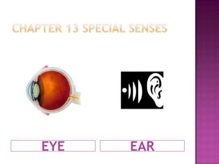
Chapter 13 Special SensesEYEEAR
Sense organs carry messages about the environment to the central nervous systemTHE SENSES
The Sense organs gather information from outside the body, then send the messages to the brain.THE ENVIRONMENT(everything outside the body)
visionYour ability to see.Vision involves on of our great senses which is the eye & also involves the brain.
The eyeThe eye gathers pictures and sends them to the brain.The eyeball is the incredible organ of sight that transmit an external image by the way of the nervous system-the optic nerve to the brain.The actual eyeball is composed of three layers:The ScleraThe ChoroidThe Retina
The Sclera:1st layerAnother term is the “white of the eye”The sclera is the outer layer that provides a tough protective coating for the inner structures of the eye.The anterior portion of the sclera is called the “cornea”. This clear transparent area of the sclera allows light to enter the interior of the eyeball. The cornea actually bends, or retracts the light rays.
Choroid:2nd layerThe middle layer of the eyeball: the opaque layer provides the blood supply for the eye.The anterior portion of the choroid layer consists of the:Iris-colored portion of the eye; contains smooth musclePupil-opening in the center of the iris that allows light rays to enter the eyeball.Ciliary Body
The Retina: 3rd layerInnermost layer of the eyeballContains the sensory receptors called:  Rods-active in dim light & help us see in gray tones.Cones-active only in bright light and responsible for color vision.
Muscles of the eyeSix muscles connect the actual eyeball of the skull:(4) RECTUS: (meaning straight) pull the eye up, down, left or right in a straight line.(2) OBLIQUE MUSCLES: on an angle and produce diagonal eye movement.These muscles allow for change in the direction of each eye’s sightline.
The eyelidsA pair of eyelids over each eyeball provides protection against foreign particles, injury from the sun and intense light and trauma.Both the upper and lower edges of the eyelids have eyelashes and cilia that protect the eye.In addition, sebaceous glands located in the eyelids secrete lubricating oil onto the eyeball.
how we seeVision requires proper functioning of four mechanisms:Coordination of the external eye muscles so that both eyes move together.The correct amount of light admitted by the pupil.The correct focus of light on the retina of the lens.The optic nerve transmitting sensory images to the brain.(The study of the eye is known as ophthalmology (OPTH))
hearingStarts when some of the sound waves go into the ear.
the earThe ear works with the brain to control your balance.The ear is subdivided into three areas:External earMiddle earInner ear
EXTERNAL EARConsists of three parts:The auricle or pinna is what is commonly referred to as the ear because this is the only visible portion.Auditory canalTympanic membrane
Middle earLocated in a small cavity in the temporal bone of the skull.This air filled cavity contain three tiny bones called ossicles.These three bones, the malleus, incus and stapes are vital to the hearing process, b/c they amplify vibrations in the middle ear and transmit them to the inner ear from the malleus to the incus and finally the stapes.
Inner earLocated in a cavity within the temporal boneThis fluid filled cavity is referred to as the labyrinth because of its shape.The labyrinth contains the hearing and equilibrium sensory organs: the cochlea for hearing and the semicircular canals, utricle and saccule for equilibrium.
Important noteOur eyes and ears are very detrimental sense organs that some might take for granted, it is important to maintain these important senses and keep them accurate by making sure to get them checked annually.

Chapter 13 Special Senses

  • 1.
    Chapter 13 SpecialSensesEYEEAR
  • 2.
    Sense organs carrymessages about the environment to the central nervous systemTHE SENSES
  • 3.
    The Sense organsgather information from outside the body, then send the messages to the brain.THE ENVIRONMENT(everything outside the body)
  • 4.
    visionYour ability tosee.Vision involves on of our great senses which is the eye & also involves the brain.
  • 5.
    The eyeThe eyegathers pictures and sends them to the brain.The eyeball is the incredible organ of sight that transmit an external image by the way of the nervous system-the optic nerve to the brain.The actual eyeball is composed of three layers:The ScleraThe ChoroidThe Retina
  • 6.
    The Sclera:1st layerAnotherterm is the “white of the eye”The sclera is the outer layer that provides a tough protective coating for the inner structures of the eye.The anterior portion of the sclera is called the “cornea”. This clear transparent area of the sclera allows light to enter the interior of the eyeball. The cornea actually bends, or retracts the light rays.
  • 7.
    Choroid:2nd layerThe middlelayer of the eyeball: the opaque layer provides the blood supply for the eye.The anterior portion of the choroid layer consists of the:Iris-colored portion of the eye; contains smooth musclePupil-opening in the center of the iris that allows light rays to enter the eyeball.Ciliary Body
  • 8.
    The Retina: 3rdlayerInnermost layer of the eyeballContains the sensory receptors called: Rods-active in dim light & help us see in gray tones.Cones-active only in bright light and responsible for color vision.
  • 9.
    Muscles of theeyeSix muscles connect the actual eyeball of the skull:(4) RECTUS: (meaning straight) pull the eye up, down, left or right in a straight line.(2) OBLIQUE MUSCLES: on an angle and produce diagonal eye movement.These muscles allow for change in the direction of each eye’s sightline.
  • 10.
    The eyelidsA pairof eyelids over each eyeball provides protection against foreign particles, injury from the sun and intense light and trauma.Both the upper and lower edges of the eyelids have eyelashes and cilia that protect the eye.In addition, sebaceous glands located in the eyelids secrete lubricating oil onto the eyeball.
  • 11.
    how we seeVisionrequires proper functioning of four mechanisms:Coordination of the external eye muscles so that both eyes move together.The correct amount of light admitted by the pupil.The correct focus of light on the retina of the lens.The optic nerve transmitting sensory images to the brain.(The study of the eye is known as ophthalmology (OPTH))
  • 12.
    hearingStarts when someof the sound waves go into the ear.
  • 13.
    the earThe earworks with the brain to control your balance.The ear is subdivided into three areas:External earMiddle earInner ear
  • 14.
    EXTERNAL EARConsists ofthree parts:The auricle or pinna is what is commonly referred to as the ear because this is the only visible portion.Auditory canalTympanic membrane
  • 15.
    Middle earLocated ina small cavity in the temporal bone of the skull.This air filled cavity contain three tiny bones called ossicles.These three bones, the malleus, incus and stapes are vital to the hearing process, b/c they amplify vibrations in the middle ear and transmit them to the inner ear from the malleus to the incus and finally the stapes.
  • 16.
    Inner earLocated ina cavity within the temporal boneThis fluid filled cavity is referred to as the labyrinth because of its shape.The labyrinth contains the hearing and equilibrium sensory organs: the cochlea for hearing and the semicircular canals, utricle and saccule for equilibrium.
  • 17.
    Important noteOur eyesand ears are very detrimental sense organs that some might take for granted, it is important to maintain these important senses and keep them accurate by making sure to get them checked annually.