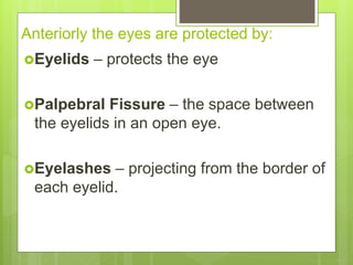
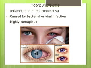
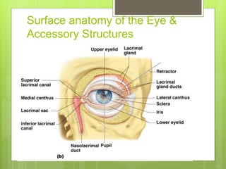
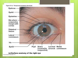
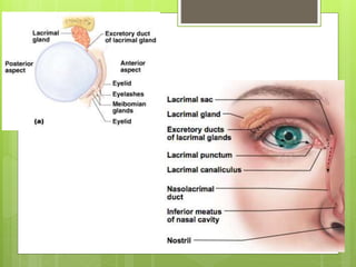
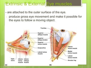
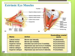
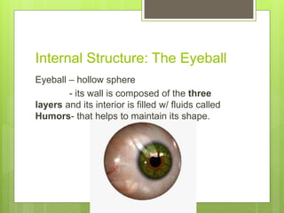
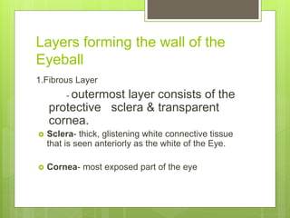
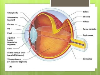
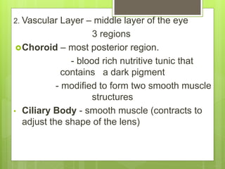
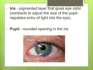
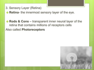
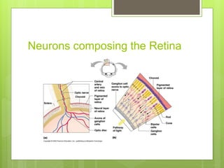
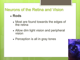
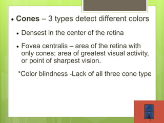
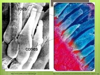
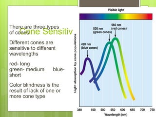
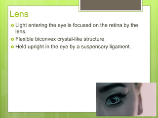
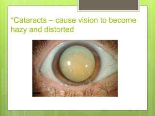
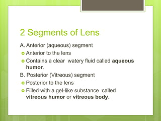
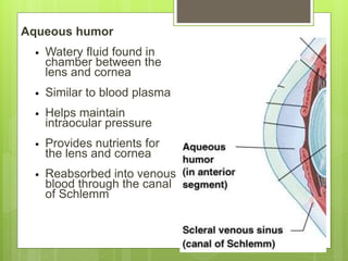
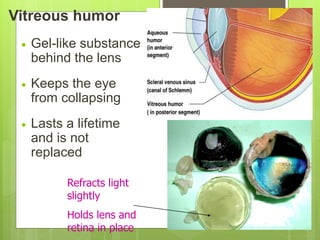
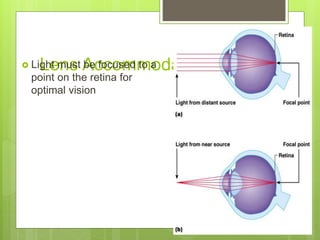
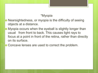
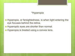
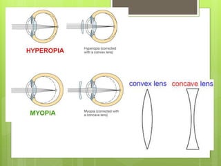
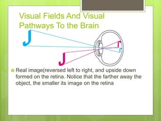
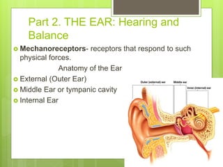
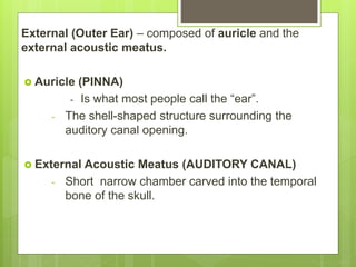
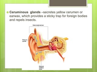
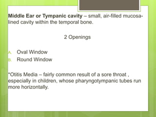
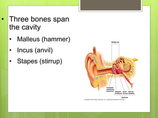
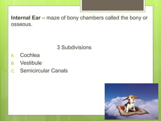
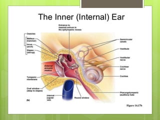
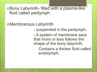
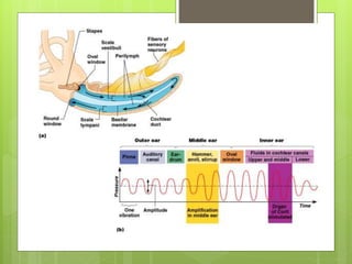
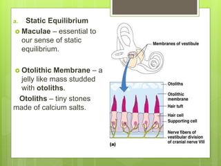
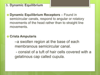
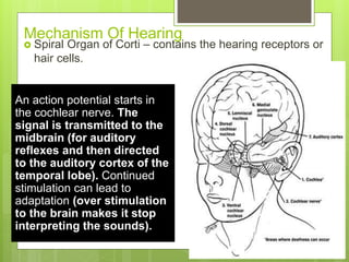
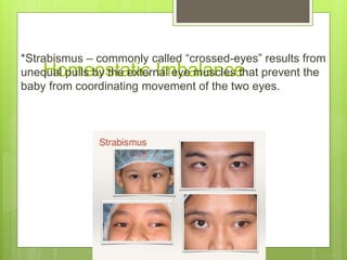
4 Traditional Senses
 Smell
 Taste
 Sight
 Hearing
* Also called Special Senses
Part 1. THE EYE & VISION
 All of the sensory receptors in the body 70%
are the EYES.
Optic Tracts - Carry information from the eyes
to the brain.
- are massive bundles, containing
over a million nerve fibers.
Anatomy of the Eye
External and Accessory
structures
 The Accessory Structures of the Eye include:
-Extrinsic eye muscle
-Eye lids
-Conjunctiva
-Lacrimal Apparatus
Anteriorly the eyes are protected by:
Eyelids – protects the eye
Palpebral Fissure – the space between
the eyelids in an open eye.
Eyelashes – projecting from the border of
each eyelid.
Tarsal glands – modified sebaceous
associated with the eyelid edges.
- produce an oily secretion
that lubricates the eye.
Ciliary glands – modified sweat glands, lie
between eyelashes. (cilium = eyelash)
Conjunctiva – lines the eyelids and covers
part of the outer surface of the eyeball. It
secretes mucus, which helps to lubricate the
eyeball and keep it moist.
*CONJUNCTIVITIS
- Inflammation of the conjunctiva
- Caused by bacterial or viral infection
- Highly contagious
Surface anatomy of the Eye &
Accessory Structures
 Lacrimal apparatus- consists of the lacrimal gland and
a number of ducts that drain the lacrimal secretions into
the nasal cavity.
 Lacrimal glands – located above the lateral end of each
eye.
-release a dilute salt solution(tears)
Lacrimal glands Lacrimal canaliculi Lacrimal sac nasolacrimal
duct
 Lacrimal secretion
-contains antibodies & lysozyme
*Lysozyme – enzyme that destroys bacteria
- cleanses and protects the eye surface as it
moistens and lubricates it.
*When lacrimal secretion increases substantially, tears spill
over the eyelids and fill the nasal cavities,causing
congestion and the “sniffles”.
Extrinsic & External Eye muscles
- are attached to the outer surface of the eye.
- produce gross eye movement and make it possible for
the eyes to follow a moving object.
Internal Structure: The Eyeball
Eyeball – hollow sphere
- its wall is composed of the three
layers and its interior is filled w/ fluids called
Humors- that helps to maintain its shape.
Layers forming the wall of the
Eyeball
1.Fibrous Layer
- outermost layer consists of the
protective sclera & transparent
cornea.
 Sclera- thick, glistening white connective tissue
that is seen anteriorly as the white of the Eye.
 Cornea- most exposed part of the eye
2. Vascular Layer – middle layer of the eye
3 regions
Choroid – most posterior region.
- blood rich nutritive tunic that
contains a dark pigment
- modified to form two smooth muscle
structures
• Ciliary Body - smooth muscle (contracts to
adjust the shape of the lens)
• Iris - pigmented layer that gives eye color
(contracts to adjust the size of the pupil-
regulates entry of light into the eye).
• Pupil - rounded opening in the iris
3. Sensory Layer (Retina)
 Retina- the innermost sensory layer of the eye.
 Rods & Cons – transparent inner neural layer of the
retina that contains millions of receptors cells
Also called Photoreceptors
Neurons composing the Retina
Neurons of the Retina and Vision
 Rods
 Most are found towards the edges of
the retina
 Allow dim light vision and peripheral
vision
 Perception is all in gray tones
 Cones – 3 types detect different colors
 Densest in the center of the retina
 Fovea centralis – area of the retina with
only cones; area of greatest visual activity,
or point of sharpest vision.
*Color blindness -Lack of all three cone type
http://www.yorku.ca/eye/rod-cone.gif http://www.secretbeyondmatter.com/ourbrains/theworldinourbrains_files/11-1.jpg
Cone Sensitivity
There are three types
of cones
Different cones are
sensitive to different
wavelengths
red- long
green- medium blue-
short
Color blindness is the
result of lack of one or
more cone type
Lens
 Light entering the eye is focused on the retina by the
lens.
 Flexible biconvex crystal-like structure
 Held upright in the eye by a suspensory ligament.
*Cataracts – cause vision to become
hazy and distorted
2 Segments of Lens
A. Anterior (aqueous) segment
 Anterior to the lens
 Contains a clear watery fluid called aqueous
humor.
B. Posterior (Vitreous) segment
 Posterior to the lens
 Filled with a gel-like substance called
vitreous humor or vitreous body.
Aqueous humor
• Watery fluid found in
chamber between the
lens and cornea
• Similar to blood plasma
• Helps maintain
intraocular pressure
• Provides nutrients for
the lens and cornea
• Reabsorbed into venous
blood through the canal
of Schlemm
Vitreous humor
 Gel-like substance
behind the lens
 Keeps the eye
from collapsing
 Lasts a lifetime
and is not
replaced
Refracts light
slightly
Holds lens and
retina in place
Lens Accommodation Light must be focused to a
point on the retina for
optimal vision
*Myopia
 Nearsightedness, or myopia is the difficulty of seeing
objects at a distance.
 Myopia occurs when the eyeball is slightly longer than
usual from front to back. This causes light rays to
focus at a point in front of the retina, rather than directly
on its surface.
 Concave lenses are used to correct the problem.
*Hyperopia
 Hyperopia, or farsightedness, is when light entering the
eye focuses behind the retina.
 Hyperoptic eyes are shorter than normal.
 Hyperopia is treated using a convex lens.
Visual Fields And Visual
Pathways To the Brain
 Real image(reversed left to right, and upside down
formed on the retina. Notice that the farther away the
object, the smaller its image on the retina
 The retinal sites at which a
real image would be
focused when both eyes are
fixed on a close, point-like
object.
Part 2. THE EAR: Hearing and
Balance
 Mechanoreceptors- receptors that respond to such
physical forces.
Anatomy of the Ear
 External (Outer Ear)
 Middle Ear or tympanic cavity
 Internal Ear
External (Outer Ear) – composed of auricle and the
external acoustic meatus.
 Auricle (PINNA)
- Is what most people call the “ear”.
- The shell-shaped structure surrounding the
auditory canal opening.
 External Acoustic Meatus (AUDITORY CANAL)
- Short narrow chamber carved into the temporal
bone of the skull.
 Ceruminous glands –secretes yellow cerumen or
earwax, which provides a sticky trap for foreign bodies
and repels insects.
Middle Ear or Tympanic cavity – small, air-filled mucosa-
lined cavity within the temporal bone.
2 Openings
A. Oval Window
B. Round Window
*Otitis Media – fairly common result of a sore throat ,
especially in children, whose pharyngotympanic tubes run
more horizontally.
• Three bones span
the cavity
• Malleus (hammer)
• Incus (anvil)
• Stapes (stirrup)
Internal Ear – maze of bony chambers called the bony or
osseous.
3 Subdivisions
A. Cochlea
B. Vestibule
C. Semicircular Canals
Bony Labyrinth- filled with a plasma-like
fluid called perilymph.
Membranous Labyrinth
- suspended in the perilymph.
- A system of membrane sacs
that more or less follows the
shape of the bony labyrinth.
- Contains a thicker fluid called
endolymph.
Vestibular Apparatus – equilibrium receptors of the inner
ear.
Divided into two:
a. Static Equilibrium
b. Dynamic Equilibrium
a. Static Equilibrium
 Maculae – essential to
our sense of static
equilibrium.
 Otolithic Membrane – a
jelly like mass studded
with otoliths.
Otoliths – tiny stones
made of calcium salts.
b. Dynamic Equilibrium
 Dynamic Equilibrium Receptors – Found in
semicircular canals, respond to angular or rotatory
movements of the head rather than to straight line
movements.
 Crista Ampularis
–a swollen region at the base of each
membranous semicircular canal.
- consist of a tuft of hair cells covered with a
gelatinous cap called cupula.
Mechanism Of Hearing
 Spiral Organ of Corti – contains the hearing receptors or
hair cells.
An action potential starts in
the cochlear nerve. The
signal is transmitted to the
midbrain (for auditory
reflexes and then directed
to the auditory cortex of the
temporal lobe). Continued
stimulation can lead to
adaptation (over stimulation
to the brain makes it stop
interpreting the sounds).
Hemeostatic Imbalance
*Deafness- defined as hearing loss of any degree – from a
slight loss to a total inability to hear sound.
*Sensorineural Deafness - occurs when there is
degeneration or damage to the receptor cells in the spiral
organ of Corti.
*Meniere’s Syndrome – serious pathology of the inner ear
*Vertigo – (a spinning of spinning)
- so severe they cannot stand up without
extreme discomfort.
Part 3. Chemical Senses: Taste
and Smell
Chemoreceptors
- receptors for taste and
olfaction
- They respond to chemicals
in solution.
Olfactory Receptors & Sense of
Smell
 Olfactory Receptors – receptors for the sense
of the smell, occupy a postage stamp-size
area in the roof of each nasal cavity.
 Olfactory receptor cells- neurons equipped
with olfactory hairs.
 Olfactory Filaments – bundled axons of axons of
olfactory neurons that collectively make up the olfactory
nerve.
*Anosmias
-olfactory disoder
-result from head injuries, the aftereffects of nasal
cavity inflammation or aging.
Taste Buds and the Sense of
Taste
 Taste – comes from Latin word “taxare”, which means to
touch , estimate or judge.
 Taste Buds - specific receptors for the sense of taste.
- 10,000 taste buds
 Papillae – small peg-like projections thatcovers the
dorsal tongue surface.
 Circumvallate Papillae – where taste buds found
 Gustatory Cells – specific cells that respond to chemicals
dissolved in the saliva.
 Facial Nerve – serves that anterior part of the tongue.
2 Cranial Nerves
a. Glossopharyngeal
b. Vagus
- Serve the other taste-bud containing areas.
Developmental Aspects of the
Special Senses
 Formed early in embryonic development
 Eyes are outgrowths of the brain
 All special senses are functional at birth
Homeostatic Imbalance
*Strabismus – commonly called “crossed-eyes” results from
unequal pulls by the external eye muscles that prevent the
baby from coordinating movement of the two eyes.
*Rubella (measles)
– occur during early pregnancy may
lead to congenital blindness or
cataracts.
*Presbyopia
– results from decreasing lens
elasticity that accompanies aging.
- Difficulty to focus for close vision.
* Presbycusis – Type of sensorineural deafness.
And That’s the End Of Special
Senses ! :D

Special Senses

  • 2.
    4 Traditional Senses Smell  Taste  Sight  Hearing * Also called Special Senses
  • 3.
    Part 1. THEEYE & VISION  All of the sensory receptors in the body 70% are the EYES. Optic Tracts - Carry information from the eyes to the brain. - are massive bundles, containing over a million nerve fibers.
  • 4.
    Anatomy of theEye External and Accessory structures  The Accessory Structures of the Eye include: -Extrinsic eye muscle -Eye lids -Conjunctiva -Lacrimal Apparatus
  • 5.
    Anteriorly the eyesare protected by: Eyelids – protects the eye Palpebral Fissure – the space between the eyelids in an open eye. Eyelashes – projecting from the border of each eyelid.
  • 6.
    Tarsal glands –modified sebaceous associated with the eyelid edges. - produce an oily secretion that lubricates the eye. Ciliary glands – modified sweat glands, lie between eyelashes. (cilium = eyelash) Conjunctiva – lines the eyelids and covers part of the outer surface of the eyeball. It secretes mucus, which helps to lubricate the eyeball and keep it moist.
  • 7.
    *CONJUNCTIVITIS - Inflammation ofthe conjunctiva - Caused by bacterial or viral infection - Highly contagious
  • 8.
    Surface anatomy ofthe Eye & Accessory Structures
  • 10.
     Lacrimal apparatus-consists of the lacrimal gland and a number of ducts that drain the lacrimal secretions into the nasal cavity.  Lacrimal glands – located above the lateral end of each eye. -release a dilute salt solution(tears) Lacrimal glands Lacrimal canaliculi Lacrimal sac nasolacrimal duct
  • 12.
     Lacrimal secretion -containsantibodies & lysozyme *Lysozyme – enzyme that destroys bacteria - cleanses and protects the eye surface as it moistens and lubricates it. *When lacrimal secretion increases substantially, tears spill over the eyelids and fill the nasal cavities,causing congestion and the “sniffles”.
  • 13.
    Extrinsic & ExternalEye muscles - are attached to the outer surface of the eye. - produce gross eye movement and make it possible for the eyes to follow a moving object.
  • 15.
    Internal Structure: TheEyeball Eyeball – hollow sphere - its wall is composed of the three layers and its interior is filled w/ fluids called Humors- that helps to maintain its shape.
  • 16.
    Layers forming thewall of the Eyeball 1.Fibrous Layer - outermost layer consists of the protective sclera & transparent cornea.  Sclera- thick, glistening white connective tissue that is seen anteriorly as the white of the Eye.  Cornea- most exposed part of the eye
  • 18.
    2. Vascular Layer– middle layer of the eye 3 regions Choroid – most posterior region. - blood rich nutritive tunic that contains a dark pigment - modified to form two smooth muscle structures • Ciliary Body - smooth muscle (contracts to adjust the shape of the lens)
  • 19.
    • Iris -pigmented layer that gives eye color (contracts to adjust the size of the pupil- regulates entry of light into the eye). • Pupil - rounded opening in the iris
  • 20.
    3. Sensory Layer(Retina)  Retina- the innermost sensory layer of the eye.  Rods & Cons – transparent inner neural layer of the retina that contains millions of receptors cells Also called Photoreceptors
  • 21.
  • 22.
    Neurons of theRetina and Vision  Rods  Most are found towards the edges of the retina  Allow dim light vision and peripheral vision  Perception is all in gray tones
  • 23.
     Cones –3 types detect different colors  Densest in the center of the retina  Fovea centralis – area of the retina with only cones; area of greatest visual activity, or point of sharpest vision. *Color blindness -Lack of all three cone type
  • 24.
  • 25.
    Cone Sensitivity There arethree types of cones Different cones are sensitive to different wavelengths red- long green- medium blue- short Color blindness is the result of lack of one or more cone type
  • 26.
    Lens  Light enteringthe eye is focused on the retina by the lens.  Flexible biconvex crystal-like structure  Held upright in the eye by a suspensory ligament.
  • 27.
    *Cataracts – causevision to become hazy and distorted
  • 28.
    2 Segments ofLens A. Anterior (aqueous) segment  Anterior to the lens  Contains a clear watery fluid called aqueous humor. B. Posterior (Vitreous) segment  Posterior to the lens  Filled with a gel-like substance called vitreous humor or vitreous body.
  • 29.
    Aqueous humor • Wateryfluid found in chamber between the lens and cornea • Similar to blood plasma • Helps maintain intraocular pressure • Provides nutrients for the lens and cornea • Reabsorbed into venous blood through the canal of Schlemm
  • 30.
    Vitreous humor  Gel-likesubstance behind the lens  Keeps the eye from collapsing  Lasts a lifetime and is not replaced Refracts light slightly Holds lens and retina in place
  • 31.
    Lens Accommodation Lightmust be focused to a point on the retina for optimal vision
  • 32.
    *Myopia  Nearsightedness, ormyopia is the difficulty of seeing objects at a distance.  Myopia occurs when the eyeball is slightly longer than usual from front to back. This causes light rays to focus at a point in front of the retina, rather than directly on its surface.  Concave lenses are used to correct the problem.
  • 33.
    *Hyperopia  Hyperopia, orfarsightedness, is when light entering the eye focuses behind the retina.  Hyperoptic eyes are shorter than normal.  Hyperopia is treated using a convex lens.
  • 35.
    Visual Fields AndVisual Pathways To the Brain  Real image(reversed left to right, and upside down formed on the retina. Notice that the farther away the object, the smaller its image on the retina
  • 36.
     The retinalsites at which a real image would be focused when both eyes are fixed on a close, point-like object.
  • 37.
    Part 2. THEEAR: Hearing and Balance  Mechanoreceptors- receptors that respond to such physical forces. Anatomy of the Ear  External (Outer Ear)  Middle Ear or tympanic cavity  Internal Ear
  • 38.
    External (Outer Ear)– composed of auricle and the external acoustic meatus.  Auricle (PINNA) - Is what most people call the “ear”. - The shell-shaped structure surrounding the auditory canal opening.  External Acoustic Meatus (AUDITORY CANAL) - Short narrow chamber carved into the temporal bone of the skull.
  • 39.
     Ceruminous glands–secretes yellow cerumen or earwax, which provides a sticky trap for foreign bodies and repels insects.
  • 40.
    Middle Ear orTympanic cavity – small, air-filled mucosa- lined cavity within the temporal bone. 2 Openings A. Oval Window B. Round Window *Otitis Media – fairly common result of a sore throat , especially in children, whose pharyngotympanic tubes run more horizontally.
  • 41.
    • Three bonesspan the cavity • Malleus (hammer) • Incus (anvil) • Stapes (stirrup)
  • 42.
    Internal Ear –maze of bony chambers called the bony or osseous. 3 Subdivisions A. Cochlea B. Vestibule C. Semicircular Canals
  • 44.
    Bony Labyrinth- filledwith a plasma-like fluid called perilymph. Membranous Labyrinth - suspended in the perilymph. - A system of membrane sacs that more or less follows the shape of the bony labyrinth. - Contains a thicker fluid called endolymph.
  • 46.
    Vestibular Apparatus –equilibrium receptors of the inner ear. Divided into two: a. Static Equilibrium b. Dynamic Equilibrium
  • 47.
    a. Static Equilibrium Maculae – essential to our sense of static equilibrium.  Otolithic Membrane – a jelly like mass studded with otoliths. Otoliths – tiny stones made of calcium salts.
  • 48.
    b. Dynamic Equilibrium Dynamic Equilibrium Receptors – Found in semicircular canals, respond to angular or rotatory movements of the head rather than to straight line movements.  Crista Ampularis –a swollen region at the base of each membranous semicircular canal. - consist of a tuft of hair cells covered with a gelatinous cap called cupula.
  • 49.
    Mechanism Of Hearing Spiral Organ of Corti – contains the hearing receptors or hair cells. An action potential starts in the cochlear nerve. The signal is transmitted to the midbrain (for auditory reflexes and then directed to the auditory cortex of the temporal lobe). Continued stimulation can lead to adaptation (over stimulation to the brain makes it stop interpreting the sounds).
  • 50.
    Hemeostatic Imbalance *Deafness- definedas hearing loss of any degree – from a slight loss to a total inability to hear sound. *Sensorineural Deafness - occurs when there is degeneration or damage to the receptor cells in the spiral organ of Corti. *Meniere’s Syndrome – serious pathology of the inner ear *Vertigo – (a spinning of spinning) - so severe they cannot stand up without extreme discomfort.
  • 51.
    Part 3. ChemicalSenses: Taste and Smell Chemoreceptors - receptors for taste and olfaction - They respond to chemicals in solution.
  • 52.
    Olfactory Receptors &Sense of Smell  Olfactory Receptors – receptors for the sense of the smell, occupy a postage stamp-size area in the roof of each nasal cavity.  Olfactory receptor cells- neurons equipped with olfactory hairs.
  • 54.
     Olfactory Filaments– bundled axons of axons of olfactory neurons that collectively make up the olfactory nerve. *Anosmias -olfactory disoder -result from head injuries, the aftereffects of nasal cavity inflammation or aging.
  • 55.
    Taste Buds andthe Sense of Taste  Taste – comes from Latin word “taxare”, which means to touch , estimate or judge.  Taste Buds - specific receptors for the sense of taste. - 10,000 taste buds  Papillae – small peg-like projections thatcovers the dorsal tongue surface.
  • 57.
     Circumvallate Papillae– where taste buds found  Gustatory Cells – specific cells that respond to chemicals dissolved in the saliva.  Facial Nerve – serves that anterior part of the tongue.
  • 59.
    2 Cranial Nerves a.Glossopharyngeal b. Vagus - Serve the other taste-bud containing areas.
  • 60.
    Developmental Aspects ofthe Special Senses  Formed early in embryonic development  Eyes are outgrowths of the brain  All special senses are functional at birth
  • 61.
    Homeostatic Imbalance *Strabismus –commonly called “crossed-eyes” results from unequal pulls by the external eye muscles that prevent the baby from coordinating movement of the two eyes.
  • 62.
    *Rubella (measles) – occurduring early pregnancy may lead to congenital blindness or cataracts. *Presbyopia – results from decreasing lens elasticity that accompanies aging. - Difficulty to focus for close vision. * Presbycusis – Type of sensorineural deafness.
  • 63.
    And That’s theEnd Of Special Senses ! :D