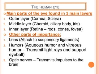
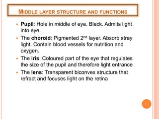
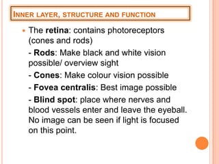
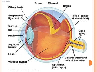
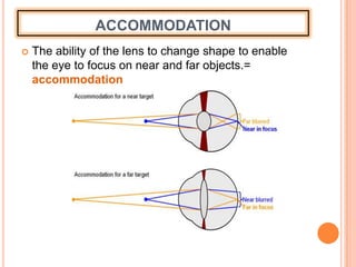
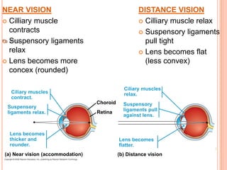
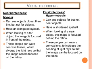
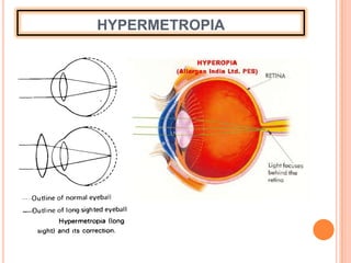
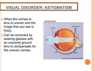
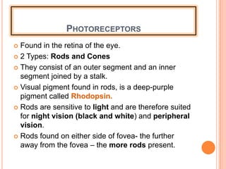
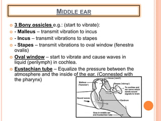
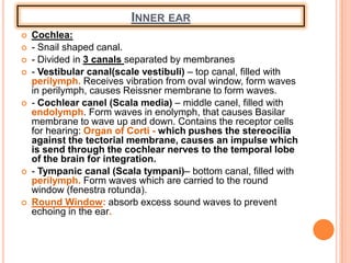
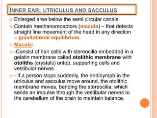
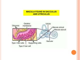
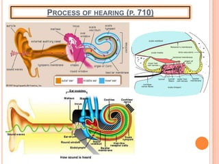
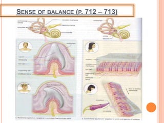
Call Girls Cuttack Just Call 9907093804 Top Class Call Girl Service Available
Sense organs
1. UNIT 8:
SENSE ORGANS
• Chemical senses
• Sense of vision
• Sense of hearing and balance
Refer to MADER, 2010: CHAPTER 38 P. 702 - 716
2. CHEMICAL SENSES
The receptors responsible for taste and
smell – CHEMORECEPTORS
2 Types of chemical senses:
1. Sense of Taste
2. Sense of Smell
3. SENSE OF TASTE
Taste receptors = TASTE BUDS
Taste buds are located on tongue(mainly), hard
palate, pharynx and epiglottis.
The tongue very rough, due to numerous papillae.
3 Types of papillae:
- Fungiform- found on front and sides of
tongue.
- Foliate found on front 2/3 of tongue
- Curcumvallate- found on the back of the
tongue.
The taste buds are located on the papillae
6. FUNCTIONS OF TASTE BUDS
Chemical molecules in food, dissolve in the saliva.
The molecules bind to the receptor proteins in the
microvilli of the taste buds.
This stimulates the taste cells to send an impulse
through the sensory nerve fibers.
Impulse move to the parietal lobe of cerebrum.
Taste is interpreted
7. 5 PRIMARY TYPES OF TASTE
Sweet
Sour
Bitter
Salty
Umami (Japanese
savory taste)
- Certain regions of the
tongue are more
sensitive to particular
tastes.
8. SENSE OF SMELL
Receptor cells of smell are OLFACTORY CELLS
Olfactory cells are located within olfactory epithelium
high in the roof of the nasal cavity.
9. PERCEPTION OF SMELL
The gas molecules in
the air dissolves in the
mucus of the nasal
cavity.
It stimulates the
microvilli of olfactory
cells.
This cause an impulse to
be send from olfactory
cell through the sensory
nerve fibers, to the
olfactory bulb in the
temporal lobe of the
cerebrum.
Smell is integrated and
perceived.
10. SENSE OF VISION
Photoreceptors are sensory receptors that are
sensitive to light.
Animals and humans with 2 eyes facing forward
have 3D or stereoscopic vision, visual fields
overlap and each eye is able to view and object
from a different angle.
11. THE HUMAN EYE
Main parts of the eye found in 3 main layers
1. Outer layer (Cornea, Sclera)
2. Middle layer (Choroid, ciliary body, iris)
3. Inner layer (Retina – rods, cones, fovea)
Other parts of importance:
1. Lens (Attach to suspensory ligaments)
2. Humors (Aqueous humor and vitreous
humor – Transmit light rays and support
eyeball)
3. Optic nerves – Transmits impulses to the
brain
12. OUTER LAYER (STRUCTURE AND FUNCTION)
The sclera: white outer layer, protects
and supports eyeball
Cornea: Front transparent part of eye.
Refracts light rays
Conjunctiva: Transparent membrane
(front). Moistens eye surface.
13. MIDDLE LAYER STRUCTURE AND FUNCTIONS
Pupil: Hole in middle of eye. Black. Admits light
into eye.
The choroid: Pigmented 2nd layer. Absorb stray
light. Contain blood vessels for nutrition and
oxygen.
The iris: Coloured part of the eye that regulates
the size of the pupil and therefore light entrance
The lens: Transparent biconvex structure that
refract and focuses light on the retina
14. INNER LAYER, STRUCTURE AND FUNCTION
The retina: contains photoreceptors
(cones and rods)
- Rods: Make black and white vision
possible/ overview sight
- Cones: Make colour vision possible
- Fovea centralis: Best image possible
- Blind spot: place where nerves and
blood vessels enter and leave the eyeball.
No image can be seen if light is focused
on this point.
15. The eye is divided into two cavities
separated by the lens and cilliary body:
The anterior cavity is filled with watery aqueous humor
The posterior cavity is filled with jellylike vitreous
humor
The cilliary body produces the aqueous
humor
16. Fig. 50-18
Optic
nerve
Fovea (center
of visual field)
Lens
Vitreous humor
Optic disk
(blind spot)
Central artery and
vein of the retina
Iris
Retina
ChoroidSclera
Ciliary body
Suspensory
ligament
Cornea
Pupil
Aqueous
humor
17. ACCOMMODATION
The ability of the lens to change shape to enable
the eye to focus on near and far objects.=
accommodation
19. VISUAL DISORDERS
Nearsightedness/
Myopia
Can see objects closer than
6m, but not far objects.
Have an elongated eyeball.
When looking at a far
object, the image is focused
in front of the retina.
These people can wear
concave lenses, which
diverge the light rays so that
the image can be focused
on the retina
Farsightedness/
Hypermetropic
Can see objects far but not
near objects.
Have a shortened eyeball.
When looking at a near
object, the image is focused
behind the retina.
These people can wear a
convex lens, to increase the
bending of light rays so that
the image can be focused on
the retina
22. VISUAL DISORDER: ASTIGMATISM
When the cornea or
lens is uneven and the
image that you see is
fuzzy.
Can be corrected by
wearing glasses with
an unevenly ground
lens to compensate for
the uneven cornea.
23. PHOTORECEPTORS
Found in the retina of the eye.
2 Types: Rods and Cones
They consist of an outer segment and an inner
segment joined by a stalk.
Visual pigment found in rods, is a deep-purple
pigment called Rhodopsin.
Rods are sensitive to light and are therefore suited
for night vision (black and white) and peripheral
vision.
Rods found on either side of fovea- the further
away from the fovea – the more rods present.
24. The cones, are primary located in the fovea
centralis, the further away from the fovea the less
the cones.
Activated by bright light
Cones allow us to detect fine detail and colour.
Cones contain 3 pigments –B (blue), G (green) and
R (red)
In colour blindness, a person lack certain of these
pigments.
25. INTEGRATION OF VISUAL SIGNALS IN THE
RETINA
Light enter the eye through the cornea, aqueous
humor, pupil, lens, vitreous humor and focuses
on the retina.
Light stimulates the photoreceptors (rods and
cones) in the retina.
Retina sends an impulse to the optic nerves
Optic nerves send an impulse to the occipital lobe
of the cerebrum
Where sight is integrated.
26. THE SENSE OF HEARING AND BALANCE
The ear has two sensory functions: Hearing and
Balance.
The sensory receptors for both of these is located
in the inner ear, and each consist of hair cells and
cillia which are sensitive to mechanical stimulation.
They are called machanoreceptors.
29. THE OUTER EAR
Pinna – Concentrate sound waves in the direction of the
external auditory canal.
External Auditory canal
– Transport sound waves from the pinna to the tympanic
membrane.
- Contain fine hairs and cerumin glands that secrete cerumin
(earwax) to help guard the ear against foreign material and
insects. (smell)
Tympanic membrane
– A thin membrane that covers the opening between the inner-
and middle ear.
- Converts soundwaves into vibrations. (starts to vibrate)
30. MIDDLE EAR
3 Bony ossicles e.g.: (start to vibrate):
- Malleus – transmit vibration to incus
- Incus – transmit vibrations to stapes
- Stapes – transmit vibrations to oval window (fenestra
ovalis)
Oval window – start to vibrate and cause waves in
liquid (perilymph) in cochlea.
Eustachian tube – Equalize the pressure between the
atmosphere and the inside of the ear. (Connested with
the pharynx)
31. INNER EAR
Cochlea:
- Snail shaped canal.
- Divided in 3 canals separated by membranes
- Vestibular canal(scale vestibuli) – top canal, filled with
perilymph. Receives vibration from oval window, form waves
in perilymph, causes Reissner membrane to form waves.
- Cochlear canel (Scala media) – middle canel, filled with
endolymph. Form waves in enolymph, that causes Basilar
membrane to wave up and down. Contains the receptor cells
for hearing: Organ of Corti - which pushes the stereocilia
against the tectorial membrane, causes an impulse which
is send through the cochlear nerves to the temporal lobe
of the brain for integration.
- Tympanic canal (Scala tympani)– bottom canal, filled with
perilymph. Form waves which are carried to the round
window (fenestra rotunda).
Round Window: absorb excess sound waves to prevent
echoing in the ear.
33. INNER EAR: SEMI CIRCULAR CANALS
Contain machanorecepters (cristae) – detect
rotational or angular movement of the head.
Cristae- located in the ampulla (enlarged base of
semi circular canals)in the endolymph found in the
semi circular canals.
- Consist of hair cells, supporting cells, stereocillia
imbedded in a gelatin capsule called cupula, and
nerve fibers.
Movement of the head causes the endolymph to
move around in the ampulla, the cupula moves,
bending the stereocilia, causing an impulse send
through the vestibular nerve to the cerebellum of
the brain for integration.
35. INNER EAR: UTRICULUS AND SACCULUS
Enlarged area below the semi circular canals.
Contain mechanoreceptors (macula) – that detects
straight line movement of the head in any direction
– gravitational equilibrium.
Macula:
-Consist of hair cells with stereocilia embedded in a
gelatin membrane called otolithic membrane with
otoliths (crystals) ontop, supporting cells and
vestibular nerves.
- If a person stops suddenly, the endolymph in the
utriculus and sacculus move around, the otolithic
membrane moves, bending the stereocilia, which
sends an impulse through the vestibular nerves to
the cerebellum of the brain to maintain balance.