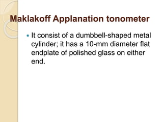
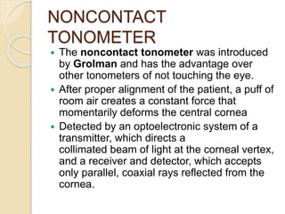
SlideShare a Scribd company logo
TONOMETRY & ITS
RECENT ADVANCES
Tonometry
 Tonometry is the procedure performed to
determine the intraocular pressure (IOP)
HISTORY
 1826: William Bowman used digital tonometry
as a routine examination test.
 1863: Albrecht von Grafe designed the first instrument to
attempt to measure intraocular pressure.
 Further instruments followed, notably by Donders in 1865
and Preistly-Smith in 1880.
All were of the indentation type and rested on the sclera
 1885: Maklakov designed an applanation tonometer. Used
for a number of years in Russia and Eastern Europe.
 1905: Hjalmar Schiotz produced his indentation tonometer.
This made tonometry a simple and routine clinical test.
Albrecht von Grafe tonometer
Donders tonometer
Ideal tonometer
 Should give accurate and reasonable
IOP measurement
 Convenient to use
 Simple to calibrate
 Stable from day to day
 Easier to standardise
 Free of maintenance problems
 All clinical tonometers measure the
IOP by relating a deformation of the
globe to the force responsible for the
deformation.
 The two basic types of tonometers
differ according to the shape of the
deformation: indentation and
applanation (flattening).
INDENTATION TONOMETER
 The shape of the deformation with this
type of tonometer is a truncated
cone.
 These instruments displace a
relatively large intraocular volume. As
a result, conversion tables based on
empirical data from in vitro and in vivo
studies must be used to estimate the
IOP.
 Prototype- Schiotz tonometer
APPLANATION
TONOMETERS
 The shape of the deformation with
these tonometers is a simple
flattening, and because the shape is
constant, its relationship to the IOP
can, in most cases, be derived from
mathematical calculations.
 The applanation tonometers are
further differentiated on the basis of
the variable that is measured.
VARIABLE FORCE
 This type of tonometer measures the
force that is required to applanate
(flatten) a standard area of the corneal
surface.
 Prototype- Goldmann applanation
tonometer, which was introduced in
1954.
VARIABLE AREA
 Other applanation tonometers
measure the area of the cornea that is
flattened by a known force (weight).
 Prototype- Maklakoff tonometer
 Goldmanntype tonometers have
relatively minimal displacement of
intraocular volume, whereas that with
Maklakoff-type tonometers is
sufficiently large to require the use of
conversion tables.
Applanation Tonometer with
variable area
TONOMETER DESCRIPTION/USE
Maklakoff-Kalfa Prototype
Applanometer Ceramic endplates
Tonomat Disposable endplates
Halberg tonometer
Transparent endplate for direct reading:
multiple weights
Barraquer tonometer
Plastic tonometer for use in operating
room
Ocular tension indicator Uses Goldmann biprism and standard
weight, for screening (measures above
or below 21 mm Hg)
Glaucotest Screening tonometer with multiple
endplates for selecting different “cutoff”
pressures
NON-CONTACT TONOMETER
 A third type of tonometer uses a puff
of air to deform the cornea and
measures the time or force of the air
puff that is required to create a
standard amount of corneal
deformation.
 The prototype was introduced by
Grolman in 1972.
Schiotz Indentation Tonometry
 It consists of a footplate that rests on
the cornea and a weighted plunger
that moves freely (except for the effect
of friction) within a shaft in the
footplate with the degree to which it
indents the cornea indicated by the
movement of a needle on a scale.
 A 5.5-g weight is permanently fixed to
the plunger, which can be increased to
7.5,10, or 15 g by adding additional
weights.
Parts of schiotz tonometer
Scale
needle
Weight 5.5g
plunge
rholder
Foot
plate
lever
3mm diameter
ROC 15mm
Tonometer weight = 11g
Additional
weights
7.5,10,15g
Schiotz tonometry -
characteristics
 The extent to which cornea is indented by
plunger is measured as the distance from
the foot plate curve to the plunger base and
a lever system moves a needle on
calibrated scale.
 The indicated scale reading and the plunger
weight are converted to an IOP
measurement.
 More the plunger indents the cornea,
higher the scale reading and lower the
IOP
 Each scale unit represents 0.05 mm
protrusion of the plunger.
PRINCIPLE
 The weight of tonometer on the eye increases the
actual IOP (Po) to a higher level (Pt).
 The change in pressure from Po to Pt is an
expression of the resistance of the eye (scleral
rigidity) to the displacement of fluid.
 IOP with Tonometer in position Pt =
Actual IOP Po + Scleral Rigidity E
(P (t) = P(o) + E)
 Determination of Po from a scale reading Pt
requires conversion which is done according to
Friedenwald conversion tables.
Friedenwald formula
 Friedenwald generated formula for linear
relationship between the log function of IOP and the
ocular distension.
 Pt = log Po + C ΔV
 This formula has ‘C’ a numerical constant, the
coefficient of ocular rigidity which is an
expression of distensibility of eye. Its average value
is 0.025
 ΔV is the change in volume
 Friedenwald developed a set of
conversion tables, referred to as the
1948 and 1955 tables for IOP.
 Subsequent studies indicated that the
1948 tables agree more closely with
measurements by Goldmann
applanation tonometry.
Friedenwald conversion table

Plunger Load
Scale
Reading 5.5 g 7.5 g 10 g 15 g
3.0 24.4 35.8 50.6 81.8
3.5 22.4 33.0 46.9 76.2
4.0 20.6 30.4 43.4 71.0
4.5 18.9 28.0 40.2 66.2
5.0 17.3 25.8 37.2 61.8
5.5 15.9 23.8 34.4 57.6
6.0 14.6 21.9 31.8 53.6
6.5 13.4 20.1 29.4 49.9
7.0 12.2 18.5 27.2 46.5
7.5 11.2 17.0 25.1 43.2
8.0 10.2 15.6 23.1 40.2
8.5 9.4 14.3 21.3 38.1
9.0 8.5 13.1 19.6 34.6
9.5 7.8 12.0 18.0 32.0
10.0 7.1 10.9 16.5 29.6
TECHNIQUE
 Patient should be anasthetised with 4%lignocaine or
0.5% proparacaine
 With the patient in supine position, looking up at a
fixation target examiner separates the lids and lowers
the tonometer plate to rest on the anesthetized cornea
so that plunger is free to move vertically .
 The examiner observes a fine movement of the indicator
needle on the scale in response to the ocular pulsations.
The average between the extremes of these is taken.
 The 5.5 gm weight is initially used.
 If scale reading is 4 or less, additional weight is added
to plunger.
 Conversion table is used to derive IOP in mm Hg from
scale reading and plunger weight.
Cause of error
 Because conversion tables were based
on an “average” coefficient of ocular
rigidity (C), eyes that deviate significantly
from this C value give false IOP
measurements.
 Another variable that affects accuracy is
expulsion of intraocular blood during
indentation tonometry .
 A relatively steep or thick cornea causes
an increased displacement of fluid during
indentation tonometry, which leads to a
falsely high IOP reading
CALIBRATION
 The instrument should be calibrated
before each use by placing it on a
polished metal sphere and checking to
be sure that the scale reading is zero.
 If the reading is not zero, the
instrument must be repaired.
STERILIZATION
 The tonometer is disassembled between each
use and the barrel is cleaned with 2 pipe
cleaners, the first soaked in isopropyl alcohol
70 % or methylated spirit and the second
dry.
 The foot plate is cleaned with alcohol swab.
 All surfaces must be dried before
reassembling.
 The instrument can be sterilized with ultraviolet
radiation, steam, ethylene oxide.
 As with other tonometer tips, the Schiotz can
be damaged by some disinfecting solutions
Differential tonometry
 It is done to get rid of ocular rigidity factor.
 A reading is taken with one weight on the Plunger and then a
second reading' in taken with a different weight.
 Making a diagnosis of glaucoma in a pt. with myopia presents
unusual difficulties. The low ocular rigidity in these eyes result
in Schiotz readings within normal limits.
5.5g 10g Ocular
rigidity
IOP
18 mm Hg 15 mm Hg lower >18
18 mm Hg 21 mm Hg higher <18
18 mm Hg 18 mm Hg equal 18
Advantages of schiotz tonometer
 Simple technique
 Elegant design
 Portable
 No need for SlitLamp or power supply
 Reasonably priced
 Anodized scale mount which is highly
resistant to sterilizing water.
 Schiotz tonometer is still most widely
tonometer.
LIMITATIONS
◦ Instrumental errors
 Standardisation - testing labs for certification
 Mechanical obstruction to plunger etc.
◦ Muscular contractions
 Of extra ocular muscles increase IOP
 Accomodation decreases IOP
 Variations in volume of globe
◦ Microphthalmos
◦ High Myopia
◦ Buphthalmos
◦ It can be recorded in supine position
only
APPLANATION
TONOMETRY
Maklakoff Applanation tonometer
 It consist of a dumbbell-shaped metal
cylinder; it has a 10-mm diameter flat
endplate of polished glass on either
end.
Maklakoff tonometer
 A set of four such instruments were available,
weighing 5, 7.5, 10, and 15 g. A dye
suspension of Argyrol, glycerin, and water
was applied to either endplate and, with the
patient in a supine position and the cornea
anesthetized, the instrument rested vertically
on the cornea for 1 second.
 The resultant circular white imprint on the
endplate corresponded to the area of cornea
that was flattened. The diameter of the white
area is measured with a transparent plastic
measuring scale to 0.1 mm, and the IOP is
read from a conversion table in the column
corresponding to the weight used.
 The Perkins applanation tonometer
uses the same biprism as the Goldmann
applanation tonometer.
 The light source is powered by a battery
and the force is varied manually.
 A counter balance makes it possible to
use the instrument in either the vertical
or horizontal position.
 Being portable it is practical when
measuring IOP in infants / children, bed
ridden patients and for use in operating
rooms.
Other Applanation Tonometers
with Variable Force
Perkins Tonometer
Perkins Tonometer
•It is similar to the Perkins tonometer, but
uses a different biprism and has an electric
motor that varies the force
•Both require training to use
Draeger applanation
tonometer
 The original Mackay-Marg tonometer
had a plate diameter of 1.5 mm
surrounded by a rubber sleeve.
 The force required to keep the plate flush
with the sleeve was electronically
monitored and recorded on a paper strip.
 This instrument is useful for measuring
IOP in eyes with scarred, irregular, or
edematous corneas because the end
point does not depend on the evaluation
of a light reflex sensitive to optical
irregularity
Mackay marg tonometer
At 1.5 mm of corneal area applanation, tracing
reaches a peak and the force applied = IOP + force
required to deform the cornea.
At 3 mm flattening, force required to deform cornea
is transferred from plunger to surrounding sleeve,
creating a dip in tracing corresponding to IOP.
Flattening of >3 mm of area gives artificial elevation
of IOP.
 The most commonly used Mackay-Marg-
type tonometer today is the Tono-Pen, a
handheld instrument with a strain gauge
that creates an electrical signal as the
footplate flattens the cornea(microstrain
gauge technology).
 It averages 4 to 10 readings to give a
final digital readout. It also provides the
percentage of variability between the
lowest and highest acceptable readings
from 5% to 20%.
Tonopen
Tonopen
Pneumotonometer
 Here a central sensing device measures the IOP, while
the force required to bend the cornea is transferred to a
surrounding structure. The sensor, is air pressure.
 It has a sensing device that consists of a gas chamber
covered by a polymeric silicone diaphragm.
 A transducer converts the gas pressure in the chamber
into an electrical signal that is recorded on a paper strip.
 As the sensing nozzle touches the cornea and when the
area of contact equals that of the central chamber, an
initial inflection is recorded, which represents the IOP
and the force required to bend the cornea. With further
enlargement of the corneal contact, the bending force is
transferred to the face of the nozzle, which is
interpreted as the actual IOP.
Pneumotonometer
NONCONTACT
TONOMETER
 The noncontact tonometer was introduced
by Grolman and has the advantage over
other tonometers of not touching the eye.
 After proper alignment of the patient, a puff of
room air creates a constant force that
momentarily deforms the central cornea
 Detected by an optoelectronic system of a
transmitter, which directs a
collimated beam of light at the corneal vertex,
and a receiver and detector, which accepts
only parallel, coaxial rays reflected from the
cornea.
 At the moment that the central cornea is
flattened, the greatest number of reflected light
rays are received, which is recorded as the
peak intensity of light detected. The time from
an internal reference point to the moment of
maximum light detection is converted to IOP.
 The time interval for an average noncontact
tonometer measurement is 1 to 3 milliseconds
(1/500th of the cardiac cycle) and is random
with respect to the phase of the cardiac cycle
so that the ocular pulse becomes a
significant variable. For this reason, it is
recommended that a minimum of three
readings within 3 mm Hg be taken and
averaged as the IOP.
New NCT, Pulsair is a portable
hand held tonometer
Miscellaneous tonometers
Rebound Tonometer
 In a new handheld tonometer, the Icare tonometer,
IOP is determined by measuring the force produced by
a small plastic probe as it rebounds from the cornea.
 The device uses an induction coil to magnetise the
probe and fire it against the cornea.
 As the probe bounces against the cornea and back into
the device it creates an induction current from which the
intraocular pressure is calculated.
 As a screening tool in children. The ability to evaluate
IOP without the use of topical anesthesia potentially
provides the opportunity to monitor IOP at home.
 The rebound tonometer has been shown to have similar
accuracy to the Tono-Pen, and it is comparable with
Goldmann tonometry for IOPs over a reasonable range
in adults.
Rebound tonometer
The Ocuton tonometer
 The Ocuton™ tonometer
 Hand-held tonometer
 Works on the applanation principle
 Probe is so light that it is barely felt
 Needs no anesthetic in most patients.
 Used for home tonometry
 Useful to get some idea of the relative diurnal variation in IOP if
the patient or relative can learn to use it.
Trans palpebral tonometry
 Used in situations where other, more accurate,
devices are not practical, such as in young children,
demented patients and severely developmentally-
challenged patients.
It adds variables such as the thickness of the
eyelids, orbicularis muscle tone and potential Intra
palpebral scarring.
 Transpalpebral tonometry does not
involve contact with the cornea and
does not require sterilization of the
device or topical anesthetic during
routine use.
 Only moderate correlation with those
provided by applanation tonometry
 Home tonometer: Proview
Phosphene Tonometer(Bausch &
Lomb) is based on phosphene
perception after eyelid indentation
Diaton tonometer (BiCOM, Inc)
 Measuring intraocular
pressure through the
Eyelid
 The Diaton tonometer
calculates pressure by
measuring the response
of a free falling rod
 The principle is based on
Newton's second law, as
it rebounds against the
tarsal plate of the eyelid.
 The patient is positioned
so that the tip of the
device and lid are
overlying sclera.
Ocular Response
Analyser(ORA)
 Provides IOP measurement free from
influence of corneal biochemical
properties
 It measures corneal hysteresis &
corneal resistance factor & thus
overcomes the demerits of GAT to
some extend
Ocular Response Analyzer
It directs the air jet against the cornea and measures not one
but two pressures at which applanation occurs
 1) when the air jet flattens the cornea as the cornea is bent
inward and 2) as the air jet lessens in force and the cornea
recovers.
Ocular response analyser
 The first is the resting intraocular pressure.
 The difference between the first and the second
applanation pressure is called corneal hysteresis
 Corneal hysteresis is a measure of the viscous
dampening and, hence, the biomechanical
properties of the cornea.
 The biomechanical properties of the cornea are
related to corneal thickness and include elastic and
viscous dampening attributes.
 IOP correlate well with Goldmann tonometry but,
on average, measure a few millimeters higher.
 Further , while IOP varies over the 24-hour day,
hysteresis seems to be stable.
 Congdon et al found that a ‘low’ hysteresis
reading with the ORA correlates with
progression of glaucoma, whereas thin central
corneal thickness correlates with glaucoma
damage.
 It has practical value in the management of
glaucoma.
Pascal’s Dynamic Contour
Tonometry
 Dynamic contour tonometry (DCT) is a novel
method which uses principle of contour
matching instead of applanation.
 Principle : By surrounding and matching the
contour of a sphere (or a portion thereof ), the
pressure on the outside equals the
pressure on the inside.
 This is designed to reduce the influence of
biomechanical properties of the cornea on
measurement.
 These include corneal thickness, rigidity,
curvature, and elastic properties.
 It is less influenced by corneal thickness but
more influenced by corneal curvature than the
Goldmann tonometer
Dynamic contour tonometer
 The contour matched tip has a concave surface of
radius 10.5 mm, which approximates to the shape
of a normal cornea when the pressure on both
sides is equal.
 The probe is placed adjacent to the central cornea.
 The integrated piezoresistive pressure sensor
automatically begins to acquire data, measuring
IOP 100 times per second.
 A complete measurement cycle requires about 8
seconds of contact time.
 The device also measures the variation in pressure
that occurs with the cardiac cycle. (Ocular pulse
Amplitude)
 The concept developed from a previous contact
lens tonometer called the ‘Smart Lens”.
 It superior in accuracy to Goldmann tonometry and
pneumotonometry .
 IOP is not affected by corneal thickness.
 IOP is not altered by corneal refractive surgery that
thins the cornea.
 The DCT shows the magnitude of the difference
between maximum and minimum IOP as the ocular
pulse amplitude.
 OPA may be indicative of the status of ocular blood
flow and be differentially affected in different types
of glaucoma.
 Ocular pulse amplitude is
increased over normals in
most forms of glaucoma and
may be related to the level
of IOP.
IOP Monitoring Devices
 There is need for an IOP telemetry
device without artificially altering the
pressure. Several prototypes—based on
a contact lens, an implantable device, or
a scleral band device have been
developed. Such a lens will help us
monitor and manage individuals who are
susceptible to wide IOP fluctuations, who
have poor adherence to medical therapy,
who perhaps are “poor responders” to
medical therapy, and who have wide IOP
fluctuations in the postoperative period
 Continuous IOP monitoring devices:
Contact lenses like ‘Sensimed
Triggerfish’ & the ‘Smart’ contact
lenses measure IOP by detecting
changes in the corneoscleral
curvature induced by IOP changes
Comparison of Tonometers
 The most precise method for evaluating the accuracy of a tonometer
is to compare it with manometric measurements of the cannulated
anterior chamber. Its use in largescale human studies has been
limited.
 The alternative is to compare the tonometer in question against the
instrument that previous studies have shown to be the most
accurate.
 In eyes with regular corneas, the Goldmann applanation tonometer
is generally accepted as the standard against which other
tonometers must be compared.
 Even with this instrument, however, inherent variability must be
taken into account. When two readings were taken on the same eye
with Goldmann tonometers in a short time frame, at least 30% of the
paired readings differed by 2 and 3 mm Hg or more. In another
study, intraobserver variation was 1.5 ± 1.96 mm Hg and
interobserver variation was 1.79 ± 2.41 mm Hg, which could be
reduced by 9% and 11%, respectively, by using the median value of
three consecutive measurements
 Clinically, the most widely used methods
for measuring IOP are by Goldmann
applanation tonometry and with use of
the Tono-Pen; the noncontact tonometer,
Perkins tonometer, pneumotonometry,
and the Schiötz tonometer.
 In general, the Schiötz tonometer reads
lower than the Goldmann. The Perkins
applanation tonometer compared
favorably against the Goldmann
tonometer.
 The Tono-Pen resembles manometric
readings in human autopsy eyes.
 Most studies agree that the Tono-Pen
underestimates Goldmann IOP in the
higher range and overestimates in the
lower range
 In multiple comparative studies,
readings taken with the
pneumotonometer correlated closely
with those obtained by using
Goldmann tonometers, although the
pneumotonometer readings tended to
be higher.
 Post-LASIK IOP measurements
obtained by pneumotonometry were
more reliable than those taken by
Goldmann applanation.
Tonometry for Special Clinical
Circumstances
 The pneumatic tonometer has been
shown to be useful in eyes with
diseased or irregular corneas.
 In eyes after penetrating keratoplasty,
the Tono-Pen significantly
overestimated Goldmann readings
Tonometry on Irregular
Corneas
Tonometry over Soft Contact
Lenses
 It has been claimed that
pneumotonometry and the Tono-Pen
can measure with reasonable
accuracy the IOP through bandage
contact lenses.
Tonometry with Gas-Filled
Eyes
 Intraocular gas significantly affects
scleral rigidity. In a study with irregular
corneas after vitrectomy and air-gas-
fluid exchange, readings with the
Tono-Pen and pneumotonometer were
highly correlated.
 A manometric study with human
autopsy eyes indicated that both
instruments significantly
underestimated the IOP at pressures
greater than 30 mm Hg
Tonometry with Flat Anterior
Chamber
 In human autopsy eyes with flat
anterior chambers, IOP readings from
the Goldmann applanation tonometer,
pneumotonometer, and Tono-Pen did
not correlate well with manometrically
determined pressures
Tonometry in Eyes with
Keratoprostheses
 In patients at high risk for corneal
transplant rejection, implantation of a
keratoprosthesis is now a viable option
for vision rehabilitation. However, given
that most keratoprostheses have a rigid,
clear surface, it is impossible to measure
IOP by using applanation or indentation
instruments.
 In such eyes, tactile assessment
appears to be the most widely used
method to estimate IOP
Tonometry & its recent advances

More Related Content

What's hot

TONOMETRY
TONOMETRYTONOMETRY
Tonometry
 Tonometry Tonometry
Gonioscopy
GonioscopyGonioscopy
Gonioscopymeenank
 
Means of IOP measurement
Means of IOP measurementMeans of IOP measurement
Pachymetry
PachymetryPachymetry
Pachymetry
SSSIHMS-PG
 
Goldman applanation tonometry
 Goldman applanation tonometry Goldman applanation tonometry
Goldman applanation tonometryHossein Mirzaie
 
Biometry
BiometryBiometry
Biometry
Binny Tyagi
 
Introduction, Assessment and Management of Amblyopia
Introduction, Assessment and Management of Amblyopia Introduction, Assessment and Management of Amblyopia
Introduction, Assessment and Management of Amblyopia
Anis Suzanna Mohamad
 
Binocular Indirect OPHTHALMOSCOPY
Binocular Indirect OPHTHALMOSCOPYBinocular Indirect OPHTHALMOSCOPY
Binocular Indirect OPHTHALMOSCOPY
Kh.Towkir-Ul- Islam
 
Indirect Ophthalmoscopy and slit lamp biomicroscopy
Indirect Ophthalmoscopy and slit lamp biomicroscopyIndirect Ophthalmoscopy and slit lamp biomicroscopy
Indirect Ophthalmoscopy and slit lamp biomicroscopy
Lakshmi Murthy
 
Assessment of Anterior Chamber Angle
Assessment of Anterior Chamber AngleAssessment of Anterior Chamber Angle
Assessment of Anterior Chamber Angle
Sujay Chauhan
 
Corneal topography wavefront analysis
Corneal topography wavefront analysisCorneal topography wavefront analysis
Corneal topography wavefront analysis
ikramdr01
 
Amblyopia : classification & Occlusion therapy
Amblyopia : classification & Occlusion therapy Amblyopia : classification & Occlusion therapy
Amblyopia : classification & Occlusion therapy
Aayush Chandan
 
Photostress recovery time
Photostress recovery timePhotostress recovery time
Photostress recovery time
OPTOM FASLU MUHAMMED
 
A scan biometry | How to Use A-scan? Types of A-Scan Biometry?
A scan biometry | How to Use A-scan? Types of A-Scan Biometry?A scan biometry | How to Use A-scan? Types of A-Scan Biometry?
A scan biometry | How to Use A-scan? Types of A-Scan Biometry?
Naeem Ahmad
 
Tonometry
TonometryTonometry
Specular microscopy
Specular microscopySpecular microscopy
Specular microscopy
Ruchi sood
 
A scan ultrasonography
A scan ultrasonographyA scan ultrasonography
A scan ultrasonography
Samuel Ponraj
 
Corneal pachymetry by ben okeeffe
Corneal pachymetry by ben okeeffeCorneal pachymetry by ben okeeffe
Corneal pachymetry by ben okeeffe
OphthalmicDocs Chiong
 
Objective refraction
Objective refractionObjective refraction
Objective refraction
sneha_thaps
 

What's hot (20)

TONOMETRY
TONOMETRYTONOMETRY
TONOMETRY
 
Tonometry
 Tonometry Tonometry
Tonometry
 
Gonioscopy
GonioscopyGonioscopy
Gonioscopy
 
Means of IOP measurement
Means of IOP measurementMeans of IOP measurement
Means of IOP measurement
 
Pachymetry
PachymetryPachymetry
Pachymetry
 
Goldman applanation tonometry
 Goldman applanation tonometry Goldman applanation tonometry
Goldman applanation tonometry
 
Biometry
BiometryBiometry
Biometry
 
Introduction, Assessment and Management of Amblyopia
Introduction, Assessment and Management of Amblyopia Introduction, Assessment and Management of Amblyopia
Introduction, Assessment and Management of Amblyopia
 
Binocular Indirect OPHTHALMOSCOPY
Binocular Indirect OPHTHALMOSCOPYBinocular Indirect OPHTHALMOSCOPY
Binocular Indirect OPHTHALMOSCOPY
 
Indirect Ophthalmoscopy and slit lamp biomicroscopy
Indirect Ophthalmoscopy and slit lamp biomicroscopyIndirect Ophthalmoscopy and slit lamp biomicroscopy
Indirect Ophthalmoscopy and slit lamp biomicroscopy
 
Assessment of Anterior Chamber Angle
Assessment of Anterior Chamber AngleAssessment of Anterior Chamber Angle
Assessment of Anterior Chamber Angle
 
Corneal topography wavefront analysis
Corneal topography wavefront analysisCorneal topography wavefront analysis
Corneal topography wavefront analysis
 
Amblyopia : classification & Occlusion therapy
Amblyopia : classification & Occlusion therapy Amblyopia : classification & Occlusion therapy
Amblyopia : classification & Occlusion therapy
 
Photostress recovery time
Photostress recovery timePhotostress recovery time
Photostress recovery time
 
A scan biometry | How to Use A-scan? Types of A-Scan Biometry?
A scan biometry | How to Use A-scan? Types of A-Scan Biometry?A scan biometry | How to Use A-scan? Types of A-Scan Biometry?
A scan biometry | How to Use A-scan? Types of A-Scan Biometry?
 
Tonometry
TonometryTonometry
Tonometry
 
Specular microscopy
Specular microscopySpecular microscopy
Specular microscopy
 
A scan ultrasonography
A scan ultrasonographyA scan ultrasonography
A scan ultrasonography
 
Corneal pachymetry by ben okeeffe
Corneal pachymetry by ben okeeffeCorneal pachymetry by ben okeeffe
Corneal pachymetry by ben okeeffe
 
Objective refraction
Objective refractionObjective refraction
Objective refraction
 

Similar to Tonometry & its recent advances

Tonometry by dr. mamta meena
Tonometry by dr. mamta meenaTonometry by dr. mamta meena
Tonometry by dr. mamta meena
AIMS Education
 
Tonometry
TonometryTonometry
tonometry-140624223245-phpapp02-converted.pptx
tonometry-140624223245-phpapp02-converted.pptxtonometry-140624223245-phpapp02-converted.pptx
tonometry-140624223245-phpapp02-converted.pptx
MUHAMMADRASHID311088
 
TONOMETRS---11.pptx
TONOMETRS---11.pptxTONOMETRS---11.pptx
TONOMETRS---11.pptx
LavanyaMadabushi
 
OT 2016 The Changing Face of Tonometry
OT 2016 The Changing Face of TonometryOT 2016 The Changing Face of Tonometry
OT 2016 The Changing Face of TonometryJason Higginbotham
 
Iop
IopIop
Tonometer ppt
Tonometer pptTonometer ppt
Tonometer ppt
Maaz ul haq
 
tonometry-140624223245-phpapp02.pptx
tonometry-140624223245-phpapp02.pptxtonometry-140624223245-phpapp02.pptx
tonometry-140624223245-phpapp02.pptx
aiswaryavenugopalann
 
Tonometer - Basics & Types
Tonometer - Basics & TypesTonometer - Basics & Types
Tonometer - Basics & Types
Rajeswari Kesavan
 
Tonometry.pptx
Tonometry.pptxTonometry.pptx
Tonometry.pptx
Anisha Heka
 
Tonometer
TonometerTonometer
Tonometer
JESLIN JOSE
 
Tonometry and tonography
Tonometry and tonographyTonometry and tonography
Tonometry and tonography
Loknath Goswami
 
Tonometry and tonography
Tonometry and tonographyTonometry and tonography
Tonometry and tonography
Dr Samarth Mishra
 
TONOMETRY IN OPTOMETRY & OPHTHALMOLOGY .PPTX
TONOMETRY IN OPTOMETRY & OPHTHALMOLOGY .PPTXTONOMETRY IN OPTOMETRY & OPHTHALMOLOGY .PPTX
TONOMETRY IN OPTOMETRY & OPHTHALMOLOGY .PPTX
ANUJA DHAKAL
 
tonometer.pptx
tonometer.pptxtonometer.pptx
tonometer.pptx
Vizo7
 
Part_1a.ppt
Part_1a.pptPart_1a.ppt
Part_1a.ppt
Khalil Alhatab
 
IOP measurements
IOP measurementsIOP measurements
IOP measurements
Maryam Fida
 

Similar to Tonometry & its recent advances (20)

Tonometry by dr. mamta meena
Tonometry by dr. mamta meenaTonometry by dr. mamta meena
Tonometry by dr. mamta meena
 
Tonometry
TonometryTonometry
Tonometry
 
tonometry-140624223245-phpapp02-converted.pptx
tonometry-140624223245-phpapp02-converted.pptxtonometry-140624223245-phpapp02-converted.pptx
tonometry-140624223245-phpapp02-converted.pptx
 
TONOMETRS---11.pptx
TONOMETRS---11.pptxTONOMETRS---11.pptx
TONOMETRS---11.pptx
 
OT 2016 The Changing Face of Tonometry
OT 2016 The Changing Face of TonometryOT 2016 The Changing Face of Tonometry
OT 2016 The Changing Face of Tonometry
 
Iop
IopIop
Iop
 
Tonometer ppt
Tonometer pptTonometer ppt
Tonometer ppt
 
tonometry-140624223245-phpapp02.pptx
tonometry-140624223245-phpapp02.pptxtonometry-140624223245-phpapp02.pptx
tonometry-140624223245-phpapp02.pptx
 
Tonometer - Basics & Types
Tonometer - Basics & TypesTonometer - Basics & Types
Tonometer - Basics & Types
 
Tonometry.pptx
Tonometry.pptxTonometry.pptx
Tonometry.pptx
 
Tonometer
TonometerTonometer
Tonometer
 
Tonometry and tonography
Tonometry and tonographyTonometry and tonography
Tonometry and tonography
 
INTRAOCULAR PRESSURE .pdf
INTRAOCULAR PRESSURE                  .pdfINTRAOCULAR PRESSURE                  .pdf
INTRAOCULAR PRESSURE .pdf
 
INTRAOCULAR PRESSURE.pdf
INTRAOCULAR                    PRESSURE.pdfINTRAOCULAR                    PRESSURE.pdf
INTRAOCULAR PRESSURE.pdf
 
Tonometry and tonography
Tonometry and tonographyTonometry and tonography
Tonometry and tonography
 
TONOMETRY IN OPTOMETRY & OPHTHALMOLOGY .PPTX
TONOMETRY IN OPTOMETRY & OPHTHALMOLOGY .PPTXTONOMETRY IN OPTOMETRY & OPHTHALMOLOGY .PPTX
TONOMETRY IN OPTOMETRY & OPHTHALMOLOGY .PPTX
 
tonometer.pptx
tonometer.pptxtonometer.pptx
tonometer.pptx
 
Part_1a.ppt
Part_1a.pptPart_1a.ppt
Part_1a.ppt
 
IOP measurements
IOP measurementsIOP measurements
IOP measurements
 
Tonometry
TonometryTonometry
Tonometry
 

Recently uploaded

CHAPTER 1 SEMESTER V PREVENTIVE-PEDIATRICS.pdf
CHAPTER 1 SEMESTER V PREVENTIVE-PEDIATRICS.pdfCHAPTER 1 SEMESTER V PREVENTIVE-PEDIATRICS.pdf
CHAPTER 1 SEMESTER V PREVENTIVE-PEDIATRICS.pdf
Sachin Sharma
 
VERIFICATION AND VALIDATION TOOLKIT Determining Performance Characteristics o...
VERIFICATION AND VALIDATION TOOLKIT Determining Performance Characteristics o...VERIFICATION AND VALIDATION TOOLKIT Determining Performance Characteristics o...
VERIFICATION AND VALIDATION TOOLKIT Determining Performance Characteristics o...
Nguyễn Thị Vân Anh
 
Navigating Healthcare with Telemedicine
Navigating Healthcare with  TelemedicineNavigating Healthcare with  Telemedicine
Navigating Healthcare with Telemedicine
Iris Thiele Isip-Tan
 
Preventing Pickleball Injuries & Treatment
Preventing Pickleball Injuries & TreatmentPreventing Pickleball Injuries & Treatment
Preventing Pickleball Injuries & Treatment
LAB Sports Therapy
 
Dehradun ❤CALL Girls 8901183002 ❤ℂall Girls IN Dehradun ESCORT SERVICE❤
Dehradun ❤CALL Girls  8901183002 ❤ℂall  Girls IN Dehradun ESCORT SERVICE❤Dehradun ❤CALL Girls  8901183002 ❤ℂall  Girls IN Dehradun ESCORT SERVICE❤
Dehradun ❤CALL Girls 8901183002 ❤ℂall Girls IN Dehradun ESCORT SERVICE❤
aunty1x2
 
The Docs PPG - 30.05.2024.pptx..........
The Docs PPG - 30.05.2024.pptx..........The Docs PPG - 30.05.2024.pptx..........
The Docs PPG - 30.05.2024.pptx..........
TheDocs
 
Antibiotic Stewardship by Anushri Srivastava.pptx
Antibiotic Stewardship by Anushri Srivastava.pptxAntibiotic Stewardship by Anushri Srivastava.pptx
Antibiotic Stewardship by Anushri Srivastava.pptx
AnushriSrivastav
 
Medical Technology Tackles New Health Care Demand - Research Report - March 2...
Medical Technology Tackles New Health Care Demand - Research Report - March 2...Medical Technology Tackles New Health Care Demand - Research Report - March 2...
Medical Technology Tackles New Health Care Demand - Research Report - March 2...
pchutichetpong
 
ABDOMINAL COMPARTMENT SYSNDROME
ABDOMINAL COMPARTMENT SYSNDROMEABDOMINAL COMPARTMENT SYSNDROME
ABDOMINAL COMPARTMENT SYSNDROME
Rommel Luis III Israel
 
Navigating Women's Health: Understanding Prenatal Care and Beyond
Navigating Women's Health: Understanding Prenatal Care and BeyondNavigating Women's Health: Understanding Prenatal Care and Beyond
Navigating Women's Health: Understanding Prenatal Care and Beyond
Aboud Health Group
 
💘Ludhiana ℂall Girls 📞]][89011★83002][[ 📱 ❤ESCORTS service in Ludhiana💃💦Ludhi...
💘Ludhiana ℂall Girls 📞]][89011★83002][[ 📱 ❤ESCORTS service in Ludhiana💃💦Ludhi...💘Ludhiana ℂall Girls 📞]][89011★83002][[ 📱 ❤ESCORTS service in Ludhiana💃💦Ludhi...
💘Ludhiana ℂall Girls 📞]][89011★83002][[ 📱 ❤ESCORTS service in Ludhiana💃💦Ludhi...
ranishasharma67
 
R3 Stem Cells and Kidney Repair A New Horizon in Nephrology.pptx
R3 Stem Cells and Kidney Repair A New Horizon in Nephrology.pptxR3 Stem Cells and Kidney Repair A New Horizon in Nephrology.pptx
R3 Stem Cells and Kidney Repair A New Horizon in Nephrology.pptx
R3 Stem Cell
 
CANCER CANCER CANCER CANCER CANCER CANCER
CANCER  CANCER  CANCER  CANCER  CANCER CANCERCANCER  CANCER  CANCER  CANCER  CANCER CANCER
CANCER CANCER CANCER CANCER CANCER CANCER
KRISTELLEGAMBOA2
 
Nursing Care of Client With Acute And Chronic Renal Failure.ppt
Nursing Care of Client With Acute And Chronic Renal Failure.pptNursing Care of Client With Acute And Chronic Renal Failure.ppt
Nursing Care of Client With Acute And Chronic Renal Failure.ppt
Rommel Luis III Israel
 
BOWEL ELIMINATION BY ANUSHRI SRIVASTAVA.pptx
BOWEL ELIMINATION BY ANUSHRI SRIVASTAVA.pptxBOWEL ELIMINATION BY ANUSHRI SRIVASTAVA.pptx
BOWEL ELIMINATION BY ANUSHRI SRIVASTAVA.pptx
AnushriSrivastav
 
Introduction to Forensic Pathology course
Introduction to Forensic Pathology courseIntroduction to Forensic Pathology course
Introduction to Forensic Pathology course
fprxsqvnz5
 
Myopia Management & Control Strategies.pptx
Myopia Management & Control Strategies.pptxMyopia Management & Control Strategies.pptx
Myopia Management & Control Strategies.pptx
RitonDeb1
 
Secret Tantric VIP Erotic Massage London
Secret Tantric VIP Erotic Massage LondonSecret Tantric VIP Erotic Massage London
Secret Tantric VIP Erotic Massage London
Secret Tantric - VIP Erotic Massage London
 
一比一原版纽约大学毕业证(NYU毕业证)成绩单留信认证
一比一原版纽约大学毕业证(NYU毕业证)成绩单留信认证一比一原版纽约大学毕业证(NYU毕业证)成绩单留信认证
一比一原版纽约大学毕业证(NYU毕业证)成绩单留信认证
o6ov5dqmf
 
Dimensions of Healthcare Quality
Dimensions of Healthcare QualityDimensions of Healthcare Quality
Dimensions of Healthcare Quality
Naeemshahzad51
 

Recently uploaded (20)

CHAPTER 1 SEMESTER V PREVENTIVE-PEDIATRICS.pdf
CHAPTER 1 SEMESTER V PREVENTIVE-PEDIATRICS.pdfCHAPTER 1 SEMESTER V PREVENTIVE-PEDIATRICS.pdf
CHAPTER 1 SEMESTER V PREVENTIVE-PEDIATRICS.pdf
 
VERIFICATION AND VALIDATION TOOLKIT Determining Performance Characteristics o...
VERIFICATION AND VALIDATION TOOLKIT Determining Performance Characteristics o...VERIFICATION AND VALIDATION TOOLKIT Determining Performance Characteristics o...
VERIFICATION AND VALIDATION TOOLKIT Determining Performance Characteristics o...
 
Navigating Healthcare with Telemedicine
Navigating Healthcare with  TelemedicineNavigating Healthcare with  Telemedicine
Navigating Healthcare with Telemedicine
 
Preventing Pickleball Injuries & Treatment
Preventing Pickleball Injuries & TreatmentPreventing Pickleball Injuries & Treatment
Preventing Pickleball Injuries & Treatment
 
Dehradun ❤CALL Girls 8901183002 ❤ℂall Girls IN Dehradun ESCORT SERVICE❤
Dehradun ❤CALL Girls  8901183002 ❤ℂall  Girls IN Dehradun ESCORT SERVICE❤Dehradun ❤CALL Girls  8901183002 ❤ℂall  Girls IN Dehradun ESCORT SERVICE❤
Dehradun ❤CALL Girls 8901183002 ❤ℂall Girls IN Dehradun ESCORT SERVICE❤
 
The Docs PPG - 30.05.2024.pptx..........
The Docs PPG - 30.05.2024.pptx..........The Docs PPG - 30.05.2024.pptx..........
The Docs PPG - 30.05.2024.pptx..........
 
Antibiotic Stewardship by Anushri Srivastava.pptx
Antibiotic Stewardship by Anushri Srivastava.pptxAntibiotic Stewardship by Anushri Srivastava.pptx
Antibiotic Stewardship by Anushri Srivastava.pptx
 
Medical Technology Tackles New Health Care Demand - Research Report - March 2...
Medical Technology Tackles New Health Care Demand - Research Report - March 2...Medical Technology Tackles New Health Care Demand - Research Report - March 2...
Medical Technology Tackles New Health Care Demand - Research Report - March 2...
 
ABDOMINAL COMPARTMENT SYSNDROME
ABDOMINAL COMPARTMENT SYSNDROMEABDOMINAL COMPARTMENT SYSNDROME
ABDOMINAL COMPARTMENT SYSNDROME
 
Navigating Women's Health: Understanding Prenatal Care and Beyond
Navigating Women's Health: Understanding Prenatal Care and BeyondNavigating Women's Health: Understanding Prenatal Care and Beyond
Navigating Women's Health: Understanding Prenatal Care and Beyond
 
💘Ludhiana ℂall Girls 📞]][89011★83002][[ 📱 ❤ESCORTS service in Ludhiana💃💦Ludhi...
💘Ludhiana ℂall Girls 📞]][89011★83002][[ 📱 ❤ESCORTS service in Ludhiana💃💦Ludhi...💘Ludhiana ℂall Girls 📞]][89011★83002][[ 📱 ❤ESCORTS service in Ludhiana💃💦Ludhi...
💘Ludhiana ℂall Girls 📞]][89011★83002][[ 📱 ❤ESCORTS service in Ludhiana💃💦Ludhi...
 
R3 Stem Cells and Kidney Repair A New Horizon in Nephrology.pptx
R3 Stem Cells and Kidney Repair A New Horizon in Nephrology.pptxR3 Stem Cells and Kidney Repair A New Horizon in Nephrology.pptx
R3 Stem Cells and Kidney Repair A New Horizon in Nephrology.pptx
 
CANCER CANCER CANCER CANCER CANCER CANCER
CANCER  CANCER  CANCER  CANCER  CANCER CANCERCANCER  CANCER  CANCER  CANCER  CANCER CANCER
CANCER CANCER CANCER CANCER CANCER CANCER
 
Nursing Care of Client With Acute And Chronic Renal Failure.ppt
Nursing Care of Client With Acute And Chronic Renal Failure.pptNursing Care of Client With Acute And Chronic Renal Failure.ppt
Nursing Care of Client With Acute And Chronic Renal Failure.ppt
 
BOWEL ELIMINATION BY ANUSHRI SRIVASTAVA.pptx
BOWEL ELIMINATION BY ANUSHRI SRIVASTAVA.pptxBOWEL ELIMINATION BY ANUSHRI SRIVASTAVA.pptx
BOWEL ELIMINATION BY ANUSHRI SRIVASTAVA.pptx
 
Introduction to Forensic Pathology course
Introduction to Forensic Pathology courseIntroduction to Forensic Pathology course
Introduction to Forensic Pathology course
 
Myopia Management & Control Strategies.pptx
Myopia Management & Control Strategies.pptxMyopia Management & Control Strategies.pptx
Myopia Management & Control Strategies.pptx
 
Secret Tantric VIP Erotic Massage London
Secret Tantric VIP Erotic Massage LondonSecret Tantric VIP Erotic Massage London
Secret Tantric VIP Erotic Massage London
 
一比一原版纽约大学毕业证(NYU毕业证)成绩单留信认证
一比一原版纽约大学毕业证(NYU毕业证)成绩单留信认证一比一原版纽约大学毕业证(NYU毕业证)成绩单留信认证
一比一原版纽约大学毕业证(NYU毕业证)成绩单留信认证
 
Dimensions of Healthcare Quality
Dimensions of Healthcare QualityDimensions of Healthcare Quality
Dimensions of Healthcare Quality
 

Tonometry & its recent advances

  • 2. Tonometry  Tonometry is the procedure performed to determine the intraocular pressure (IOP)
  • 3. HISTORY  1826: William Bowman used digital tonometry as a routine examination test.  1863: Albrecht von Grafe designed the first instrument to attempt to measure intraocular pressure.  Further instruments followed, notably by Donders in 1865 and Preistly-Smith in 1880. All were of the indentation type and rested on the sclera  1885: Maklakov designed an applanation tonometer. Used for a number of years in Russia and Eastern Europe.  1905: Hjalmar Schiotz produced his indentation tonometer. This made tonometry a simple and routine clinical test.
  • 4. Albrecht von Grafe tonometer
  • 6. Ideal tonometer  Should give accurate and reasonable IOP measurement  Convenient to use  Simple to calibrate  Stable from day to day  Easier to standardise  Free of maintenance problems
  • 7.  All clinical tonometers measure the IOP by relating a deformation of the globe to the force responsible for the deformation.  The two basic types of tonometers differ according to the shape of the deformation: indentation and applanation (flattening).
  • 8. INDENTATION TONOMETER  The shape of the deformation with this type of tonometer is a truncated cone.  These instruments displace a relatively large intraocular volume. As a result, conversion tables based on empirical data from in vitro and in vivo studies must be used to estimate the IOP.  Prototype- Schiotz tonometer
  • 9.
  • 10. APPLANATION TONOMETERS  The shape of the deformation with these tonometers is a simple flattening, and because the shape is constant, its relationship to the IOP can, in most cases, be derived from mathematical calculations.  The applanation tonometers are further differentiated on the basis of the variable that is measured.
  • 11. VARIABLE FORCE  This type of tonometer measures the force that is required to applanate (flatten) a standard area of the corneal surface.  Prototype- Goldmann applanation tonometer, which was introduced in 1954.
  • 12. VARIABLE AREA  Other applanation tonometers measure the area of the cornea that is flattened by a known force (weight).  Prototype- Maklakoff tonometer  Goldmanntype tonometers have relatively minimal displacement of intraocular volume, whereas that with Maklakoff-type tonometers is sufficiently large to require the use of conversion tables.
  • 13. Applanation Tonometer with variable area TONOMETER DESCRIPTION/USE Maklakoff-Kalfa Prototype Applanometer Ceramic endplates Tonomat Disposable endplates Halberg tonometer Transparent endplate for direct reading: multiple weights Barraquer tonometer Plastic tonometer for use in operating room Ocular tension indicator Uses Goldmann biprism and standard weight, for screening (measures above or below 21 mm Hg) Glaucotest Screening tonometer with multiple endplates for selecting different “cutoff” pressures
  • 14. NON-CONTACT TONOMETER  A third type of tonometer uses a puff of air to deform the cornea and measures the time or force of the air puff that is required to create a standard amount of corneal deformation.  The prototype was introduced by Grolman in 1972.
  • 15. Schiotz Indentation Tonometry  It consists of a footplate that rests on the cornea and a weighted plunger that moves freely (except for the effect of friction) within a shaft in the footplate with the degree to which it indents the cornea indicated by the movement of a needle on a scale.  A 5.5-g weight is permanently fixed to the plunger, which can be increased to 7.5,10, or 15 g by adding additional weights.
  • 16. Parts of schiotz tonometer Scale needle Weight 5.5g plunge rholder Foot plate lever 3mm diameter ROC 15mm Tonometer weight = 11g Additional weights 7.5,10,15g
  • 17.
  • 18.
  • 19. Schiotz tonometry - characteristics  The extent to which cornea is indented by plunger is measured as the distance from the foot plate curve to the plunger base and a lever system moves a needle on calibrated scale.  The indicated scale reading and the plunger weight are converted to an IOP measurement.  More the plunger indents the cornea, higher the scale reading and lower the IOP  Each scale unit represents 0.05 mm protrusion of the plunger.
  • 20. PRINCIPLE  The weight of tonometer on the eye increases the actual IOP (Po) to a higher level (Pt).  The change in pressure from Po to Pt is an expression of the resistance of the eye (scleral rigidity) to the displacement of fluid.  IOP with Tonometer in position Pt = Actual IOP Po + Scleral Rigidity E (P (t) = P(o) + E)  Determination of Po from a scale reading Pt requires conversion which is done according to Friedenwald conversion tables.
  • 21. Friedenwald formula  Friedenwald generated formula for linear relationship between the log function of IOP and the ocular distension.  Pt = log Po + C ΔV  This formula has ‘C’ a numerical constant, the coefficient of ocular rigidity which is an expression of distensibility of eye. Its average value is 0.025  ΔV is the change in volume
  • 22.  Friedenwald developed a set of conversion tables, referred to as the 1948 and 1955 tables for IOP.  Subsequent studies indicated that the 1948 tables agree more closely with measurements by Goldmann applanation tonometry.
  • 23. Friedenwald conversion table  Plunger Load Scale Reading 5.5 g 7.5 g 10 g 15 g 3.0 24.4 35.8 50.6 81.8 3.5 22.4 33.0 46.9 76.2 4.0 20.6 30.4 43.4 71.0 4.5 18.9 28.0 40.2 66.2 5.0 17.3 25.8 37.2 61.8 5.5 15.9 23.8 34.4 57.6 6.0 14.6 21.9 31.8 53.6 6.5 13.4 20.1 29.4 49.9 7.0 12.2 18.5 27.2 46.5 7.5 11.2 17.0 25.1 43.2 8.0 10.2 15.6 23.1 40.2 8.5 9.4 14.3 21.3 38.1 9.0 8.5 13.1 19.6 34.6 9.5 7.8 12.0 18.0 32.0 10.0 7.1 10.9 16.5 29.6
  • 24. TECHNIQUE  Patient should be anasthetised with 4%lignocaine or 0.5% proparacaine  With the patient in supine position, looking up at a fixation target examiner separates the lids and lowers the tonometer plate to rest on the anesthetized cornea so that plunger is free to move vertically .  The examiner observes a fine movement of the indicator needle on the scale in response to the ocular pulsations. The average between the extremes of these is taken.  The 5.5 gm weight is initially used.  If scale reading is 4 or less, additional weight is added to plunger.  Conversion table is used to derive IOP in mm Hg from scale reading and plunger weight.
  • 25.
  • 26. Cause of error  Because conversion tables were based on an “average” coefficient of ocular rigidity (C), eyes that deviate significantly from this C value give false IOP measurements.  Another variable that affects accuracy is expulsion of intraocular blood during indentation tonometry .  A relatively steep or thick cornea causes an increased displacement of fluid during indentation tonometry, which leads to a falsely high IOP reading
  • 27. CALIBRATION  The instrument should be calibrated before each use by placing it on a polished metal sphere and checking to be sure that the scale reading is zero.  If the reading is not zero, the instrument must be repaired.
  • 28. STERILIZATION  The tonometer is disassembled between each use and the barrel is cleaned with 2 pipe cleaners, the first soaked in isopropyl alcohol 70 % or methylated spirit and the second dry.  The foot plate is cleaned with alcohol swab.  All surfaces must be dried before reassembling.  The instrument can be sterilized with ultraviolet radiation, steam, ethylene oxide.  As with other tonometer tips, the Schiotz can be damaged by some disinfecting solutions
  • 29. Differential tonometry  It is done to get rid of ocular rigidity factor.  A reading is taken with one weight on the Plunger and then a second reading' in taken with a different weight.  Making a diagnosis of glaucoma in a pt. with myopia presents unusual difficulties. The low ocular rigidity in these eyes result in Schiotz readings within normal limits. 5.5g 10g Ocular rigidity IOP 18 mm Hg 15 mm Hg lower >18 18 mm Hg 21 mm Hg higher <18 18 mm Hg 18 mm Hg equal 18
  • 30. Advantages of schiotz tonometer  Simple technique  Elegant design  Portable  No need for SlitLamp or power supply  Reasonably priced  Anodized scale mount which is highly resistant to sterilizing water.  Schiotz tonometer is still most widely tonometer.
  • 31. LIMITATIONS ◦ Instrumental errors  Standardisation - testing labs for certification  Mechanical obstruction to plunger etc. ◦ Muscular contractions  Of extra ocular muscles increase IOP  Accomodation decreases IOP  Variations in volume of globe ◦ Microphthalmos ◦ High Myopia ◦ Buphthalmos ◦ It can be recorded in supine position only
  • 33. Maklakoff Applanation tonometer  It consist of a dumbbell-shaped metal cylinder; it has a 10-mm diameter flat endplate of polished glass on either end.
  • 35.  A set of four such instruments were available, weighing 5, 7.5, 10, and 15 g. A dye suspension of Argyrol, glycerin, and water was applied to either endplate and, with the patient in a supine position and the cornea anesthetized, the instrument rested vertically on the cornea for 1 second.  The resultant circular white imprint on the endplate corresponded to the area of cornea that was flattened. The diameter of the white area is measured with a transparent plastic measuring scale to 0.1 mm, and the IOP is read from a conversion table in the column corresponding to the weight used.
  • 36.  The Perkins applanation tonometer uses the same biprism as the Goldmann applanation tonometer.  The light source is powered by a battery and the force is varied manually.  A counter balance makes it possible to use the instrument in either the vertical or horizontal position.  Being portable it is practical when measuring IOP in infants / children, bed ridden patients and for use in operating rooms. Other Applanation Tonometers with Variable Force Perkins Tonometer
  • 38. •It is similar to the Perkins tonometer, but uses a different biprism and has an electric motor that varies the force •Both require training to use Draeger applanation tonometer
  • 39.  The original Mackay-Marg tonometer had a plate diameter of 1.5 mm surrounded by a rubber sleeve.  The force required to keep the plate flush with the sleeve was electronically monitored and recorded on a paper strip.  This instrument is useful for measuring IOP in eyes with scarred, irregular, or edematous corneas because the end point does not depend on the evaluation of a light reflex sensitive to optical irregularity
  • 41. At 1.5 mm of corneal area applanation, tracing reaches a peak and the force applied = IOP + force required to deform the cornea. At 3 mm flattening, force required to deform cornea is transferred from plunger to surrounding sleeve, creating a dip in tracing corresponding to IOP. Flattening of >3 mm of area gives artificial elevation of IOP.
  • 42.  The most commonly used Mackay-Marg- type tonometer today is the Tono-Pen, a handheld instrument with a strain gauge that creates an electrical signal as the footplate flattens the cornea(microstrain gauge technology).  It averages 4 to 10 readings to give a final digital readout. It also provides the percentage of variability between the lowest and highest acceptable readings from 5% to 20%. Tonopen
  • 44. Pneumotonometer  Here a central sensing device measures the IOP, while the force required to bend the cornea is transferred to a surrounding structure. The sensor, is air pressure.  It has a sensing device that consists of a gas chamber covered by a polymeric silicone diaphragm.  A transducer converts the gas pressure in the chamber into an electrical signal that is recorded on a paper strip.  As the sensing nozzle touches the cornea and when the area of contact equals that of the central chamber, an initial inflection is recorded, which represents the IOP and the force required to bend the cornea. With further enlargement of the corneal contact, the bending force is transferred to the face of the nozzle, which is interpreted as the actual IOP.
  • 46. NONCONTACT TONOMETER  The noncontact tonometer was introduced by Grolman and has the advantage over other tonometers of not touching the eye.  After proper alignment of the patient, a puff of room air creates a constant force that momentarily deforms the central cornea  Detected by an optoelectronic system of a transmitter, which directs a collimated beam of light at the corneal vertex, and a receiver and detector, which accepts only parallel, coaxial rays reflected from the cornea.
  • 47.  At the moment that the central cornea is flattened, the greatest number of reflected light rays are received, which is recorded as the peak intensity of light detected. The time from an internal reference point to the moment of maximum light detection is converted to IOP.  The time interval for an average noncontact tonometer measurement is 1 to 3 milliseconds (1/500th of the cardiac cycle) and is random with respect to the phase of the cardiac cycle so that the ocular pulse becomes a significant variable. For this reason, it is recommended that a minimum of three readings within 3 mm Hg be taken and averaged as the IOP.
  • 48.
  • 49. New NCT, Pulsair is a portable hand held tonometer
  • 50. Miscellaneous tonometers Rebound Tonometer  In a new handheld tonometer, the Icare tonometer, IOP is determined by measuring the force produced by a small plastic probe as it rebounds from the cornea.  The device uses an induction coil to magnetise the probe and fire it against the cornea.  As the probe bounces against the cornea and back into the device it creates an induction current from which the intraocular pressure is calculated.  As a screening tool in children. The ability to evaluate IOP without the use of topical anesthesia potentially provides the opportunity to monitor IOP at home.  The rebound tonometer has been shown to have similar accuracy to the Tono-Pen, and it is comparable with Goldmann tonometry for IOPs over a reasonable range in adults.
  • 52. The Ocuton tonometer  The Ocuton™ tonometer  Hand-held tonometer  Works on the applanation principle  Probe is so light that it is barely felt  Needs no anesthetic in most patients.  Used for home tonometry  Useful to get some idea of the relative diurnal variation in IOP if the patient or relative can learn to use it.
  • 53. Trans palpebral tonometry  Used in situations where other, more accurate, devices are not practical, such as in young children, demented patients and severely developmentally- challenged patients. It adds variables such as the thickness of the eyelids, orbicularis muscle tone and potential Intra palpebral scarring.
  • 54.  Transpalpebral tonometry does not involve contact with the cornea and does not require sterilization of the device or topical anesthetic during routine use.  Only moderate correlation with those provided by applanation tonometry  Home tonometer: Proview Phosphene Tonometer(Bausch & Lomb) is based on phosphene perception after eyelid indentation
  • 55. Diaton tonometer (BiCOM, Inc)  Measuring intraocular pressure through the Eyelid  The Diaton tonometer calculates pressure by measuring the response of a free falling rod  The principle is based on Newton's second law, as it rebounds against the tarsal plate of the eyelid.  The patient is positioned so that the tip of the device and lid are overlying sclera.
  • 56. Ocular Response Analyser(ORA)  Provides IOP measurement free from influence of corneal biochemical properties  It measures corneal hysteresis & corneal resistance factor & thus overcomes the demerits of GAT to some extend
  • 57. Ocular Response Analyzer It directs the air jet against the cornea and measures not one but two pressures at which applanation occurs  1) when the air jet flattens the cornea as the cornea is bent inward and 2) as the air jet lessens in force and the cornea recovers.
  • 58. Ocular response analyser  The first is the resting intraocular pressure.  The difference between the first and the second applanation pressure is called corneal hysteresis  Corneal hysteresis is a measure of the viscous dampening and, hence, the biomechanical properties of the cornea.  The biomechanical properties of the cornea are related to corneal thickness and include elastic and viscous dampening attributes.
  • 59.  IOP correlate well with Goldmann tonometry but, on average, measure a few millimeters higher.  Further , while IOP varies over the 24-hour day, hysteresis seems to be stable.  Congdon et al found that a ‘low’ hysteresis reading with the ORA correlates with progression of glaucoma, whereas thin central corneal thickness correlates with glaucoma damage.  It has practical value in the management of glaucoma.
  • 60. Pascal’s Dynamic Contour Tonometry  Dynamic contour tonometry (DCT) is a novel method which uses principle of contour matching instead of applanation.  Principle : By surrounding and matching the contour of a sphere (or a portion thereof ), the pressure on the outside equals the pressure on the inside.  This is designed to reduce the influence of biomechanical properties of the cornea on measurement.  These include corneal thickness, rigidity, curvature, and elastic properties.  It is less influenced by corneal thickness but more influenced by corneal curvature than the Goldmann tonometer
  • 62.  The contour matched tip has a concave surface of radius 10.5 mm, which approximates to the shape of a normal cornea when the pressure on both sides is equal.  The probe is placed adjacent to the central cornea.  The integrated piezoresistive pressure sensor automatically begins to acquire data, measuring IOP 100 times per second.  A complete measurement cycle requires about 8 seconds of contact time.  The device also measures the variation in pressure that occurs with the cardiac cycle. (Ocular pulse Amplitude)
  • 63.  The concept developed from a previous contact lens tonometer called the ‘Smart Lens”.  It superior in accuracy to Goldmann tonometry and pneumotonometry .  IOP is not affected by corneal thickness.  IOP is not altered by corneal refractive surgery that thins the cornea.
  • 64.  The DCT shows the magnitude of the difference between maximum and minimum IOP as the ocular pulse amplitude.  OPA may be indicative of the status of ocular blood flow and be differentially affected in different types of glaucoma.  Ocular pulse amplitude is increased over normals in most forms of glaucoma and may be related to the level of IOP.
  • 65. IOP Monitoring Devices  There is need for an IOP telemetry device without artificially altering the pressure. Several prototypes—based on a contact lens, an implantable device, or a scleral band device have been developed. Such a lens will help us monitor and manage individuals who are susceptible to wide IOP fluctuations, who have poor adherence to medical therapy, who perhaps are “poor responders” to medical therapy, and who have wide IOP fluctuations in the postoperative period
  • 66.  Continuous IOP monitoring devices: Contact lenses like ‘Sensimed Triggerfish’ & the ‘Smart’ contact lenses measure IOP by detecting changes in the corneoscleral curvature induced by IOP changes
  • 67. Comparison of Tonometers  The most precise method for evaluating the accuracy of a tonometer is to compare it with manometric measurements of the cannulated anterior chamber. Its use in largescale human studies has been limited.  The alternative is to compare the tonometer in question against the instrument that previous studies have shown to be the most accurate.  In eyes with regular corneas, the Goldmann applanation tonometer is generally accepted as the standard against which other tonometers must be compared.  Even with this instrument, however, inherent variability must be taken into account. When two readings were taken on the same eye with Goldmann tonometers in a short time frame, at least 30% of the paired readings differed by 2 and 3 mm Hg or more. In another study, intraobserver variation was 1.5 ± 1.96 mm Hg and interobserver variation was 1.79 ± 2.41 mm Hg, which could be reduced by 9% and 11%, respectively, by using the median value of three consecutive measurements
  • 68.  Clinically, the most widely used methods for measuring IOP are by Goldmann applanation tonometry and with use of the Tono-Pen; the noncontact tonometer, Perkins tonometer, pneumotonometry, and the Schiötz tonometer.  In general, the Schiötz tonometer reads lower than the Goldmann. The Perkins applanation tonometer compared favorably against the Goldmann tonometer.
  • 69.  The Tono-Pen resembles manometric readings in human autopsy eyes.  Most studies agree that the Tono-Pen underestimates Goldmann IOP in the higher range and overestimates in the lower range
  • 70.  In multiple comparative studies, readings taken with the pneumotonometer correlated closely with those obtained by using Goldmann tonometers, although the pneumotonometer readings tended to be higher.  Post-LASIK IOP measurements obtained by pneumotonometry were more reliable than those taken by Goldmann applanation.
  • 71. Tonometry for Special Clinical Circumstances  The pneumatic tonometer has been shown to be useful in eyes with diseased or irregular corneas.  In eyes after penetrating keratoplasty, the Tono-Pen significantly overestimated Goldmann readings Tonometry on Irregular Corneas
  • 72. Tonometry over Soft Contact Lenses  It has been claimed that pneumotonometry and the Tono-Pen can measure with reasonable accuracy the IOP through bandage contact lenses.
  • 73. Tonometry with Gas-Filled Eyes  Intraocular gas significantly affects scleral rigidity. In a study with irregular corneas after vitrectomy and air-gas- fluid exchange, readings with the Tono-Pen and pneumotonometer were highly correlated.  A manometric study with human autopsy eyes indicated that both instruments significantly underestimated the IOP at pressures greater than 30 mm Hg
  • 74. Tonometry with Flat Anterior Chamber  In human autopsy eyes with flat anterior chambers, IOP readings from the Goldmann applanation tonometer, pneumotonometer, and Tono-Pen did not correlate well with manometrically determined pressures
  • 75. Tonometry in Eyes with Keratoprostheses  In patients at high risk for corneal transplant rejection, implantation of a keratoprosthesis is now a viable option for vision rehabilitation. However, given that most keratoprostheses have a rigid, clear surface, it is impossible to measure IOP by using applanation or indentation instruments.  In such eyes, tactile assessment appears to be the most widely used method to estimate IOP