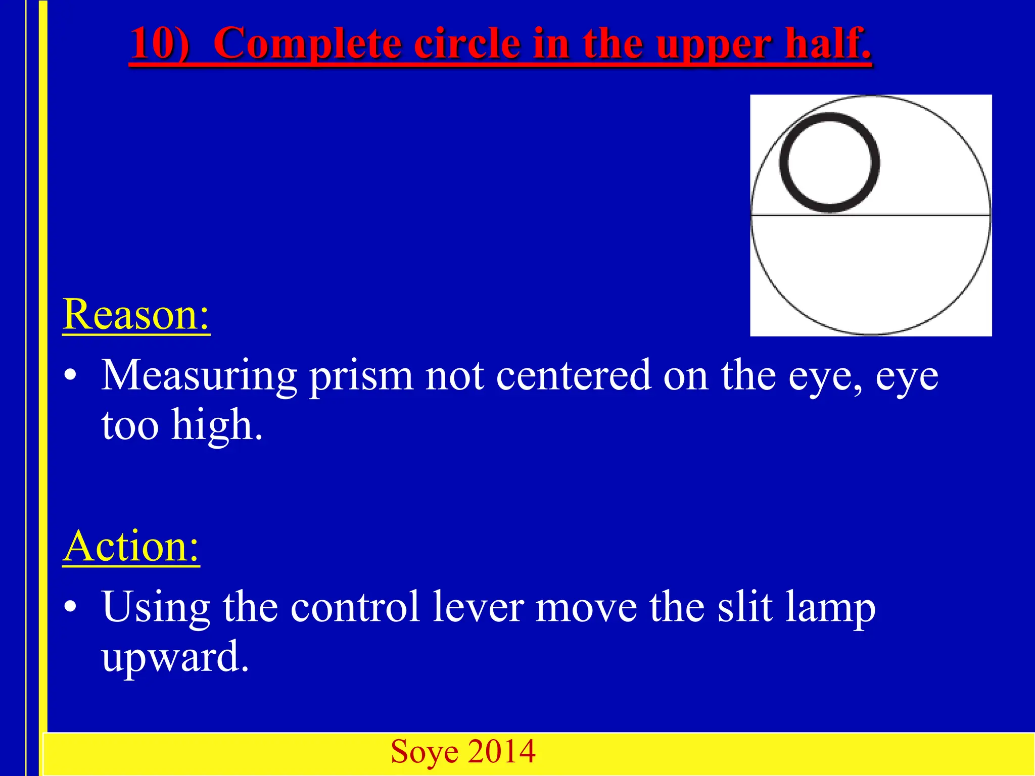
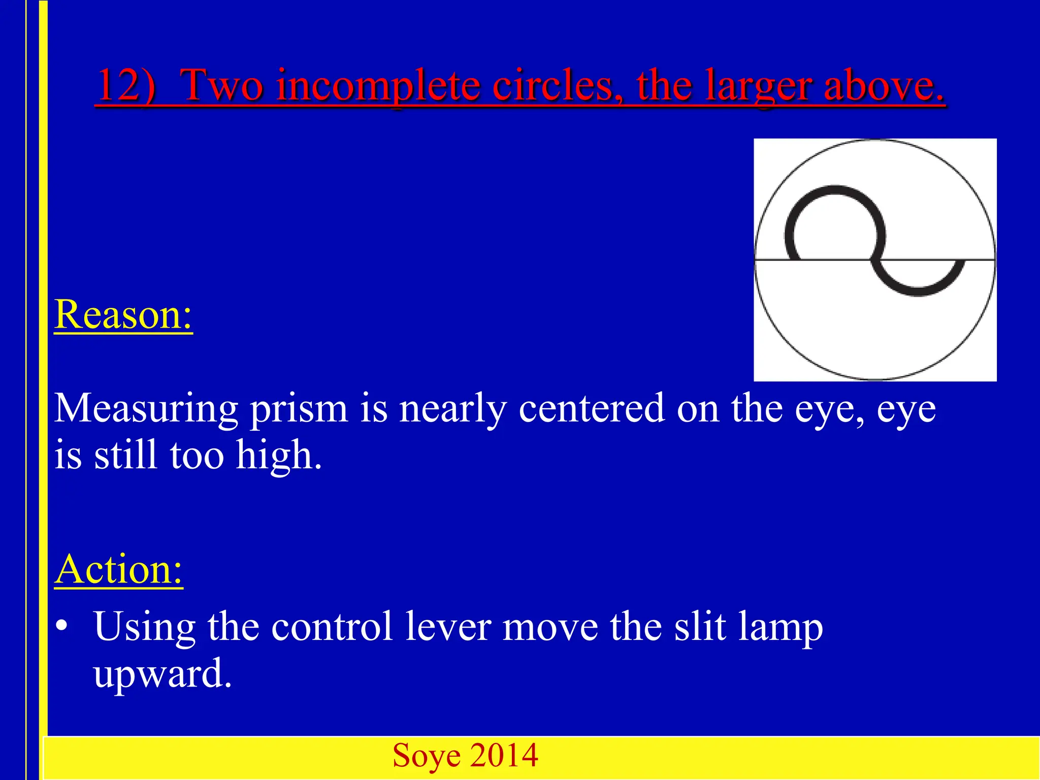
E.M.B. Sept. 2000
HT-5000 Digital Applanation
Tonometry
By
Dr. Felix Olafisoye
For NOA Abuja Intern Mentoring
Programme
May 2014
Soye
2014
E.M.B. Sept. 2000
Introduction
In recent years, Applanation tonometry has become the
gold standard in the assessment of intraocular
pressure.
Variable applanation tonometers (Huvitz Digital,
Goldmann, Perkins, Draeger, MacKay-Marg, and Tono-
Pen and Pneumatonometer):
• Applanation tonometry: measures IOP by
providing force which flattens the cornea.
Soye
2014
Soye 2014
E.M.B. Sept. 2000
Soye 2014
Principles
E.M.B. Sept. 2000
Imbert–Fick principle, which states that the pressure (P) inside
an ideal sphere is equal to the force (F) necessary to flatten the
surface divided by the area (A) which is flattened:
(P = F/A).
The eye is not an ideal sphere, primarily because corneal rigidity
resists the force and the capillary action of the tear film attracts
the tonometer prism.
The design of the Applanation tonometer (Goldmann or Huvitz)
exploits these two opposing forces, as they approximately
cancel each other when the applanated area is of 3.06 mm
diameter (the diameter of the tonometer prism).
Soye 2014
E.M.B. Sept. 2000
Technique of measurement
• plastic bi-prism which contacts cornea creates two
semicircles
• edge of corneal contact is visible after placing
fluorescein into tear film & viewing with cobalt
blue light
• manually rotate the dial calibrated in grams, force
is adjusted by changing the length of a spring
within the device.
• inner margins of semicircles touch when 3.06 mm
of cornea is applanated.
Soye
2014
Soye 2014
E.M.B. Sept. 2000
Configuration
Measuring prism
Feeler arm
Control drum
Connection arm
Adapter
Function
button
Digital
display
. Control
weight
insertion
Soye
2014
E.M.B. Sept. 2000
Soye 2014
E.M.B. Sept. 2000
Measuring prism
Soye 2014
Conical cylinder, plastic device.
Total diameter of 7 mm
Applanating surface of 3.06 mm in diameter.
A doubling prism.
Has two rings (mires).
E.M.B. Sept. 2000
IMPORTANT NOTICE
• Digital applanation tonometer may malfunction due
to electromagnetic waves caused by portable
personal telephones, transceivers, radio-controlled
toys, etc. Be sure to avoid having objects such as,
which affect this product, brought near the product.
• Do not examine in case of eye infections or injured
corneas.
• Do not use damaged measuring prisms. Fluorescein
paper should always be used because pathogenic
exciters thrive well in Fluorescein solutions.
Soye 2014
E.M.B. Sept. 2000
Pre-procedure evaluation
Soye 2014
E.M.B. Sept. 2000
• A. Slit lamp biomicroscopy to evaluate cornea for
abnormalities that may affect IOP measurement
accuracy
• 1. Corneal edema
• 2. Corneal scars
• 3. Band keratopathy
• 4. Epithelial irregularity or defects
• 5. Keratoconus
• 6. Corneal prosthesis
• B. Pachymetry to measure corneal thickness
Soye 2014
E.M.B. Sept. 2000
• C. Assess patient for things that might affect
tonometry accuracy
• 1. Abdominal or thoracic obesity
• 2. Tight collar or necktie
• 3. High astigmatism
• 4. Breath holding or Valsalva
• 5. Contact lens
• 6. Extraocular muscles acting on restricted globe
• 7. Lid squeezing
• 8. Narrow intrapalpebral fissure
Soye 2014
E.M.B. Sept. 2000
Patient
and
Instrument Preparation
Soye 2014
E.M.B. Sept. 2000
• Remove the tonometer prism from the disinfectant solution
(some units use
alcohol wipes), rinse and dry it.
• Insert the prism into the tonometer bracket holder, ensuring the
0° or 180°
markings line up with the white line on the bracket.
• Anaesthetize both eyes within half a minute. Always
anaesthetize both eyes as otherwise blinking is
unavoidable.
• Place a fluorescein paper strip in the outer external canthus in
the lower conjunctival sac. After a couple of seconds the tear
fluid has taken on enough sufficient fluorescein. The paper
strip can be removed.
Soye 2014
E.M.B. Sept. 2000
• Immediately before taking the measurements, the patient should
be made to close the eyes briefly so that the cornea becomes
sufficiently moistened with the lacrimal fluid and fluorescein.
• The correct eye height of the patient can be set at the chin rest.
• The focus of the eyepiece is to be checked before the examination.
•
• Set the magnification to 10x.
•
• Set blue filter and open the slit diaphragm fully.
• Position the Feeler arm into place so that the axis of the
measuring device and the microscope coincide.
Soye 2014
E.M.B. Sept. 2000
 When using drops, a 0.25 % to 0.5 % sodium fluorescein solution
is recommended. Should a 1 % or a 2 % fluorescein solution be
instilled, a small drop is introduced to the conjunctival sac using
a glass rod.
 Turn the tonometer on and adjust it to a value between 5 and 10.
 Bring the illumination device from the left into contact with the
tonometer bearer arm. This is the only illumination position in
which both the patient’s left and right eye can be examined (no
60° position). This arrangement simplifies the splaying of the
patient’s eyelids should this be necessary to make measurements.
Soye 2014
E.M.B. Sept. 2000
Patient instructions
• Press the head firmly on the chin rest and forehead support. If
necessary use a holding band to fix the head’s position.
• The patient must look straight forward. Fixation light or
examiners opposite ear can be used as target.
• It is recommended that the patient should be repeatedly
requested to open the eyes wide during the examination. The
examiner must perhaps keep the eye open by splaying the
eyelids with thumb and index finger. This must be done
without pressure to the eye.
• Ask patient to breathe normally, do not hold your breath
• Blink immediately prior to measurement to moisten cornea.
Soye 2014
E.M.B. Sept. 2000
Note that the prism and patient
are at the same height and that the slitlamp
joystick is pointing backwards, ready to
carefully place the prism on to the cornea
using a fine movement.
Carefully holding the lids
against the orbital rims allows correct
measurement of pressure in patients who
are otherwise unable to keep eyes open
E.M.B. Sept. 2000
IOP Measurement
• Now with maximal illumination of bi-prism, move the Slitlamp
toward the eye until the tip of bi-prism contacts the apex of the
cornea
• Stop moving forward when limbus shines with blue light, best
observed with naked eye. This illumination is best observed by
direct sight from the opposite side of the illumination unit by
the examiner.
• After contact, visible semicircles appear through left (or right)
ocular.
• Adjust vertically until semicircles equal in size. The width of
the fluorescein ring should be 1/10 approximately of the
diameter of the applanation surface.
• Adjust tension knob until inner edge of upper and lower
semicircles are aligned.
Soye 2014
E.M.B. Sept. 2000
Soye 2014
Turn the force knob until the inside edges of the
end of each split ring just touch, forming a “lazy S”
pattern, which is the measurement endpoint.
Typically, the pulse pressure causes an oscillation of
the mires. The force knob is adjusted until these
oscillations are centered about the endpoint. When
the endpoint is reached, the applanated area has a
diameter of 3.06 mm.
E.M.B. Sept. 2000
Soye 2014
E.M.B. Sept. 2000
• In Huvtz, simply read the display
in mmHg.
• E.g. If the number of display is
10.0, = It means 10mmHg.
Note: In Goldman 1 = 10mmHg
Soye 2014
E.M.B. Sept. 2000
Take Note
• Blue central area represents applanated cornea,
green semicircles are fluorescein-stained tears,
inner border of ring is demarcation between
flattened and non-flattened cornea.
• Without staining of tears, bright reflection from
air-cornea interface is seen; leads to
underestimation of IOP.
• Mires should be approximately 10% of circle
width.
Soye 2014
E.M.B. Sept. 2000
• The measurements should be carried out as quickly
as possible
• The corneal epithelia can dry out when the
measurement takes a long time. If this occurs, dry
irregular fluorescein patches will be seen. These are
are unsuitable for taking measurements.
Note:
• Extensive dryness disappears quickly without any
particular treatment. The visual acuity is influenced
by this light epithelium defect.
Soye 2014
E.M.B. Sept. 2000
Sources of error
Soye 2014
E.M.B. Sept. 2000
(1 ) Fluorescein band too thick or wide.
• Reason:
A. The measuring prism was not dried after cleaning or the
eyelids came into contact with the measuring prism.
B. Too much fluorescein dye, which yield an over-estimation
of the intraocular pressure.
• Action:
Pull the Slitlamp back. Dry the measuring prism with
a cotton-wool ball. Excess fluorescein should be removed
from the lower canthus with a tissue prior to applanating
Incorrect Fluorescein band
Soye 2014
E.M.B. Sept. 2000
2. Fluorescein band too narrow.
• Reason: A) The tear fluid has dried up during a longer
lasting measuring.
B)Too little fluorescein dye produces thin mires,
which can lead to an under-estimation of the
intraocular pressure.
• Action:
Ask the patient to blink well or close the eyes
few times and then repeat the measurement
Soye 2014
E.M.B. Sept. 2000
3. No semi circular image visible,
only markings
Reason:
• Wrong distance to patient .
• The measuring prism does not contact the cornea. If the patient
retreats the head a little, irregular pulsations will appear
because the measuring prism touches the eye partially.
• If the patient retreats still further, then the Fluorescein rings
disappear completely.
Action:
• Reposition patient’s head firmly against the headrest.
Soye 2014
E.M.B. Sept. 2000
4. Only parts of both oversize semicircles are visible:
Reason:
• Slitlamp pushed too far against cornea.
• Patient moves towards the slit lamp.
(This error will create too large applanation
surface)
Action:
• Pull the slit lamp back until uniform mires appears.
Soye 2014
E.M.B. Sept. 2000
5. Only part of the upper semicircle.
Reason:
• Measuring prism not centered on
the eye and eye much too far to the right.
Action:
• Using the control lever, move the slit lamp to the
right.
Soye 2014
E.M.B. Sept. 2000
6) Only part of the lower semicircle
Reason:
• Measuring prism not centered on the eye, eye
too far to the left.
Action:
• Using the control lever, move the slit lamp to
the left.
Soye 2014
E.M.B. Sept. 2000
7) All of the lower semicircle - part of the upper
semicircle.
Reason:
• Measuring prism not centered on the eye, eye
is too far to the left.
Action:
• Using the control lever, move the slit lamp to
the left
Soye 2014
E.M.B. Sept. 2000
8) All of the upper semicircle - part of the lower
semicircle
Reason:
Measuring prism not centered on the
eye, eye is too far to the right.
Action:
• Using the control lever, move the slit lamp to
the right.
Soye 2014
E.M.B. Sept. 2000
Correct setting
Two semicircles appear exactly in the middle of
the ocular.
Soye 2014
E.M.B. Sept. 2000
Soye 2014
E.M.B. Sept. 2000
9) Only part of a semicircle in the upper half
Reason:
• Measuring prism not centered on the eye, eye
too high.
Action:
• Using the control lever move the slit lamp
upward.
Soye 2014
E.M.B. Sept. 2000
10) Complete circle in the upper half.
Reason:
• Measuring prism not centered on the eye, eye
too high.
Action:
• Using the control lever move the slit lamp
upward.
Soye 2014
E.M.B. Sept. 2000
11) Nearly complete circles above,
incomplete circles below
Reason:
• Measuring prism not centered on the eye, eye
still too high.
Action:
• Using the control lever move the slit lamp
upward.
Soye 2014
E.M.B. Sept. 2000
12) Two incomplete circles, the larger above.
Reason:
Measuring prism is nearly centered on the eye, eye
is still too high.
Action:
• Using the control lever move the slit lamp
upward.
Soye 2014
E.M.B. Sept. 2000
13) Contact with the outside borders of the
fluorescein bands
Reason:
Not enough pressure
Action:
Increase the pressure slightly by turning the
knob on the tonometer.
Soye 2014
E.M.B. Sept. 2000
14) Fluorescein bands are superimposed to
form a band
Reason:
Pressure slightly too little
Action:
Increase the pressure slightly by turning the
knob on the tonometer.
Soye 2014
E.M.B. Sept. 2000
15) Bands are not in contact.
Reason:
Pressure is clearly too high
Action:
Lower the pressure by turning the tonometer
adjustment knob in the opposite direction.
Soye 2014
E.M.B. Sept. 2000
Correct setting
Two semicircles appear exactly in the middle of
the ocular.
Soye 2014
E.M.B. Sept. 2000
Soye 2014
C. Too High
A. Too Low B. Normal
A. The rings of fluorescein are too wide apart and the pressure will
be measured low. B. Rings just touching. Correct reading. C. Rings
overlap. Pressure will be measured high.
E.M.B. Sept. 2000
Applanation tonometry in
Astigmatism
Soye 2014
E.M.B. Sept. 2000
• In patients with astigmatism of greater than 3 D,
the applanated area will be elliptical, not circular.
This error can be avoided by applanation at 43°
to the meridian of the greater radius or axis of
minus cylinder
• If the cornea is spherical, measurements can be
made on any meridian, but it is most convenient
to do it on the 0° or 180° meridian.
This is not so when eyes with higher corneal
astigmatism than 3 diopters are examined, as the
flattened areas are not circular but elliptic.
Soye 2014
E.M.B. Sept. 2000
When regular astigmatism > 3.00D is present, an
elliptical contact with tonometer head occurs. This
results in an under estimation of IOP in with-the-rule
astigmatism and an over estimation with against-the-
rule astigmatism, with an error range of about
-2.5 to +2.5 mmHg.
Soye 2014
E.M.B. Sept. 2000
• In Huvitz HT 5000, One options exist to counteract this source
of error:
Example 1:
If the corneal astigmatism is:
6.5 mm x 30° = 52.0 D x 30°
8.5 mm x 120° = 40.0 D x 120°
• The graduation value 120° of the prism is set at the
red 43° mark of the prism holder.
Soye 2014
E.M.B. Sept. 2000
Example 2:
If there is a corneal astigmatism of
8.5 mm x 30° = 40.0 D x 30° and
6.5 mm x 120° = 52.0 D x 120°
The graduation value 30° is set at the red 43° mark.
• In other words, set the axial position of the greatest
radius, which is the axis of a minus cylinder, on the
prism graduation at the red mark on the prism holder.
Soye 2014
E.M.B. Sept. 2000
• Notice that the tip has been rotated so that the "120" on the
scale has aligned with the red mark on the tip holder. This
is the proper position for measuring this cornea. You will
now see an image that resembles the image below. You
will have to measure using the image on the slant, but the
mires will be the same size and the measurement will be
more accurate.
Soye 2014
E.M.B. Sept. 2000
Mire in high astigmatism
Soye 2014
E.M.B. Sept. 2000
Complications of the procedure,
their prevention and management
Soye 2014
E.M.B. Sept. 2000
Complication 1: Corneal abrasion
• a. Prevent by slow careful applanation and encouraging
patient to maintain steady head and eye position
• b. Most applanation induced abrasions heal overnight
without treatment
Complication 2: Epithelial Antiseptic toxicity.
• a. Tonometer tip should be allow to fully dry between
patients
• Complication 3: Anesthetic toxicity to epithelium
• a. Use minimal anesthetic drop on cornea.
•
• Complication 4: Potential for transmission of infection
• a. Ensure proper sterilization of measuring probe
Soye 2014
E.M.B. Sept. 2000
Technician or clinician influences
on IOP measurement
Soye 2014
E.M.B. Sept. 2000
• 1. Pressure from fingers holding lids may be
transmitted to globe and elevate IOP
• 2. Excess fluorescein (thick mires) may cause
overestimation of IOP
• 3. Inadequate fluorescein (thin mires) may cause
underestimation of IOP
• 4. Improper vertical alignment of mires may cause
overestimation of IOP
• 5. Inadequate tonometer calibration
• 6. Repeated applanation tonometry reduces IOP
readings
Soye 2014
E.M.B. Sept. 2000
Care of measuring prism
Soye 2014
E.M.B. Sept. 2000
Disinfection of Prism
1. Measuring prisms must be cleaned and disinfected
after each use.
2. Prior to disinfection the measuring prisms must be
rinsed under cold running water for 30 to 60 seconds.
3. Extremely dirty measuring prisms can additionally
be cleaned using a soap and a cotton-wool ball.
4. For an optimal cleaning and disinfection, the
measuring prisms must be submerged and must
move freely in the disinfectant fluid.
Soye 2014
E.M.B. Sept. 2000
• 4. Measuring prisms to end disinfection are
rinsed cold running water.
• - Caution : Time to rinse in water is 10 to a
maximum 60 minutes.
• 5. The measuring prisms should be dried after
cleaning, and stored in closed container.
• 6. To the measuring prisms must not be a
residue. Residues can injure to irritation of the
patient’s eye or chemical burns.
Soye 2014
E.M.B. Sept. 2000
Cleaning procedure
• 1. Remove measuring prism carefully from
holder.
• 2. Clean : Wipe prisms clean before rinsing for
30-60 seconds in running cold water.
• 3. Disinfect :
• Hydrogen Peroxide(10 minutes) or Sodium
Hypochlorite(10 minutes)
• 4. Rinse
• Rinse thoroughly in running, cold drinking
water.
• 5. Dry : With a one-way tissue, clean and soft
• 6. Store : Place into container clean and dry.
Soye 2014
E.M.B. Sept. 2000
Sterilization
• CDC recommendation (HSV, and
adenovirus): wipe tip clean and disinfect tip
only with bleach (1:10 dilution x 5”, changed
once daily).
• Alternative is 3% H2O2, changed at least
twice daily (affects tip less than bleach or
ETOH).
• Alternative #2: wiping tip with 70% ETOH
Soye 2014
E.M.B. Sept. 2000
Thank you for
listening
Soye 2014

APPLANATION TONOMETRY (2) for Optometry .pptx

  • 1.
    E.M.B. Sept. 2000 HT-5000Digital Applanation Tonometry By Dr. Felix Olafisoye For NOA Abuja Intern Mentoring Programme May 2014 Soye 2014
  • 2.
    E.M.B. Sept. 2000 Introduction Inrecent years, Applanation tonometry has become the gold standard in the assessment of intraocular pressure. Variable applanation tonometers (Huvitz Digital, Goldmann, Perkins, Draeger, MacKay-Marg, and Tono- Pen and Pneumatonometer): • Applanation tonometry: measures IOP by providing force which flattens the cornea. Soye 2014 Soye 2014
  • 3.
    E.M.B. Sept. 2000 Soye2014 Principles
  • 4.
    E.M.B. Sept. 2000 Imbert–Fickprinciple, which states that the pressure (P) inside an ideal sphere is equal to the force (F) necessary to flatten the surface divided by the area (A) which is flattened: (P = F/A). The eye is not an ideal sphere, primarily because corneal rigidity resists the force and the capillary action of the tear film attracts the tonometer prism. The design of the Applanation tonometer (Goldmann or Huvitz) exploits these two opposing forces, as they approximately cancel each other when the applanated area is of 3.06 mm diameter (the diameter of the tonometer prism). Soye 2014
  • 5.
    E.M.B. Sept. 2000 Techniqueof measurement • plastic bi-prism which contacts cornea creates two semicircles • edge of corneal contact is visible after placing fluorescein into tear film & viewing with cobalt blue light • manually rotate the dial calibrated in grams, force is adjusted by changing the length of a spring within the device. • inner margins of semicircles touch when 3.06 mm of cornea is applanated. Soye 2014 Soye 2014
  • 6.
    E.M.B. Sept. 2000 Configuration Measuringprism Feeler arm Control drum Connection arm Adapter Function button Digital display . Control weight insertion Soye 2014
  • 7.
  • 8.
    E.M.B. Sept. 2000 Measuringprism Soye 2014 Conical cylinder, plastic device. Total diameter of 7 mm Applanating surface of 3.06 mm in diameter. A doubling prism. Has two rings (mires).
  • 9.
    E.M.B. Sept. 2000 IMPORTANTNOTICE • Digital applanation tonometer may malfunction due to electromagnetic waves caused by portable personal telephones, transceivers, radio-controlled toys, etc. Be sure to avoid having objects such as, which affect this product, brought near the product. • Do not examine in case of eye infections or injured corneas. • Do not use damaged measuring prisms. Fluorescein paper should always be used because pathogenic exciters thrive well in Fluorescein solutions. Soye 2014
  • 10.
    E.M.B. Sept. 2000 Pre-procedureevaluation Soye 2014
  • 11.
    E.M.B. Sept. 2000 •A. Slit lamp biomicroscopy to evaluate cornea for abnormalities that may affect IOP measurement accuracy • 1. Corneal edema • 2. Corneal scars • 3. Band keratopathy • 4. Epithelial irregularity or defects • 5. Keratoconus • 6. Corneal prosthesis • B. Pachymetry to measure corneal thickness Soye 2014
  • 12.
    E.M.B. Sept. 2000 •C. Assess patient for things that might affect tonometry accuracy • 1. Abdominal or thoracic obesity • 2. Tight collar or necktie • 3. High astigmatism • 4. Breath holding or Valsalva • 5. Contact lens • 6. Extraocular muscles acting on restricted globe • 7. Lid squeezing • 8. Narrow intrapalpebral fissure Soye 2014
  • 13.
  • 14.
    E.M.B. Sept. 2000 •Remove the tonometer prism from the disinfectant solution (some units use alcohol wipes), rinse and dry it. • Insert the prism into the tonometer bracket holder, ensuring the 0° or 180° markings line up with the white line on the bracket. • Anaesthetize both eyes within half a minute. Always anaesthetize both eyes as otherwise blinking is unavoidable. • Place a fluorescein paper strip in the outer external canthus in the lower conjunctival sac. After a couple of seconds the tear fluid has taken on enough sufficient fluorescein. The paper strip can be removed. Soye 2014
  • 15.
    E.M.B. Sept. 2000 •Immediately before taking the measurements, the patient should be made to close the eyes briefly so that the cornea becomes sufficiently moistened with the lacrimal fluid and fluorescein. • The correct eye height of the patient can be set at the chin rest. • The focus of the eyepiece is to be checked before the examination. • • Set the magnification to 10x. • • Set blue filter and open the slit diaphragm fully. • Position the Feeler arm into place so that the axis of the measuring device and the microscope coincide. Soye 2014
  • 16.
    E.M.B. Sept. 2000 When using drops, a 0.25 % to 0.5 % sodium fluorescein solution is recommended. Should a 1 % or a 2 % fluorescein solution be instilled, a small drop is introduced to the conjunctival sac using a glass rod.  Turn the tonometer on and adjust it to a value between 5 and 10.  Bring the illumination device from the left into contact with the tonometer bearer arm. This is the only illumination position in which both the patient’s left and right eye can be examined (no 60° position). This arrangement simplifies the splaying of the patient’s eyelids should this be necessary to make measurements. Soye 2014
  • 17.
    E.M.B. Sept. 2000 Patientinstructions • Press the head firmly on the chin rest and forehead support. If necessary use a holding band to fix the head’s position. • The patient must look straight forward. Fixation light or examiners opposite ear can be used as target. • It is recommended that the patient should be repeatedly requested to open the eyes wide during the examination. The examiner must perhaps keep the eye open by splaying the eyelids with thumb and index finger. This must be done without pressure to the eye. • Ask patient to breathe normally, do not hold your breath • Blink immediately prior to measurement to moisten cornea. Soye 2014
  • 18.
    E.M.B. Sept. 2000 Notethat the prism and patient are at the same height and that the slitlamp joystick is pointing backwards, ready to carefully place the prism on to the cornea using a fine movement. Carefully holding the lids against the orbital rims allows correct measurement of pressure in patients who are otherwise unable to keep eyes open
  • 19.
    E.M.B. Sept. 2000 IOPMeasurement • Now with maximal illumination of bi-prism, move the Slitlamp toward the eye until the tip of bi-prism contacts the apex of the cornea • Stop moving forward when limbus shines with blue light, best observed with naked eye. This illumination is best observed by direct sight from the opposite side of the illumination unit by the examiner. • After contact, visible semicircles appear through left (or right) ocular. • Adjust vertically until semicircles equal in size. The width of the fluorescein ring should be 1/10 approximately of the diameter of the applanation surface. • Adjust tension knob until inner edge of upper and lower semicircles are aligned. Soye 2014
  • 20.
    E.M.B. Sept. 2000 Soye2014 Turn the force knob until the inside edges of the end of each split ring just touch, forming a “lazy S” pattern, which is the measurement endpoint. Typically, the pulse pressure causes an oscillation of the mires. The force knob is adjusted until these oscillations are centered about the endpoint. When the endpoint is reached, the applanated area has a diameter of 3.06 mm.
  • 21.
  • 22.
    E.M.B. Sept. 2000 •In Huvtz, simply read the display in mmHg. • E.g. If the number of display is 10.0, = It means 10mmHg. Note: In Goldman 1 = 10mmHg Soye 2014
  • 23.
    E.M.B. Sept. 2000 TakeNote • Blue central area represents applanated cornea, green semicircles are fluorescein-stained tears, inner border of ring is demarcation between flattened and non-flattened cornea. • Without staining of tears, bright reflection from air-cornea interface is seen; leads to underestimation of IOP. • Mires should be approximately 10% of circle width. Soye 2014
  • 24.
    E.M.B. Sept. 2000 •The measurements should be carried out as quickly as possible • The corneal epithelia can dry out when the measurement takes a long time. If this occurs, dry irregular fluorescein patches will be seen. These are are unsuitable for taking measurements. Note: • Extensive dryness disappears quickly without any particular treatment. The visual acuity is influenced by this light epithelium defect. Soye 2014
  • 25.
    E.M.B. Sept. 2000 Sourcesof error Soye 2014
  • 26.
    E.M.B. Sept. 2000 (1) Fluorescein band too thick or wide. • Reason: A. The measuring prism was not dried after cleaning or the eyelids came into contact with the measuring prism. B. Too much fluorescein dye, which yield an over-estimation of the intraocular pressure. • Action: Pull the Slitlamp back. Dry the measuring prism with a cotton-wool ball. Excess fluorescein should be removed from the lower canthus with a tissue prior to applanating Incorrect Fluorescein band Soye 2014
  • 27.
    E.M.B. Sept. 2000 2.Fluorescein band too narrow. • Reason: A) The tear fluid has dried up during a longer lasting measuring. B)Too little fluorescein dye produces thin mires, which can lead to an under-estimation of the intraocular pressure. • Action: Ask the patient to blink well or close the eyes few times and then repeat the measurement Soye 2014
  • 28.
    E.M.B. Sept. 2000 3.No semi circular image visible, only markings Reason: • Wrong distance to patient . • The measuring prism does not contact the cornea. If the patient retreats the head a little, irregular pulsations will appear because the measuring prism touches the eye partially. • If the patient retreats still further, then the Fluorescein rings disappear completely. Action: • Reposition patient’s head firmly against the headrest. Soye 2014
  • 29.
    E.M.B. Sept. 2000 4.Only parts of both oversize semicircles are visible: Reason: • Slitlamp pushed too far against cornea. • Patient moves towards the slit lamp. (This error will create too large applanation surface) Action: • Pull the slit lamp back until uniform mires appears. Soye 2014
  • 30.
    E.M.B. Sept. 2000 5.Only part of the upper semicircle. Reason: • Measuring prism not centered on the eye and eye much too far to the right. Action: • Using the control lever, move the slit lamp to the right. Soye 2014
  • 31.
    E.M.B. Sept. 2000 6)Only part of the lower semicircle Reason: • Measuring prism not centered on the eye, eye too far to the left. Action: • Using the control lever, move the slit lamp to the left. Soye 2014
  • 32.
    E.M.B. Sept. 2000 7)All of the lower semicircle - part of the upper semicircle. Reason: • Measuring prism not centered on the eye, eye is too far to the left. Action: • Using the control lever, move the slit lamp to the left Soye 2014
  • 33.
    E.M.B. Sept. 2000 8)All of the upper semicircle - part of the lower semicircle Reason: Measuring prism not centered on the eye, eye is too far to the right. Action: • Using the control lever, move the slit lamp to the right. Soye 2014
  • 34.
    E.M.B. Sept. 2000 Correctsetting Two semicircles appear exactly in the middle of the ocular. Soye 2014
  • 35.
  • 36.
    E.M.B. Sept. 2000 9)Only part of a semicircle in the upper half Reason: • Measuring prism not centered on the eye, eye too high. Action: • Using the control lever move the slit lamp upward. Soye 2014
  • 37.
    E.M.B. Sept. 2000 10)Complete circle in the upper half. Reason: • Measuring prism not centered on the eye, eye too high. Action: • Using the control lever move the slit lamp upward. Soye 2014
  • 38.
    E.M.B. Sept. 2000 11)Nearly complete circles above, incomplete circles below Reason: • Measuring prism not centered on the eye, eye still too high. Action: • Using the control lever move the slit lamp upward. Soye 2014
  • 39.
    E.M.B. Sept. 2000 12)Two incomplete circles, the larger above. Reason: Measuring prism is nearly centered on the eye, eye is still too high. Action: • Using the control lever move the slit lamp upward. Soye 2014
  • 40.
    E.M.B. Sept. 2000 13)Contact with the outside borders of the fluorescein bands Reason: Not enough pressure Action: Increase the pressure slightly by turning the knob on the tonometer. Soye 2014
  • 41.
    E.M.B. Sept. 2000 14)Fluorescein bands are superimposed to form a band Reason: Pressure slightly too little Action: Increase the pressure slightly by turning the knob on the tonometer. Soye 2014
  • 42.
    E.M.B. Sept. 2000 15)Bands are not in contact. Reason: Pressure is clearly too high Action: Lower the pressure by turning the tonometer adjustment knob in the opposite direction. Soye 2014
  • 43.
    E.M.B. Sept. 2000 Correctsetting Two semicircles appear exactly in the middle of the ocular. Soye 2014
  • 44.
    E.M.B. Sept. 2000 Soye2014 C. Too High A. Too Low B. Normal A. The rings of fluorescein are too wide apart and the pressure will be measured low. B. Rings just touching. Correct reading. C. Rings overlap. Pressure will be measured high.
  • 45.
    E.M.B. Sept. 2000 Applanationtonometry in Astigmatism Soye 2014
  • 46.
    E.M.B. Sept. 2000 •In patients with astigmatism of greater than 3 D, the applanated area will be elliptical, not circular. This error can be avoided by applanation at 43° to the meridian of the greater radius or axis of minus cylinder • If the cornea is spherical, measurements can be made on any meridian, but it is most convenient to do it on the 0° or 180° meridian. This is not so when eyes with higher corneal astigmatism than 3 diopters are examined, as the flattened areas are not circular but elliptic. Soye 2014
  • 47.
    E.M.B. Sept. 2000 Whenregular astigmatism > 3.00D is present, an elliptical contact with tonometer head occurs. This results in an under estimation of IOP in with-the-rule astigmatism and an over estimation with against-the- rule astigmatism, with an error range of about -2.5 to +2.5 mmHg. Soye 2014
  • 48.
    E.M.B. Sept. 2000 •In Huvitz HT 5000, One options exist to counteract this source of error: Example 1: If the corneal astigmatism is: 6.5 mm x 30° = 52.0 D x 30° 8.5 mm x 120° = 40.0 D x 120° • The graduation value 120° of the prism is set at the red 43° mark of the prism holder. Soye 2014
  • 49.
    E.M.B. Sept. 2000 Example2: If there is a corneal astigmatism of 8.5 mm x 30° = 40.0 D x 30° and 6.5 mm x 120° = 52.0 D x 120° The graduation value 30° is set at the red 43° mark. • In other words, set the axial position of the greatest radius, which is the axis of a minus cylinder, on the prism graduation at the red mark on the prism holder. Soye 2014
  • 50.
    E.M.B. Sept. 2000 •Notice that the tip has been rotated so that the "120" on the scale has aligned with the red mark on the tip holder. This is the proper position for measuring this cornea. You will now see an image that resembles the image below. You will have to measure using the image on the slant, but the mires will be the same size and the measurement will be more accurate. Soye 2014
  • 51.
    E.M.B. Sept. 2000 Mirein high astigmatism Soye 2014
  • 52.
    E.M.B. Sept. 2000 Complicationsof the procedure, their prevention and management Soye 2014
  • 53.
    E.M.B. Sept. 2000 Complication1: Corneal abrasion • a. Prevent by slow careful applanation and encouraging patient to maintain steady head and eye position • b. Most applanation induced abrasions heal overnight without treatment Complication 2: Epithelial Antiseptic toxicity. • a. Tonometer tip should be allow to fully dry between patients • Complication 3: Anesthetic toxicity to epithelium • a. Use minimal anesthetic drop on cornea. • • Complication 4: Potential for transmission of infection • a. Ensure proper sterilization of measuring probe Soye 2014
  • 54.
    E.M.B. Sept. 2000 Technicianor clinician influences on IOP measurement Soye 2014
  • 55.
    E.M.B. Sept. 2000 •1. Pressure from fingers holding lids may be transmitted to globe and elevate IOP • 2. Excess fluorescein (thick mires) may cause overestimation of IOP • 3. Inadequate fluorescein (thin mires) may cause underestimation of IOP • 4. Improper vertical alignment of mires may cause overestimation of IOP • 5. Inadequate tonometer calibration • 6. Repeated applanation tonometry reduces IOP readings Soye 2014
  • 56.
    E.M.B. Sept. 2000 Careof measuring prism Soye 2014
  • 57.
    E.M.B. Sept. 2000 Disinfectionof Prism 1. Measuring prisms must be cleaned and disinfected after each use. 2. Prior to disinfection the measuring prisms must be rinsed under cold running water for 30 to 60 seconds. 3. Extremely dirty measuring prisms can additionally be cleaned using a soap and a cotton-wool ball. 4. For an optimal cleaning and disinfection, the measuring prisms must be submerged and must move freely in the disinfectant fluid. Soye 2014
  • 58.
    E.M.B. Sept. 2000 •4. Measuring prisms to end disinfection are rinsed cold running water. • - Caution : Time to rinse in water is 10 to a maximum 60 minutes. • 5. The measuring prisms should be dried after cleaning, and stored in closed container. • 6. To the measuring prisms must not be a residue. Residues can injure to irritation of the patient’s eye or chemical burns. Soye 2014
  • 59.
    E.M.B. Sept. 2000 Cleaningprocedure • 1. Remove measuring prism carefully from holder. • 2. Clean : Wipe prisms clean before rinsing for 30-60 seconds in running cold water. • 3. Disinfect : • Hydrogen Peroxide(10 minutes) or Sodium Hypochlorite(10 minutes) • 4. Rinse • Rinse thoroughly in running, cold drinking water. • 5. Dry : With a one-way tissue, clean and soft • 6. Store : Place into container clean and dry. Soye 2014
  • 60.
    E.M.B. Sept. 2000 Sterilization •CDC recommendation (HSV, and adenovirus): wipe tip clean and disinfect tip only with bleach (1:10 dilution x 5”, changed once daily). • Alternative is 3% H2O2, changed at least twice daily (affects tip less than bleach or ETOH). • Alternative #2: wiping tip with 70% ETOH Soye 2014
  • 61.
    E.M.B. Sept. 2000 Thankyou for listening Soye 2014