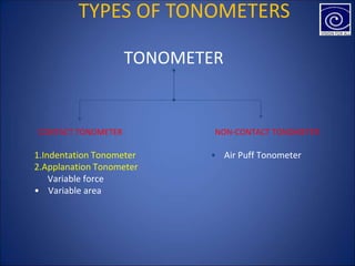
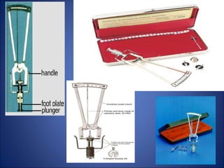
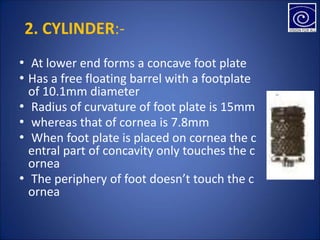
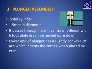
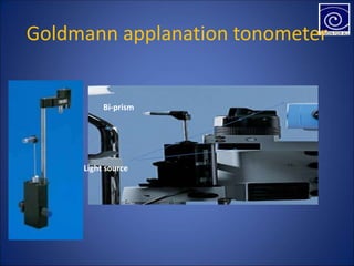
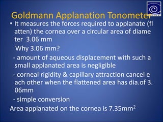
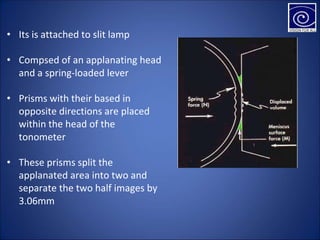
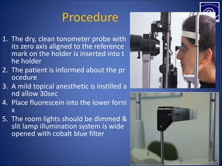
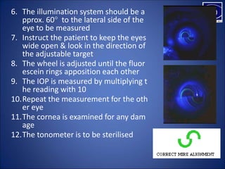
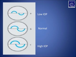
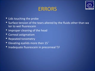
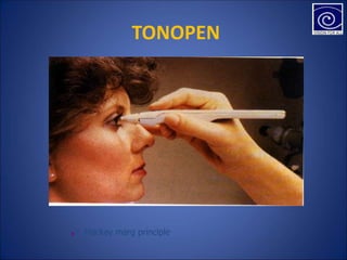
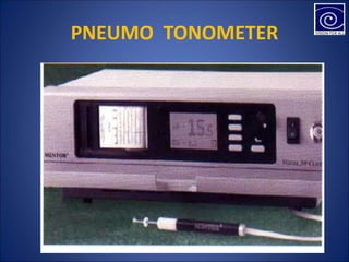
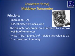
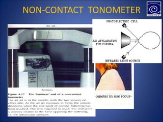
TONOMETERS
MAAZ UL HAQ
OPTOMETRIST
INTRODUCTION
• Intraocular Pressure & its significance
• Factors influencing IOP level
• WHAT IS A TONOMETER???
TYPES OF TONOMETERS
TONOMETER
CONTACT TONOMETER
1.Indentation Tonometer
2.Applanation Tonometer
• Variable force
• Variable area
NON-CONTACT TONOMETER
• Air Puff Tonometer
Indentation Tonometer
• Indentation tonometer measures the depth of the impression
or indentation by a small plunger carrying a known weight
• Principle: the degree of indentation is inversely proportional to
intraocular pressure
• The degree of indentation is depend on the flexibility of the co
rnea, surface tension and elasticity of the walls of the eyeball
SCHIOTZ TONOMETER
Invented in 1905 & modified by Schiotz in 1924.
Parts:-
1. Handle 5. Scale with markings
2. Cylinder 6. Weights
3. Plunger assembly 7. Metal sphere
4. Bend Lever
PARTS
1. HANDLE:-
• For holding the instrument in vertical position
on cornea
2. CYLINDER:-
• At lower end forms a concave foot plate
• Has a free floating barrel with a footplate
of 10.1mm diameter
• Radius of curvature of foot plate is 15mm
• whereas that of cornea is 7.8mm
• When foot plate is placed on cornea the c
entral part of concavity only touches the c
ornea
• The periphery of foot doesn’t touch the c
ornea
3. PLUNGER ASSEMBLY:-
• Solid cylinder
• 1.5mm in diameter
• It passes through hole in centre of cylinder wit
h foot plate & can be moved up & down
• Lower end of plunger has a slightly convex surf
ace which indents the cornea when placed ov
er it
4. BEND LEVER:-
• Has a shorter arm resting on upper edge of
plunger
• Long arm that acts as a pointer moves agai
nst scale
5. SCALE WITH MARKINGS:-
• Pointer glides against scale according to mo
vement of plunger
• Interval between consecutive marking on sc
ale represent displacement 0.05mm of top
of plunger relative to foot plate
6. WEIGHT:-
• Three types of weights are used
• Additional weights are 7.5 & 10gms
• Plunger itself weighs about 5.5gms which is marked on circ
ular disc above upper part of plunger
7. METAL SPHERE:-
• Used as dummy cornea
• Radius of curvature is 1.5mm
PROCEDURE
1. The tonometer should be sterilized
2. The patient is informed about the procedure
3. Topical anesthetic (0.5 % Proparacaine or 0.4%
Benoxinate) is instilled
4. Patient is seated in semi supine position with f
ace turned upwards
5. Instruct the patient to keep his both eyes open
6. Place 7.5g on the plunger and hold the instrum
ent with thumb and first finger
PROCEDURE CONT..
7. Hold patient’s lids apart gently, not applying press
ure on the globe
8. Place the tonometer so that the foot plate rests c
entrally on cornea and the instrument is vertical
9. The pointer should show slight fluctuations and t
he mean is recorded
10.Convert the reading in mmHg
11.Repeat the procedure for the other eye
12.Inspect the cornea for any abrasion
• Errors inherent in instrument:-
- Difference in weight of different part of instru
ment
- Friction arising in working of plunger
- Shape & curvature of foot plate
- Smoothness of gliding movement of the point
er on the scale
ERRORS
- During accommodation there is contraction of
ciliary muscles thus increasing aqueous flow d
ue to pull on trabecular meshwork at angle of
A/C & thus causing lowering of IOP
• Errors due to contraction of EOM’s
• Errors due to rigidity of outer coats of eyeball
• Errors due to corneal curvature
Errors due to accommodation
Advantages of Schiotz Tonometer
• Availability (portable)
• Can measure IOP of the eye with scarred cornea
• Simplicity of the design
• Can be used on opaque corneas
• low cost
Applanation Tonometer
• The first applanation tonometers were designed by Makla
kow(1885) and Weber(1867)
• It is based on Imbert Fick law
• It states that pressure within a sphere(P) is equal to the ex
ternal force(F) needed to flatten a portion of the sphere di
vided by the area(A) of the sphere which is flattened
• IOP = tonometer weight (g)/ applanated area (mm )
• P = F/A
• P = Pressure
• F = Force
• A = Area of sphere
• It is correct only for spherical container, with a thin li
miting membrane, perfectly flexible, elastic and dry
• Cornea satisfies none of these criteria
• It is thick and has some structural rigidity and is wet a
llowing tear meniscus to form between the tonomete
r head and the cornea
• Two types of Applanation tonometers
• Variable weight tonometer
• Variable area tonometer
EXAMPLES
Variable force
• Goldmann
• Perkins
• Draeger
• Mackay-marg
Variable area
• Maklakow
• Applanaometer
• Tonomat
• Barraquer
Two parts
• Mechanical device - It exerts force against the
cornea
• Contact element-It indicate the degree of flatte
ning of cornea
Goldmann applanation tonometer
Bi-prism
Light source
Goldmann Applanation Tonometer
• It measures the forces required to applanate (fl
atten) the cornea over a circular area of diame
ter 3.06 mm
�Why 3.06 mm?
- amount of aqueous displacement with such a
small applanated area is negligible
- corneal rigidity & capillary attraction cancel e
ach other when the flattened area has dia.of 3.
06mm
- simple conversion
Area applanated on the cornea is 7.35mm2
• Its is attached to slit lamp
• Compsed of an applanating head
and a spring-loaded lever
• Prisms with their based in
opposite directions are placed
within the head of the
tonometer
• These prisms split the
applanated area into two and
separate the two half images by
3.06mm
Procedure
1. The dry, clean tonometer probe with
its zero axis aligned to the reference
mark on the holder is inserted into t
he holder
2. The patient is informed about the pr
ocedure
3. A mild topical anesthetic is instilled a
nd allow 30sec
4. Place fluorescein into the lower forni
x
5. The room lights should be dimmed &
slit lamp illumination system is wide
opened with cobalt blue filter
6. The illumination system should be a
pprox. 60° to the lateral side of the
eye to be measured
7. Instruct the patient to keep the eyes
wide open & look in the direction of
the adjustable target
8. The wheel is adjusted until the fluor
escein rings apposition each other
9. The IOP is measured by multiplying t
he reading with 10
10.Repeat the measurement for the oth
er eye
11.The cornea is examined for any dam
age
12.The tonometer is to be sterilised
Low IOP
Normal
High IOP
ERRORS
• Lids touching the probe
• Surface tension of the tears altered by the fluids other than wa
ter to wet fluorescein
• Improper cleaning of the head
• Corneal astigmatism
• Repeated tonometry
• Elevating eyelids more then 15°
• Inadequate fluorescein in precorneal T.F
ADVANTAGES
• It displaces only 0.56 ul of aqueous humour
and hence the factor of scleral rigidity is
almost negligible
• Displacement of 0.56 ul aqueous increases
IOP by about 2.5 %
Checking the calibration of
applanation tonometer
PERKINS
TONOMETER
MACKAY-MARG TONOMETER
TONOPEN
■ Mackay marg principle
PNEUMO TONOMETER
(constant force)
Maklakov Tonometer
▪ Principle:
▪ Impression – AT
▪ IOP estimated by measuring
the diameter of corneal area flattened by a known
weight of tonometer.
▪ P=W/∏(d/2)2 grams/cm2 - divide this value by 1.3
6, a conversion to mm hg.
NON-CONTACT TONOMETER
OTHER TYPES OF TONOMETERS
• Dynamic contour tonometer
• Electronic indentation tonometer
• Rebound tonometer
• Non-corneal and Transpalpebral tonometry
• Ocular response analyzer
• OCT Tonometer
Transpalpebral(through eyelid) diaton
tonometry
• It measures IOP through eyelid
• Requires no contact with cornea, therefore ste
rilizationRequires no contact with cornea, ther
efore sterilization of the device and topical ane
sthetic drops are not required and there is very
little risk of infection
• It is useful in post operatives, childrens,
• and in corneal pathology cases
Ocular response analyzer
The difference between the pressures at which the
cornea flattens inward and outward is measured by
the machine

Tonometer ppt

  • 1.
  • 2.
    INTRODUCTION • Intraocular Pressure& its significance • Factors influencing IOP level • WHAT IS A TONOMETER???
  • 3.
    TYPES OF TONOMETERS TONOMETER CONTACTTONOMETER 1.Indentation Tonometer 2.Applanation Tonometer • Variable force • Variable area NON-CONTACT TONOMETER • Air Puff Tonometer
  • 4.
    Indentation Tonometer • Indentationtonometer measures the depth of the impression or indentation by a small plunger carrying a known weight • Principle: the degree of indentation is inversely proportional to intraocular pressure • The degree of indentation is depend on the flexibility of the co rnea, surface tension and elasticity of the walls of the eyeball
  • 5.
    SCHIOTZ TONOMETER Invented in1905 & modified by Schiotz in 1924. Parts:- 1. Handle 5. Scale with markings 2. Cylinder 6. Weights 3. Plunger assembly 7. Metal sphere 4. Bend Lever
  • 7.
    PARTS 1. HANDLE:- • Forholding the instrument in vertical position on cornea
  • 8.
    2. CYLINDER:- • Atlower end forms a concave foot plate • Has a free floating barrel with a footplate of 10.1mm diameter • Radius of curvature of foot plate is 15mm • whereas that of cornea is 7.8mm • When foot plate is placed on cornea the c entral part of concavity only touches the c ornea • The periphery of foot doesn’t touch the c ornea
  • 9.
    3. PLUNGER ASSEMBLY:- •Solid cylinder • 1.5mm in diameter • It passes through hole in centre of cylinder wit h foot plate & can be moved up & down • Lower end of plunger has a slightly convex surf ace which indents the cornea when placed ov er it
  • 10.
    4. BEND LEVER:- •Has a shorter arm resting on upper edge of plunger • Long arm that acts as a pointer moves agai nst scale
  • 11.
    5. SCALE WITHMARKINGS:- • Pointer glides against scale according to mo vement of plunger • Interval between consecutive marking on sc ale represent displacement 0.05mm of top of plunger relative to foot plate
  • 12.
    6. WEIGHT:- • Threetypes of weights are used • Additional weights are 7.5 & 10gms • Plunger itself weighs about 5.5gms which is marked on circ ular disc above upper part of plunger
  • 13.
    7. METAL SPHERE:- •Used as dummy cornea • Radius of curvature is 1.5mm
  • 14.
    PROCEDURE 1. The tonometershould be sterilized 2. The patient is informed about the procedure 3. Topical anesthetic (0.5 % Proparacaine or 0.4% Benoxinate) is instilled 4. Patient is seated in semi supine position with f ace turned upwards 5. Instruct the patient to keep his both eyes open 6. Place 7.5g on the plunger and hold the instrum ent with thumb and first finger
  • 15.
    PROCEDURE CONT.. 7. Holdpatient’s lids apart gently, not applying press ure on the globe 8. Place the tonometer so that the foot plate rests c entrally on cornea and the instrument is vertical 9. The pointer should show slight fluctuations and t he mean is recorded 10.Convert the reading in mmHg 11.Repeat the procedure for the other eye 12.Inspect the cornea for any abrasion
  • 16.
    • Errors inherentin instrument:- - Difference in weight of different part of instru ment - Friction arising in working of plunger - Shape & curvature of foot plate - Smoothness of gliding movement of the point er on the scale ERRORS
  • 17.
    - During accommodationthere is contraction of ciliary muscles thus increasing aqueous flow d ue to pull on trabecular meshwork at angle of A/C & thus causing lowering of IOP • Errors due to contraction of EOM’s • Errors due to rigidity of outer coats of eyeball • Errors due to corneal curvature Errors due to accommodation
  • 18.
    Advantages of SchiotzTonometer • Availability (portable) • Can measure IOP of the eye with scarred cornea • Simplicity of the design • Can be used on opaque corneas • low cost
  • 19.
    Applanation Tonometer • Thefirst applanation tonometers were designed by Makla kow(1885) and Weber(1867) • It is based on Imbert Fick law • It states that pressure within a sphere(P) is equal to the ex ternal force(F) needed to flatten a portion of the sphere di vided by the area(A) of the sphere which is flattened • IOP = tonometer weight (g)/ applanated area (mm ) • P = F/A • P = Pressure • F = Force • A = Area of sphere
  • 20.
    • It iscorrect only for spherical container, with a thin li miting membrane, perfectly flexible, elastic and dry • Cornea satisfies none of these criteria • It is thick and has some structural rigidity and is wet a llowing tear meniscus to form between the tonomete r head and the cornea • Two types of Applanation tonometers • Variable weight tonometer • Variable area tonometer
  • 21.
    EXAMPLES Variable force • Goldmann •Perkins • Draeger • Mackay-marg Variable area • Maklakow • Applanaometer • Tonomat • Barraquer
  • 22.
    Two parts • Mechanicaldevice - It exerts force against the cornea • Contact element-It indicate the degree of flatte ning of cornea
  • 23.
  • 24.
    Goldmann Applanation Tonometer •It measures the forces required to applanate (fl atten) the cornea over a circular area of diame ter 3.06 mm �Why 3.06 mm? - amount of aqueous displacement with such a small applanated area is negligible - corneal rigidity & capillary attraction cancel e ach other when the flattened area has dia.of 3. 06mm - simple conversion Area applanated on the cornea is 7.35mm2
  • 25.
    • Its isattached to slit lamp • Compsed of an applanating head and a spring-loaded lever • Prisms with their based in opposite directions are placed within the head of the tonometer • These prisms split the applanated area into two and separate the two half images by 3.06mm
  • 26.
    Procedure 1. The dry,clean tonometer probe with its zero axis aligned to the reference mark on the holder is inserted into t he holder 2. The patient is informed about the pr ocedure 3. A mild topical anesthetic is instilled a nd allow 30sec 4. Place fluorescein into the lower forni x 5. The room lights should be dimmed & slit lamp illumination system is wide opened with cobalt blue filter
  • 27.
    6. The illuminationsystem should be a pprox. 60° to the lateral side of the eye to be measured 7. Instruct the patient to keep the eyes wide open & look in the direction of the adjustable target 8. The wheel is adjusted until the fluor escein rings apposition each other 9. The IOP is measured by multiplying t he reading with 10 10.Repeat the measurement for the oth er eye 11.The cornea is examined for any dam age 12.The tonometer is to be sterilised
  • 28.
  • 29.
    ERRORS • Lids touchingthe probe • Surface tension of the tears altered by the fluids other than wa ter to wet fluorescein • Improper cleaning of the head • Corneal astigmatism • Repeated tonometry • Elevating eyelids more then 15° • Inadequate fluorescein in precorneal T.F
  • 30.
    ADVANTAGES • It displacesonly 0.56 ul of aqueous humour and hence the factor of scleral rigidity is almost negligible • Displacement of 0.56 ul aqueous increases IOP by about 2.5 %
  • 31.
    Checking the calibrationof applanation tonometer
  • 32.
  • 33.
  • 34.
  • 35.
  • 36.
    (constant force) Maklakov Tonometer ▪Principle: ▪ Impression – AT ▪ IOP estimated by measuring the diameter of corneal area flattened by a known weight of tonometer. ▪ P=W/∏(d/2)2 grams/cm2 - divide this value by 1.3 6, a conversion to mm hg.
  • 37.
  • 38.
    OTHER TYPES OFTONOMETERS • Dynamic contour tonometer • Electronic indentation tonometer • Rebound tonometer • Non-corneal and Transpalpebral tonometry • Ocular response analyzer • OCT Tonometer
  • 39.
    Transpalpebral(through eyelid) diaton tonometry •It measures IOP through eyelid • Requires no contact with cornea, therefore ste rilizationRequires no contact with cornea, ther efore sterilization of the device and topical ane sthetic drops are not required and there is very little risk of infection • It is useful in post operatives, childrens, • and in corneal pathology cases
  • 40.
    Ocular response analyzer Thedifference between the pressures at which the cornea flattens inward and outward is measured by the machine