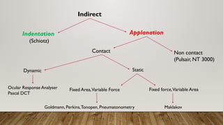
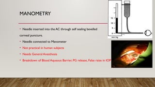
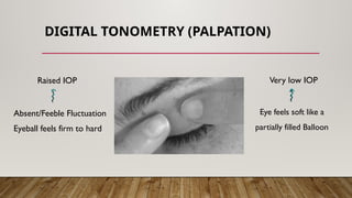
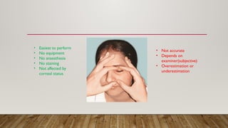
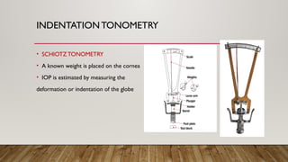
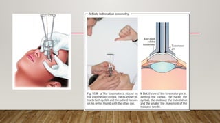
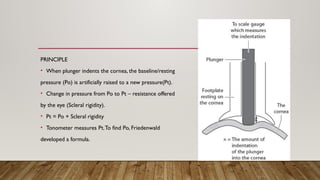
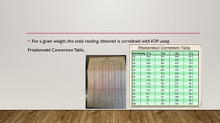
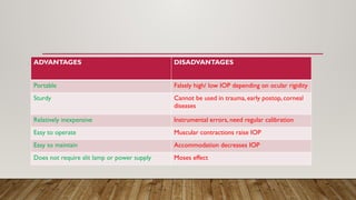
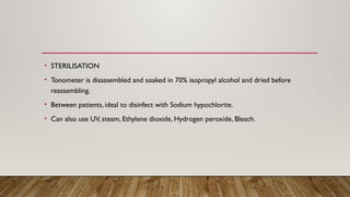
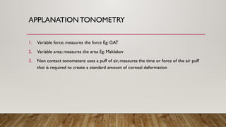
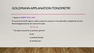
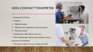
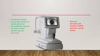
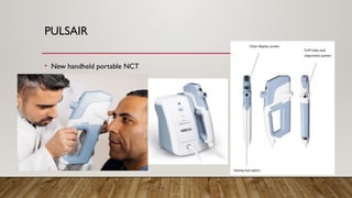
TONOMETRY
DR LAVANYA
INTRODUCTION
• Tonometry is the procedure performed to determine the intraocular pressure.
• Tonometers measure the IOP by relating a deformation of the globe to the force
responsible for the deformation.
HISTORY
1826 William
Bowman
Digital Tonometry
1865 Von Graefe First instrument to measure IOP
1885 Maklakov First Applanation Tonometer
1905 Schiotz First Indentation Tonometer
1954 Goldmann Prototype Applanation Tonometer
1972 Grolman NCT
TONOMETRY
Indirect
Direct
Manometry Indentation Applanation
Digital
Tonometry
Indirect
Indentation
(Schiotz)
Applanation
Non contact
(Pulsair, NT 3000)
Contact
Dynamic Static
Ocular Response Analyser
Pascal DCT
Fixed Area,Variable Force Fixed force,Variable Area
Maklakov
Goldmann, Perkins,Tonopen, Pneumatonometry
MANOMETRY
• Needle inserted into the AC through self sealing bevelled
corneal puncture.
• Needle connected to Manometer
• Not practical in human subjects
• Needs General Anesthesia
• Breakdown of Blood Aqueous Barrier, PG release, False raise in IOP
DIGITAL TONOMETRY (PALPATION)
Raised IOP Very low IOP
Absent/Feeble Fluctuation
Eyeball feels firm to hard
Eye feels soft like a
partially filled Balloon
• Easiest to perform
• No equipment
• No anaesthesia
• No staining
• Not affected by
corneal status
• Not accurate
• Depends on
examiner(subjective)
• Overestimation or
underestimation
INDENTATION TONOMETRY
• SCHIOTZTONOMETRY
• A known weight is placed on the cornea
• IOP is estimated by measuring the
deformation or indentation of the globe
PRINCIPLE
• When plunger indents the cornea, the baseline/resting
pressure (Po) is artificially raised to a new pressure(Pt).
• Change in pressure from Po to Pt – resistance offered
by the eye (Scleral rigidity).
• Pt = Po + Scleral rigidity
• Tonometer measures Pt.To find Po, Friedenwald
developed a formula.
• For a given weight, the scale reading obtained is correlated with IOP using
Friedenwald Conversion Table.
ADVANTAGES DISADVANTAGES
Portable Falsely high/ low IOP depending on ocular rigidity
Sturdy Cannot be used in trauma, early postop, corneal
diseases
Relatively inexpensive Instrumental errors, need regular calibration
Easy to operate Muscular contractions raise IOP
Easy to maintain Accommodation decreases IOP
Does not require slit lamp or power supply Moses effect
• STERILISATION
• Tonometer is disassembled and soaked in 70% isopropyl alcohol and dried before
reassembling.
• Between patients, ideal to disinfect with Sodium hypochlorite.
• Can also use UV, steam, Ethylene dioxide, Hydrogen peroxide, Bleach.
APPLANATIONTONOMETRY
1. Variable force; measures the force Eg: GAT
2. Variable area; measures the area Eg: Maklakov
3. Non contact tonometers: uses a puff of air, measures the time or force of the air puff
that is required to create a standard amount of corneal deformation
GOLDMANN APPLANATION TONOMETRY
• Based on IMBERT FICK LAW :
An external force(W) against a sphere equals the pressure in the sphere(Pt) multiplied by the area
flattened(applanated) by the external force(A).
W= Pt X A
• The sphere should be a) perfectly spherical
b) dry
c) perfectly flexible
d) infinitely thin
• Moisture creates surface tension(S)
• Lack of flexibility requires a force to bend the cornea(B)
• As cornea has a central thickness of 550 micrometers,
outer area of flattening(A) is not same as the inner area(A1).
• MODIFIED IMBERT FICK LAW:
W+ S = PtA1 + B
• W+ S = PtA1+ B
• When A1= 7.35sqmm, S balances B, then W= Pt
• This internal area of applanation is obtained when the diameter of the external area of
corneal applanation is 3.06mm.
• Then ,Volume of displacement ~ 0.50cumm, so that Pt is very close to Po, ocular rigidity
does not significantly influence the measurement.
TECHNIQUE
Cornea anesthetized, tear film stained with sodium fluorescein.
Tonometer tip cleaned
Tension knob is se at 1gm
Widest beam, maximum illumination, cobalt blue filter
Prism is advanced until it just touches the apex of cornea
2 Fluorescent semicircles are viewed through the biprism, one above and below the
horizontal midline
Force against the cornea is adjusted by rotating the
Tension knob until the inner edges overlap
The reading on the dial multiplied by 10 gives the
IOP in mmHg
SOURCES OF ERROR
• Inappropriate fluorescein pattern
• Pressure on the globe- anomalous high reading
• CCT- assumed to be 520micrometer; thin cornea- underestimation, thick- overestimation
• Corneal edema- artificial lowering of IOP
• Astigmatism- distorted mires
• Incorrect calibration
• Wide pulse pressure
• Repeated readings over a short period
FALSELY LOW IOP
1. Too little fluorescein
2. Thin cornea
3. Corneal edema
4. WTR Astigmatism(1mmHg per 4D)
5. Repeated tonometry
FALSELY HIGH IOP
1. Too much fluorescein
2. Thick cornea
3. Steep cornea
4. ATR Astigmatism(1mmHg per3D)
5. Excessive tearing
6. Pressure on globe
7. Obstruction of venous return
ADVANTAGES DISADVANTAGES
Most accurate Needs slit lamp
Unlike indentation, not much pressure is applied
on the cornea
Needs Anaesthesia
Does not get affected by corneoscleral rigidity Staining
IOP readings are derived from the knob Dark room
Can be done in postop/ trauma cases
DISINFECTION
• Applanation tip soaked for 5-15min in diluted sodium hypochlorite, 3% H2O2 or 70%
isopropyl alcohol
• Wiping with alcohol, H2O2, povidone iodine or 1:1000 Merthiolate
• Other methods: 10min rinsing in running tap water, wash with soap and water, cover the
tip with disposable film, exposure to UV light
PERKINS TONOMETER
• Same prisms as Goldmann Tonometry
• Can be performed in any position
• Force adjusted manually
• Illuminated by battery powered bulbs
• Portable–IOP in infants/children, operating rooms
DRAEGERTONOMETER
• Similar to Perkins
• Uses different set of prisms
• It has an electric motor that varies the force
MACKAY MARG TONOMETER
• No longer available
• A plunger with diameter 1.5mm surrounded by a rubber sleeve
• The plunger indents the cornea, movement of the plunger electronically monitored by a
transducer, recorded on a moving paper strip
• Specific utility – scarred, irregular & edematous corneas
TONOPEN
• Same principle as Mackay Marg
• Hand held instrument
• Built in single chip microprocessor senses the proper force
curves & averages 4-10 readings to give a final digital reading
• Battery operated
• Most accurate between10-50mmHg
• Useful in irregular surface, amniotic membrane graft
• Disposable latex cover
PNEUMOTONOMETER
• It is like Mackay Marg tonometer
• Plunger is replaced by a column of air
• It has a sensing device that consists of gas chamber
covered by a polymeric silicone diaphragm
• A transducer converts the gas pressure in the chamber
into an electrical signal that is recorded on a paper strip
• Used for continuous monitoring of IOP
• Scarred, irregular, edematous cornea
MAKLAKOV TONOMETER
• IOP is estimated by measuring the area of cornea flattened by a known weight
• Here, the volume displacement is large enough to consider ocular rigidity in calculating
IOP – elastometric rise
• Dumb bell shaped metal cylinder, 10mm diameter flat end plate of polished glass on
either end
• A set of 4 such instruments weighing 5, 7.5, 10 and 15gm
• A dye suspension of argyrol, glycerin & water applied to either end plate.
• Patient in supine position
• Cornea anaesthetized, instrument rested vertically on the cornea for 1sec
• Resultant circular white imprint on the end plate corresponds to the area of cornea that
was flattened.
• Diameter of white area is measured with transparent plastic measuring scale to 0.1mm
• IOP is read from a conversion table corresponding to the weight used.
NON CONTACT TONOMETER
• Introduced by Grolman
• 3 systems
1) Alignment system
2) Optoelectronic applanation monitoring system
3) Pneumatic system
• Corneal apex is deformed by a jet of air
• When reflected light reaches peak intensity, cornea is
presumed to be flattened.
• Time taken to flatten cornea correlates with IOP
• Screening procedure
• Can be operated by non
medical personnel
• No anaesthesia required
• No direct contact
between eye and
instrument
• Accuracy
decreases with
increase in IOP &
with poor corneal
fixation
PULSAIR
• New handheld portable NCT
REBOUND TONOMETER
• Eg: iCare
• 1.8mm plastic ball attached to a wire
• Deceleration of the probe upon contact with the cornea is
proportional to IOP
• Probe bounces faster as the IOP increases
• No anaesthesia required
• For self monitoring of IOP
• For screening in community
• Repeatable & reliable readings
NEWER TONOMETERS
1. Transpalpebral Tonometer
2. Disposable Tonometer: Tonosafe(acrylic biprism),Tonojet
3. Dynamic ContourTonometer
4. Ocular Response Analyser
5. ImplantableTonometer
TRANSPALPEBRAL TONOMETRY
• Pressure on the eyelid required to induce retinal phosphene is proportional to IOP
• Young children
• Demented or
developmentally
challenged patients
• Home monitoring
of IOP
• Thickness of eyelids
• Higher effect of scleral
rigidity
• Tone of orbicularis
• Potential intrapalpebral
scarring
DIATONTONOMETER
• Estimates IOP by rebound tonometry through the upper eyelid
• It calculates pressure by measuring the response of a free falling rod, based on Newton’s
second law, as it rebounds against the tarsal plate of the eyelid.
• The patient is positioned so that the tip of the device and lid are overlying the sclera
DYNAMIC CONTOURTONOMETRY (PASCAL)
• Based on PASCAL’S LAW OF PRESSURE
• Concave contour of probe tip generate minimal
corneal distortion, thereby eliminating errors arising
due to ocular rigidity when the cornea is applanated
with Flat Tip Tonometer
• Operated like GAT
• Pascal DCT utilises the principle that when the contours of
the cornea & tonometer tip match, the pressure measured at
the surface of the eye equals pressure inside the eye.
• Microchip enabled, solid- state sensor embedded within the
tip records 100 IOP measurements per second and averages
them.
• Digital display shows the final average IOP
and Q value(ie. Ocular pulse amplitude -
difference between diastolic& systolic IOP)
• Studies comparing DCT & GAT IOP
readings reveal that DCT provides more
physiological measurements.
DRAWBACKS OF DCT
• More time consuming than GAT as 5 cardiac cycles need to be recorded
• Doesn’t seem as useful in diseased corneas or after corneal transplant
• Tends to read a little higher, in general than AT in people with thin or average thickness
corneas
• Recurrent cost of disposable tip
OCULAR RESPONSE ANALYSER
• Eg: ReichertTonometer
• Non contact tonometer
• Utilises air-puff technology to record two Applanation
measurements: one while the cornea is moving inward and
one when the cornea returns to its normal position.
• Average of these two gives Goldmann correlated IOP
• Difference between the two – Corneal Hysteresis
• Corneal Hysteresis is a measure of the viscous dampening and hence the biochemical
properties of the cornea.
• Biochemical properties of cornea are related to corneal thickness and include elastic and
viscous dampening attributes
• The machine uses this value to correct for the effects
of cornea on the IOP measurement
• Provides IOP measurement less
affected by factors such as previous
Laser Refractive Surgery
• Low Hysteresis value associated with
greater risk of Glaucoma Progression
• Biomarker to aid Glaucoma case detection
CONTINUOUS IOP MONITORING
• Contact lenses like SENSIMED TRIGGER FISH measure IOP by detecting changes in the
corneoscleral curvature induced by IOP changes
• Used for 24 hour IOP recording including sleeping hours
• It uses a sensor- soft hydrophilic single use contact lens, containing passive & active strain
gauges embedded in the silicone- fluctuations in diameter of corneoscleral junction.
• Output signal sent wirelessly to the antenna
• Adhesive antenna, worn around the eye is connected to a portable recorder through a thin
flexible data cable.
TONOMETRY ON IRREGULAR CORNEAS
• Scarred or edematous corneas, the Mackay MargTonometer is considered to be the
most accurate
• Pneumatic tonometers are also useful in patients with diseased corneas
• Tonopen also compared favourably with Mackay Marg on irregular corneas
TONOMETRY OVER SOFT CONTACT LENSES
• The Mackay Marg tonometer, Pneumotonometer &Tonopen can measure IOP through
bandage contact lenses with reasonable accuracy.
• Applanation tonometers are affected by the power of the contact lens with high water
content & correction tables are developed to compensate it.
TONOMETRY WITH GAS FILLED EYES
• Intraocular gas significantly influences scleral rigidity rendering indentation tonometry
unsatisfactory.
• Pneumotonometer underestimates GAT readings in gas filled eyes whileTonopen
compared favorably with GAT readings.
• Both instruments significantly underestimated the IOP at pressures greater than
30mmHg
TONOMETRY WITH FLAT ANTERIOR CHAMBER
• IOP readings from the GAT, Pneumotonometer andTonopen do not correlate well with
manometrically determined pressures
TONOMETRY IN EYESWITH KERATOPROSTHESES
• In patients at high risk for corneal transplant rejection, implantation of a keratoprosthesis
is now viable option for vision rehabilitation
• Most keratoprosthesis have a rigid, clear surface, it is impossible to measure IOP by using
applanation or indentation instruments
• In such eyes, tactile assessment appears to be the most widely used method to estimate
IOP.
THANKYOU…

TONOMETRY and its basic principles in ophthalmology

  • 1.
  • 2.
    INTRODUCTION • Tonometry isthe procedure performed to determine the intraocular pressure. • Tonometers measure the IOP by relating a deformation of the globe to the force responsible for the deformation.
  • 3.
    HISTORY 1826 William Bowman Digital Tonometry 1865Von Graefe First instrument to measure IOP 1885 Maklakov First Applanation Tonometer 1905 Schiotz First Indentation Tonometer 1954 Goldmann Prototype Applanation Tonometer 1972 Grolman NCT
  • 4.
  • 5.
    Indirect Indentation (Schiotz) Applanation Non contact (Pulsair, NT3000) Contact Dynamic Static Ocular Response Analyser Pascal DCT Fixed Area,Variable Force Fixed force,Variable Area Maklakov Goldmann, Perkins,Tonopen, Pneumatonometry
  • 6.
    MANOMETRY • Needle insertedinto the AC through self sealing bevelled corneal puncture. • Needle connected to Manometer • Not practical in human subjects • Needs General Anesthesia • Breakdown of Blood Aqueous Barrier, PG release, False raise in IOP
  • 7.
    DIGITAL TONOMETRY (PALPATION) RaisedIOP Very low IOP Absent/Feeble Fluctuation Eyeball feels firm to hard Eye feels soft like a partially filled Balloon
  • 8.
    • Easiest toperform • No equipment • No anaesthesia • No staining • Not affected by corneal status • Not accurate • Depends on examiner(subjective) • Overestimation or underestimation
  • 9.
    INDENTATION TONOMETRY • SCHIOTZTONOMETRY •A known weight is placed on the cornea • IOP is estimated by measuring the deformation or indentation of the globe
  • 11.
    PRINCIPLE • When plungerindents the cornea, the baseline/resting pressure (Po) is artificially raised to a new pressure(Pt). • Change in pressure from Po to Pt – resistance offered by the eye (Scleral rigidity). • Pt = Po + Scleral rigidity • Tonometer measures Pt.To find Po, Friedenwald developed a formula.
  • 12.
    • For agiven weight, the scale reading obtained is correlated with IOP using Friedenwald Conversion Table.
  • 13.
    ADVANTAGES DISADVANTAGES Portable Falselyhigh/ low IOP depending on ocular rigidity Sturdy Cannot be used in trauma, early postop, corneal diseases Relatively inexpensive Instrumental errors, need regular calibration Easy to operate Muscular contractions raise IOP Easy to maintain Accommodation decreases IOP Does not require slit lamp or power supply Moses effect
  • 14.
    • STERILISATION • Tonometeris disassembled and soaked in 70% isopropyl alcohol and dried before reassembling. • Between patients, ideal to disinfect with Sodium hypochlorite. • Can also use UV, steam, Ethylene dioxide, Hydrogen peroxide, Bleach.
  • 15.
    APPLANATIONTONOMETRY 1. Variable force;measures the force Eg: GAT 2. Variable area; measures the area Eg: Maklakov 3. Non contact tonometers: uses a puff of air, measures the time or force of the air puff that is required to create a standard amount of corneal deformation
  • 16.
    GOLDMANN APPLANATION TONOMETRY •Based on IMBERT FICK LAW : An external force(W) against a sphere equals the pressure in the sphere(Pt) multiplied by the area flattened(applanated) by the external force(A). W= Pt X A • The sphere should be a) perfectly spherical b) dry c) perfectly flexible d) infinitely thin
  • 17.
    • Moisture createssurface tension(S) • Lack of flexibility requires a force to bend the cornea(B) • As cornea has a central thickness of 550 micrometers, outer area of flattening(A) is not same as the inner area(A1). • MODIFIED IMBERT FICK LAW: W+ S = PtA1 + B
  • 18.
    • W+ S= PtA1+ B • When A1= 7.35sqmm, S balances B, then W= Pt • This internal area of applanation is obtained when the diameter of the external area of corneal applanation is 3.06mm. • Then ,Volume of displacement ~ 0.50cumm, so that Pt is very close to Po, ocular rigidity does not significantly influence the measurement.
  • 20.
    TECHNIQUE Cornea anesthetized, tearfilm stained with sodium fluorescein. Tonometer tip cleaned Tension knob is se at 1gm Widest beam, maximum illumination, cobalt blue filter Prism is advanced until it just touches the apex of cornea
  • 21.
    2 Fluorescent semicirclesare viewed through the biprism, one above and below the horizontal midline Force against the cornea is adjusted by rotating the Tension knob until the inner edges overlap The reading on the dial multiplied by 10 gives the IOP in mmHg
  • 22.
    SOURCES OF ERROR •Inappropriate fluorescein pattern • Pressure on the globe- anomalous high reading • CCT- assumed to be 520micrometer; thin cornea- underestimation, thick- overestimation • Corneal edema- artificial lowering of IOP • Astigmatism- distorted mires • Incorrect calibration • Wide pulse pressure • Repeated readings over a short period
  • 23.
    FALSELY LOW IOP 1.Too little fluorescein 2. Thin cornea 3. Corneal edema 4. WTR Astigmatism(1mmHg per 4D) 5. Repeated tonometry FALSELY HIGH IOP 1. Too much fluorescein 2. Thick cornea 3. Steep cornea 4. ATR Astigmatism(1mmHg per3D) 5. Excessive tearing 6. Pressure on globe 7. Obstruction of venous return
  • 24.
    ADVANTAGES DISADVANTAGES Most accurateNeeds slit lamp Unlike indentation, not much pressure is applied on the cornea Needs Anaesthesia Does not get affected by corneoscleral rigidity Staining IOP readings are derived from the knob Dark room Can be done in postop/ trauma cases
  • 25.
    DISINFECTION • Applanation tipsoaked for 5-15min in diluted sodium hypochlorite, 3% H2O2 or 70% isopropyl alcohol • Wiping with alcohol, H2O2, povidone iodine or 1:1000 Merthiolate • Other methods: 10min rinsing in running tap water, wash with soap and water, cover the tip with disposable film, exposure to UV light
  • 26.
    PERKINS TONOMETER • Sameprisms as Goldmann Tonometry • Can be performed in any position • Force adjusted manually • Illuminated by battery powered bulbs • Portable–IOP in infants/children, operating rooms
  • 27.
    DRAEGERTONOMETER • Similar toPerkins • Uses different set of prisms • It has an electric motor that varies the force
  • 28.
    MACKAY MARG TONOMETER •No longer available • A plunger with diameter 1.5mm surrounded by a rubber sleeve • The plunger indents the cornea, movement of the plunger electronically monitored by a transducer, recorded on a moving paper strip • Specific utility – scarred, irregular & edematous corneas
  • 29.
    TONOPEN • Same principleas Mackay Marg • Hand held instrument • Built in single chip microprocessor senses the proper force curves & averages 4-10 readings to give a final digital reading • Battery operated • Most accurate between10-50mmHg • Useful in irregular surface, amniotic membrane graft • Disposable latex cover
  • 30.
    PNEUMOTONOMETER • It islike Mackay Marg tonometer • Plunger is replaced by a column of air • It has a sensing device that consists of gas chamber covered by a polymeric silicone diaphragm • A transducer converts the gas pressure in the chamber into an electrical signal that is recorded on a paper strip • Used for continuous monitoring of IOP • Scarred, irregular, edematous cornea
  • 31.
    MAKLAKOV TONOMETER • IOPis estimated by measuring the area of cornea flattened by a known weight • Here, the volume displacement is large enough to consider ocular rigidity in calculating IOP – elastometric rise • Dumb bell shaped metal cylinder, 10mm diameter flat end plate of polished glass on either end • A set of 4 such instruments weighing 5, 7.5, 10 and 15gm • A dye suspension of argyrol, glycerin & water applied to either end plate.
  • 32.
    • Patient insupine position • Cornea anaesthetized, instrument rested vertically on the cornea for 1sec • Resultant circular white imprint on the end plate corresponds to the area of cornea that was flattened. • Diameter of white area is measured with transparent plastic measuring scale to 0.1mm • IOP is read from a conversion table corresponding to the weight used.
  • 34.
    NON CONTACT TONOMETER •Introduced by Grolman • 3 systems 1) Alignment system 2) Optoelectronic applanation monitoring system 3) Pneumatic system • Corneal apex is deformed by a jet of air • When reflected light reaches peak intensity, cornea is presumed to be flattened. • Time taken to flatten cornea correlates with IOP
  • 35.
    • Screening procedure •Can be operated by non medical personnel • No anaesthesia required • No direct contact between eye and instrument • Accuracy decreases with increase in IOP & with poor corneal fixation
  • 36.
  • 37.
    REBOUND TONOMETER • Eg:iCare • 1.8mm plastic ball attached to a wire • Deceleration of the probe upon contact with the cornea is proportional to IOP • Probe bounces faster as the IOP increases • No anaesthesia required • For self monitoring of IOP • For screening in community • Repeatable & reliable readings
  • 38.
    NEWER TONOMETERS 1. TranspalpebralTonometer 2. Disposable Tonometer: Tonosafe(acrylic biprism),Tonojet 3. Dynamic ContourTonometer 4. Ocular Response Analyser 5. ImplantableTonometer
  • 39.
    TRANSPALPEBRAL TONOMETRY • Pressureon the eyelid required to induce retinal phosphene is proportional to IOP • Young children • Demented or developmentally challenged patients • Home monitoring of IOP • Thickness of eyelids • Higher effect of scleral rigidity • Tone of orbicularis • Potential intrapalpebral scarring
  • 40.
    DIATONTONOMETER • Estimates IOPby rebound tonometry through the upper eyelid • It calculates pressure by measuring the response of a free falling rod, based on Newton’s second law, as it rebounds against the tarsal plate of the eyelid. • The patient is positioned so that the tip of the device and lid are overlying the sclera
  • 41.
    DYNAMIC CONTOURTONOMETRY (PASCAL) •Based on PASCAL’S LAW OF PRESSURE • Concave contour of probe tip generate minimal corneal distortion, thereby eliminating errors arising due to ocular rigidity when the cornea is applanated with Flat Tip Tonometer • Operated like GAT
  • 42.
    • Pascal DCTutilises the principle that when the contours of the cornea & tonometer tip match, the pressure measured at the surface of the eye equals pressure inside the eye. • Microchip enabled, solid- state sensor embedded within the tip records 100 IOP measurements per second and averages them.
  • 43.
    • Digital displayshows the final average IOP and Q value(ie. Ocular pulse amplitude - difference between diastolic& systolic IOP) • Studies comparing DCT & GAT IOP readings reveal that DCT provides more physiological measurements.
  • 45.
    DRAWBACKS OF DCT •More time consuming than GAT as 5 cardiac cycles need to be recorded • Doesn’t seem as useful in diseased corneas or after corneal transplant • Tends to read a little higher, in general than AT in people with thin or average thickness corneas • Recurrent cost of disposable tip
  • 46.
    OCULAR RESPONSE ANALYSER •Eg: ReichertTonometer • Non contact tonometer • Utilises air-puff technology to record two Applanation measurements: one while the cornea is moving inward and one when the cornea returns to its normal position. • Average of these two gives Goldmann correlated IOP • Difference between the two – Corneal Hysteresis
  • 47.
    • Corneal Hysteresisis a measure of the viscous dampening and hence the biochemical properties of the cornea. • Biochemical properties of cornea are related to corneal thickness and include elastic and viscous dampening attributes • The machine uses this value to correct for the effects of cornea on the IOP measurement
  • 48.
    • Provides IOPmeasurement less affected by factors such as previous Laser Refractive Surgery • Low Hysteresis value associated with greater risk of Glaucoma Progression • Biomarker to aid Glaucoma case detection
  • 49.
    CONTINUOUS IOP MONITORING •Contact lenses like SENSIMED TRIGGER FISH measure IOP by detecting changes in the corneoscleral curvature induced by IOP changes • Used for 24 hour IOP recording including sleeping hours • It uses a sensor- soft hydrophilic single use contact lens, containing passive & active strain gauges embedded in the silicone- fluctuations in diameter of corneoscleral junction. • Output signal sent wirelessly to the antenna • Adhesive antenna, worn around the eye is connected to a portable recorder through a thin flexible data cable.
  • 51.
    TONOMETRY ON IRREGULARCORNEAS • Scarred or edematous corneas, the Mackay MargTonometer is considered to be the most accurate • Pneumatic tonometers are also useful in patients with diseased corneas • Tonopen also compared favourably with Mackay Marg on irregular corneas
  • 52.
    TONOMETRY OVER SOFTCONTACT LENSES • The Mackay Marg tonometer, Pneumotonometer &Tonopen can measure IOP through bandage contact lenses with reasonable accuracy. • Applanation tonometers are affected by the power of the contact lens with high water content & correction tables are developed to compensate it.
  • 53.
    TONOMETRY WITH GASFILLED EYES • Intraocular gas significantly influences scleral rigidity rendering indentation tonometry unsatisfactory. • Pneumotonometer underestimates GAT readings in gas filled eyes whileTonopen compared favorably with GAT readings. • Both instruments significantly underestimated the IOP at pressures greater than 30mmHg
  • 54.
    TONOMETRY WITH FLATANTERIOR CHAMBER • IOP readings from the GAT, Pneumotonometer andTonopen do not correlate well with manometrically determined pressures
  • 55.
    TONOMETRY IN EYESWITHKERATOPROSTHESES • In patients at high risk for corneal transplant rejection, implantation of a keratoprosthesis is now viable option for vision rehabilitation • Most keratoprosthesis have a rigid, clear surface, it is impossible to measure IOP by using applanation or indentation instruments • In such eyes, tactile assessment appears to be the most widely used method to estimate IOP.
  • 56.

Editor's Notes

  • #7 Patient looks down Index finger of both hands placed over the eyelid. One finger kept stationary to feel the fluctuation produced by the indentation of globe by other finger.
  • #10 Patient in supine position. Anaesthetise cornea. Asked to look up at a fixation target (own thumb). Place tonometer plate over cornea, so that plunger is free to move vertically. Scale reading measured. IOP measurement repeated until 3 consecutive readings come within 0.5 scale units. Use conversion table. Calibration check to be done at the start of everyday. Place footplate on test block, a correctly calibrated instrument will show a scale reading of zero.
  • #13 Moses effect- false high IOP if the cornea gets sucked into the space between the plunger & hole in the footplate
  • #16 Cornea fails to satisfy this requirements as it is aspherical, and wet, neither perfectly flexible nor infinitely thin
  • #34 OPTOELECTRONIC APPLANATION MONITORING SYSTEM : Transmitter, Receiver & detector, Timer
  • #39 Pressure on the eyelid in most eyes produces retinal phosphenes. Drawback – patients with field loss may not perceive phosphenes.
  • #41 Mounted on a slit lamp, ROC-10.5mm, contact surface-7mm, sensor diameter- 1.2mm, constant force- 1gm
  • #43 Ocular pulse amplitude- indirect indicator of choroidal perfusion