Downloaded 122 times
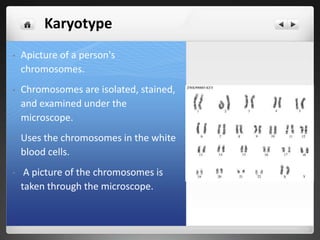
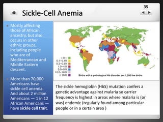
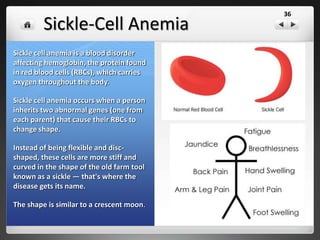
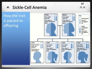

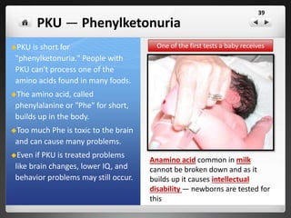
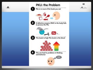
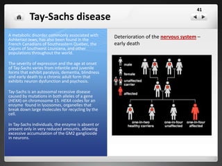
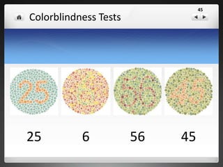
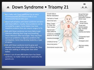
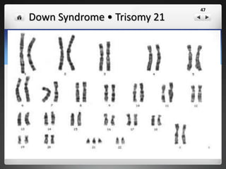
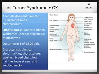
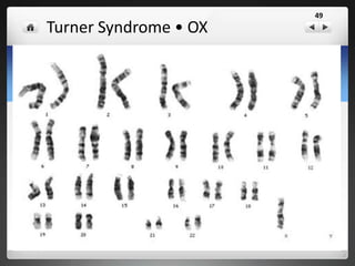

The document discusses genetics topics including multiple alleles, sex chromosomes, sex-linked traits, human genetic disorders, and karyotypes. It provides examples of genetic disorders such as sickle cell anemia caused by a mutation that provides resistance to malaria. It also discusses disorders like phenylketonuria (PKU) where an amino acid builds up and causes mental retardation without treatment. Finally, it addresses sex-linked genetic disorders like colorblindness.


![Ch. 4 human inheritance and genetic disorders [new]](https://cdn.slidesharecdn.com/ss_thumbnails/ch-4humaninheritanceandgeneticdisordersnew-130515015408-phpapp01-thumbnail.jpg?width=640&height=640&fit=bounds)


