This document discusses various fungal diseases that affect banana crops, including the causal organisms, symptoms, and control methods. It describes 33 different diseases including anthracnose, black sigatoka, Panama disease, and leaf spot diseases caused by fungi such as Colletotrichum musae and Mycosphaerella fijiensis. The diseases cause symptoms like lesions, rot, wilting and discoloration of leaves, fruits and roots. Control methods mentioned are fungicides, resistant varieties, removing infected plant materials, and improving soil drainage and sanitation.
The overall description of major diseases of Rice or Paddy crop is ellustrated in presentation. The students prepairing for Agriculture can feel helpful. Thank You!
The overall description of major diseases of Rice or Paddy crop is ellustrated in presentation. The students prepairing for Agriculture can feel helpful. Thank You!
Wilt is a pernicious disease of guava in India.
In India the disease was first recorded near Allahabad in 1935 . The infection was reported 15 -30 %.
The disease is a serious threat to guava cultivation in U.P. In West Bengal it reduces the yield in affected orchard by 80% .
The disease is also prevalent in Haryana Rajasthan , A.P ,
Punjab and M.P.The exact cause of the disease is still not fully understood but the pathogens viz. Fusarium oxysporum f. sp. psidii (Prasad, Mehta & Lal), Rhizoctonia spp. (Taub.) and various pathogens are reported by different workers may be the incitant of the disease.
Survival and spread:
Through movement of plants containing sick soil in virgin areas.
Short distance spread is by water.
Root injury predisposes wilt disease.
It has forced uprooting of about 150 acre of guava orchard in Panjab and 300 acres in Haryana during 1971-81.
This was presented by one of the group of students to our Asst. professors Mr. and Mrs. Poudel (Pathology) in 2017. By B.Sc.Ag Paklihawa IAAS campus, Full phase 6th batch
This ppt will help Agricultural professionals to diagnose banana diseases and the management strategies. This is a compilation of important diseases of banana prevalent in India which contains some of my own photographs and others collected from Web. This is intended only for educating students and other agricultural field staff.
Introduction:
RNA interference (RNAi) or Post-Transcriptional Gene Silencing (PTGS) is an important biological process for modulating eukaryotic gene expression.
It is highly conserved process of posttranscriptional gene silencing by which double stranded RNA (dsRNA) causes sequence-specific degradation of mRNA sequences.
dsRNA-induced gene silencing (RNAi) is reported in a wide range of eukaryotes ranging from worms, insects, mammals and plants.
This process mediates resistance to both endogenous parasitic and exogenous pathogenic nucleic acids, and regulates the expression of protein-coding genes.
What are small ncRNAs?
micro RNA (miRNA)
short interfering RNA (siRNA)
Properties of small non-coding RNA:
Involved in silencing mRNA transcripts.
Called “small” because they are usually only about 21-24 nucleotides long.
Synthesized by first cutting up longer precursor sequences (like the 61nt one that Lee discovered).
Silence an mRNA by base pairing with some sequence on the mRNA.
Discovery of siRNA?
The first small RNA:
In 1993 Rosalind Lee (Victor Ambros lab) was studying a non- coding gene in C. elegans, lin-4, that was involved in silencing of another gene, lin-14, at the appropriate time in the
development of the worm C. elegans.
Two small transcripts of lin-4 (22nt and 61nt) were found to be complementary to a sequence in the 3' UTR of lin-14.
Because lin-4 encoded no protein, she deduced that it must be these transcripts that are causing the silencing by RNA-RNA interactions.
Types of RNAi ( non coding RNA)
MiRNA
Length (23-25 nt)
Trans acting
Binds with target MRNA in mismatch
Translation inhibition
Si RNA
Length 21 nt.
Cis acting
Bind with target Mrna in perfect complementary sequence
Piwi-RNA
Length ; 25 to 36 nt.
Expressed in Germ Cells
Regulates trnasposomes activity
MECHANISM OF RNAI:
First the double-stranded RNA teams up with a protein complex named Dicer, which cuts the long RNA into short pieces.
Then another protein complex called RISC (RNA-induced silencing complex) discards one of the two RNA strands.
The RISC-docked, single-stranded RNA then pairs with the homologous mRNA and destroys it.
THE RISC COMPLEX:
RISC is large(>500kD) RNA multi- protein Binding complex which triggers MRNA degradation in response to MRNA
Unwinding of double stranded Si RNA by ATP independent Helicase
Active component of RISC is Ago proteins( ENDONUCLEASE) which cleave target MRNA.
DICER: endonuclease (RNase Family III)
Argonaute: Central Component of the RNA-Induced Silencing Complex (RISC)
One strand of the dsRNA produced by Dicer is retained in the RISC complex in association with Argonaute
ARGONAUTE PROTEIN :
1.PAZ(PIWI/Argonaute/ Zwille)- Recognition of target MRNA
2.PIWI (p-element induced wimpy Testis)- breaks Phosphodiester bond of mRNA.)RNAse H activity.
MiRNA:
The Double-stranded RNAs are naturally produced in eukaryotic cells during development, and they have a key role in regulating gene expression .
Professional air quality monitoring systems provide immediate, on-site data for analysis, compliance, and decision-making.
Monitor common gases, weather parameters, particulates.
Cancer cell metabolism: special Reference to Lactate PathwayAADYARAJPANDEY1
Normal Cell Metabolism:
Cellular respiration describes the series of steps that cells use to break down sugar and other chemicals to get the energy we need to function.
Energy is stored in the bonds of glucose and when glucose is broken down, much of that energy is released.
Cell utilize energy in the form of ATP.
The first step of respiration is called glycolysis. In a series of steps, glycolysis breaks glucose into two smaller molecules - a chemical called pyruvate. A small amount of ATP is formed during this process.
Most healthy cells continue the breakdown in a second process, called the Kreb's cycle. The Kreb's cycle allows cells to “burn” the pyruvates made in glycolysis to get more ATP.
The last step in the breakdown of glucose is called oxidative phosphorylation (Ox-Phos).
It takes place in specialized cell structures called mitochondria. This process produces a large amount of ATP. Importantly, cells need oxygen to complete oxidative phosphorylation.
If a cell completes only glycolysis, only 2 molecules of ATP are made per glucose. However, if the cell completes the entire respiration process (glycolysis - Kreb's - oxidative phosphorylation), about 36 molecules of ATP are created, giving it much more energy to use.
IN CANCER CELL:
Unlike healthy cells that "burn" the entire molecule of sugar to capture a large amount of energy as ATP, cancer cells are wasteful.
Cancer cells only partially break down sugar molecules. They overuse the first step of respiration, glycolysis. They frequently do not complete the second step, oxidative phosphorylation.
This results in only 2 molecules of ATP per each glucose molecule instead of the 36 or so ATPs healthy cells gain. As a result, cancer cells need to use a lot more sugar molecules to get enough energy to survive.
introduction to WARBERG PHENOMENA:
WARBURG EFFECT Usually, cancer cells are highly glycolytic (glucose addiction) and take up more glucose than do normal cells from outside.
Otto Heinrich Warburg (; 8 October 1883 – 1 August 1970) In 1931 was awarded the Nobel Prize in Physiology for his "discovery of the nature and mode of action of the respiratory enzyme.
WARNBURG EFFECT : cancer cells under aerobic (well-oxygenated) conditions to metabolize glucose to lactate (aerobic glycolysis) is known as the Warburg effect. Warburg made the observation that tumor slices consume glucose and secrete lactate at a higher rate than normal tissues.
Richard's entangled aventures in wonderlandRichard Gill
Since the loophole-free Bell experiments of 2020 and the Nobel prizes in physics of 2022, critics of Bell's work have retreated to the fortress of super-determinism. Now, super-determinism is a derogatory word - it just means "determinism". Palmer, Hance and Hossenfelder argue that quantum mechanics and determinism are not incompatible, using a sophisticated mathematical construction based on a subtle thinning of allowed states and measurements in quantum mechanics, such that what is left appears to make Bell's argument fail, without altering the empirical predictions of quantum mechanics. I think however that it is a smoke screen, and the slogan "lost in math" comes to my mind. I will discuss some other recent disproofs of Bell's theorem using the language of causality based on causal graphs. Causal thinking is also central to law and justice. I will mention surprising connections to my work on serial killer nurse cases, in particular the Dutch case of Lucia de Berk and the current UK case of Lucy Letby.
Multi-source connectivity as the driver of solar wind variability in the heli...Sérgio Sacani
The ambient solar wind that flls the heliosphere originates from multiple
sources in the solar corona and is highly structured. It is often described
as high-speed, relatively homogeneous, plasma streams from coronal
holes and slow-speed, highly variable, streams whose source regions are
under debate. A key goal of ESA/NASA’s Solar Orbiter mission is to identify
solar wind sources and understand what drives the complexity seen in the
heliosphere. By combining magnetic feld modelling and spectroscopic
techniques with high-resolution observations and measurements, we show
that the solar wind variability detected in situ by Solar Orbiter in March
2022 is driven by spatio-temporal changes in the magnetic connectivity to
multiple sources in the solar atmosphere. The magnetic feld footpoints
connected to the spacecraft moved from the boundaries of a coronal hole
to one active region (12961) and then across to another region (12957). This
is refected in the in situ measurements, which show the transition from fast
to highly Alfvénic then to slow solar wind that is disrupted by the arrival of
a coronal mass ejection. Our results describe solar wind variability at 0.5 au
but are applicable to near-Earth observatories.
Seminar of U.V. Spectroscopy by SAMIR PANDASAMIR PANDA
Spectroscopy is a branch of science dealing the study of interaction of electromagnetic radiation with matter.
Ultraviolet-visible spectroscopy refers to absorption spectroscopy or reflect spectroscopy in the UV-VIS spectral region.
Ultraviolet-visible spectroscopy is an analytical method that can measure the amount of light received by the analyte.
Nutraceutical market, scope and growth: Herbal drug technologyLokesh Patil
As consumer awareness of health and wellness rises, the nutraceutical market—which includes goods like functional meals, drinks, and dietary supplements that provide health advantages beyond basic nutrition—is growing significantly. As healthcare expenses rise, the population ages, and people want natural and preventative health solutions more and more, this industry is increasing quickly. Further driving market expansion are product formulation innovations and the use of cutting-edge technology for customized nutrition. With its worldwide reach, the nutraceutical industry is expected to keep growing and provide significant chances for research and investment in a number of categories, including vitamins, minerals, probiotics, and herbal supplements.
Observation of Io’s Resurfacing via Plume Deposition Using Ground-based Adapt...Sérgio Sacani
Since volcanic activity was first discovered on Io from Voyager images in 1979, changes
on Io’s surface have been monitored from both spacecraft and ground-based telescopes.
Here, we present the highest spatial resolution images of Io ever obtained from a groundbased telescope. These images, acquired by the SHARK-VIS instrument on the Large
Binocular Telescope, show evidence of a major resurfacing event on Io’s trailing hemisphere. When compared to the most recent spacecraft images, the SHARK-VIS images
show that a plume deposit from a powerful eruption at Pillan Patera has covered part
of the long-lived Pele plume deposit. Although this type of resurfacing event may be common on Io, few have been detected due to the rarity of spacecraft visits and the previously low spatial resolution available from Earth-based telescopes. The SHARK-VIS instrument ushers in a new era of high resolution imaging of Io’s surface using adaptive
optics at visible wavelengths.
PRESENTATION ABOUT PRINCIPLE OF COSMATIC EVALUATION
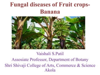
Fungal diseases of fruit crops banana
1. Fungal diseases of Fruit crops-
Banana
Vaishali S.Patil
Assosiate Professor, Department of Botany
Shri Shivaji College of Arts, Commerce & Science
Akola
2. 1.Anthracnose and Fungal scald and Stem-end rot caused by
Colletotrichum musae
Symptoms-At the initial stage, small, circular, black spots develop on
the affected fruits. Then these spots enlarge in size, turn to brown colour.
The skin of the fruit turns black and shrivels and becomes covered with
characteristic pink acervuli. Finally the whole finger is affected. Later the
disease spreads and affects the whole bunch. The disease results in
premature ripening and shriveling of the fruits which are covered with
pink spore masses. Sometimes the main stalk of the bunch may become
diseased. Infected fruits become black and rotten.
Control-Fungicides
3. 2.Armillaria corn rot caused by Armillaria mellea, Armillaria
tabescens
Symptoms- plants slowly decline, leaf yellowing, reduced leaf growth
and twig dieback. presence of cream-coloured fungal growth, sometimes
fan-shaped, just beneath the bark of the crown and large roots, and a
strong mushroom smell. Black, cord-like threads of the fungus often
occur on the surface of the roots, forming a branched network that may
extend 200-300 mm into the soil. Honey-coloured mushrooms with
widely separated gills can form at the base of an affected tree during wet,
cold weather in early winter.
Control- Prepare soil thoroughly, remove as many roots and stumps as
possible before planting, use resistant rootstocks, Fumigate infested soil,
fungicides.
4. 3.Black cross caused by Phyllachora musicola
Symptoms- The spots are black, four-pointed stars, most
clearly seen on the lower surface of older leaves. The long axis
of the star is parallel to the leaf veins, that is, at right angles to
the length of the leaf. The spots are scattered, but sometimes
occur in large groups.A velvet-like mass of spores is produced
on the lower surface of the spots.
Control- reducing shade levels or planting the bananas in
open ground, resistant varieties, fungicides.
5. 4.Black leaf streak (BLS) or Black Sigatoka and Septoria leaf spot
,Yellow Sigatoka caused by Mycosphaerella fijiensis,
Pseudocercospora (Paracercospora) fijiensis, Mycosphaerella eumusae
, Septoria eumusae, Mycosphaerella musicola
Symptoms-Early symptoms appear on the third or fourth leaf from the
top, i.e., on young leaves. Small spindle shaped spots on foliage with
greyish centre and yellowish halo running parallel to veins.If the fruit is
nearing maturity at the time of heavy infection, the flesh ripens but
evenly and individual bananas appear undersized and their flesh develops
a buff pinkish colour, and store poorly.
Control- fungicide.
6. 5.Black root rot caused by Rosellinia bunodes
Symptoms- Brown lesions on roots that progressively darken to
black. Dark discoloration of vascular bundles is visible shortly thereafter.
Once inside the vascular tissue, compromises xylem flow and transport
of nutrients to aerial parts of the host resulting in chlorosis, wilting, dry
die-back, leaf drop, and eventually host death. White, cottony growth on
roots that also progressively darken with age.As mycelia grow, they form
dense mats over roots and at the base of tree trunks that thicken into
irregular knots and aggregate into rhizomorphs. Over time mycelia
darken to form black, branching strands firmly attached to host roots;
occasionally black dots are visible before this color change.
Control- Good soil drainage and appropriate soil fertility, removal of
crop residue, fungicide, crop rotation, soil solarization.
7. 6.Brown blotch caused by Pestalotiopsis leprogena
Symptoms-Occur on leaf blade (leaflets or leaf segments) or only the
petiole and rachis; or, it can develop on both tissues at the same time.
Spots will begin as very small yellow, brown or black spots. the spots
may expand and increase in number until they merge (coalesce) to form a
leaf blight or rachis blight (larger area of affected tissue). Often, the spots
turn a grayish color that are outlined in black. The same type of lesions
occurs on the petiole or rachis
Control- sanitation and water management , diseased leaves should be
pruned and destroyed, fungicides
8. 7.Brown spot caused by Cercospora hayi
Symptoms- lesions occurs on the fruit, stalk, crown cushion,
and fingers. The spots are dark brown, have an irregular
margin, and are surrounded by a halo of water-soaked tissue
Control- Plantation sanitation, good drainage and proper
spacing, fungicides.
9. 8.Ceratocystis fruit rot and Main stalk rot caused by Ceratocystis
paradoxa,Chalara paradoxa
Symptoms- black end and finger-tip rot symptoms are produced. black
lesions on the fruit is the main infection part of the plant. If the pathogen
infects the plant while fruits are still on it, they will prematurely drop.
Discoloration of leaves as well as the seeds also take place. The lesions
on the fruit evolve to become soft rot spots that produce a heinous odor.
The fruit can even get to the point of breakdown.
Control- Heat Treatment , hot-water treated, fungicide .
10. 9.Cigar-end /Verticillium tip rot and Trachysphaera finger
rot caused by Verticillium theobromae, Trachysphaera
fructigena
Symptoms-Attack ripening fruit causing a dry rot of the
flower end that produces an ash grey wrinkled lesion similar to
the burnt end of a cigar. Rotting occurs causing a black
wrinkled necrosis with the fruit eventually becoming
mummified
Control-sanitation and avoiding damage to host tissues,
deflowering and bagging of maturing banana stems, fungicide
11. 10.Cladosporium speckle caused by Cladosporium musae
Symptoms- Pale greenish flecks, ellipsoid to oblong, forming
streaks, pale to blackish brown, turning orange or dark
brown, merging and forming large patches, occasionally
somewhat target-like.
Control- Reduce plant density to lower leaf infection,
Collect, remove and destroy heavily speckled leaves from
plants, Remove shade and weeds, desucker plants, fungicides
12. 11.Corm dry rot caused by Junghuhnia vincta
Symptoms- Yellowing and browning of foliage with older foliage dying
first. Some branches affected before others, giving tree crowns an
uneven, patchy appearance . Small pads of white or pale pink fungal
tissue develop at the base of the tree, and on roots just below the soil
surface. Fruiting bodies in the form of pink encrustations with pores may
develop on the lower stem.
Control- Improved/resistant cultivars and disease free planting stocks,
judicious use of pesticides, irrigation water, removal of diseased plants
and plant parts, proper sanitation in plantation.
13. 12.Cordana leaf spot caused by Cordana johnstonii,
Cordana musae
Symptoms- On the leaf large, pale brown, oval to fusiform
necrotic lesions with pale grey concentric ring patterns, with a
dark brown border surrounded by a bright yellow halo
separating are found.Often, lesions coalesce into large necrotic
patches. The leaves ultimately turn brown and dry out.
Control- fungicides.
14. 13.Crown rot and Peduncle rot caused by Fusarium pallidoroseum,
Colletotrichum musae, Verticillium theobromae, Fusarium spp.,
Acremonium spp. Lasiodiplodia theobromae
Symptoms- Blackening and softening of tissues and begins at or near
the cut surface of the crescent-shaped crown where the hand is detached
from the main fruit stalk. The fungus also extends through cushions and
causes finger stalk rot and finger dropping. The skin becomes soft, black,
wrinkled and encrusted with pycnidia. Infection may lead premature
ripening of the fruits.
Control- fungicides, avoid and injury to the fruits, use of resistant
varieties, storage of fruits at 100 C.
15. 14.Cylindrocladium root rot caused by
Cylindrocladium spp.
Symptoms- Chlorotic lower leaves and a wilted appearance,
onto leaves, dime sized circular brown necrotic lesions
surrounded by yellow halos will eventually form on
leaves.Reddish-brown lesions can be found on root. These
lesions grow rapidly causing total root collapse and rot.
Control- fungicide, removal of infected plants.
16. 15.Damping-off caused by Deightoniella torulosa
Symptoms- On leaves lesions were tan to black. However,
larger oval black lesions with yellow halos also occurred.
Lesions were more prevalent on older leaves. On young
leaves, lesions first appeared along the leaf margin near the tip
of the leaf on one side of the central vein. Lesions expanded to
the entire leaf as the disease progressed, but were more
prevalent along leaf margins.
Control- fungicide
17. 16.Deightoniella fruit speckle, leaf spot and tip rot caused
by Deightoniella torulosa
Symptoms- On the leaves, it causes oval, tan spots with a
black border, usually on the older leaves . On the fruit, sunken
dark brown or black spots, surrounded by a dark green halo.
Spotting of the fruit is greatest towards the tips of the fingers.
The fungus does not produce spores on the fruit. The fungus
also attacks the flower parts.
Control- Remove dead or dying leaves , fungicides
18. 17.Diamond spot caused by Cercospora hayi, Fusarium spp
Symptoms- The spots are oval to diamond-shaped, and at
right angles to the length of the leaf . They are brown on the
upper surface, zoned, with yellow margins, and grey to brown
below. The spots often merge, covering large areas of the leaf;
this occurs particularly at the margin of the leaf giving a band
of dead tissue with a zigzag yellow border between diseased
and healthy parts. Some time the spots appear greyish-brown
and hairy.
Control- Resistant varieties, fungicides.
19. 18.Dwarf Cavendish tip rot caused by Nattrassia
mangiferae= Hendersonula toruloidea
Symptoms- Lodging, partial or complete wilting of the plant,
and rotting (dry or soft) of roots. It also cause pit canker and
spot on the stem of plants or fruits, as well as internal black rot
of fruits,
Control- fungicides.
20. 19.Eyespot and Leaf spot caused by Drechslera gigantea,
Drechslera musae-sapientum
Symptoms- mature lesion has a white or grey centre and a
narrow, well‐defined, brown border.
Control-Provide good drainage, Aerate to eliminate soil
compaction, Avoid herbicide applications, Water adequately,
but not excessively, fungicide.
21. 20.Fruit freckle (freckle) caused by Guignardia musae,
Phyllosticta musarum
Symptoms- Spots clustering in lines running horizontally
across leaf which down veins of leaf. Dense aggregations of
spots cause black blemishes on skin of fruit. Although
detracting from the appearance of the fruit, the eating qualities
are not affected.
Control- cutting out infected leaves, the paper bag method,
fungicide application, and proper sanitation techniques.
22. 21.Fruit rot caused by Botryosphaeria ribis
Symptoms- elongated black spots and the entire fruit became
rotten.
Control-Soil treatment involving fumigation, soil solarization
or sanitation, Soil preparation, fungicide
23. 22.Fungal root-rot caused by Fusarium solani, Nectria
haematococca, Fusarium oxysporum, Rhizoctonia spp.
Symptoms-Wilting, stunting and chlorosis or lesions on the
stem and/or leaves.Narrow, long, red to brown lesions on the
stems, and lengthwise cracks often develop. Lesions extend
down the main taproot, which may shrivel, decay and die.
Control- crop rotation and timing of planting, resistant
varieties.
24. 23.Leaf rust caused by Uredo musae, Uromyces musae
Symptoms-Dark brown to black streaks appear on the leaves,
often surrounded by yellow halos. The streaks are more
numerous on the lower surface of the leaves. They are slightly
raised, and feel rough to the touch due to the spore masses of
the fungus. If the disease is severe, the leaves may turn yellow.
Mainly the older leaves are affected.
Control-fungicides.
25. 24.Leaf speckle caused by Acrodontium simplex
Symptoms- The lesions first appear as brown to dark brown
tiny specks with elongate into black fine streaks parallel to the
veinlets. The affected areas become necrotic and the infected
leaves eventually die.
Control-fungicides
26. 25.Leaf spot caused by Curvularia eragrostidis
Symptoms-leaf spot starts as very small round tan lesions on
leaves. Lesions often have a brown border and can be
surrounded by a yellow halo . A few lesions scattered across
leaves to lesions densely covering large sections of leaves.
Symptoms can be observed at any growth stage.
Control-residue decomposition, crop rotating, hybrid
resistance.
27. 26.Leaf spot caused by Leptosphaeria musarum
Symptoms-Light greenish-brown, somewhat indistinct,
narrow streaks or less in diameter. The brown stage, in which
the spot expands laterally to become elliptical and turns first
light and then dark brown to almost black; the leaf has dried
out at severity. the grey centre stage, in which the elliptical
lesion dries out to a light grey, with the bases of the old
fructifications showing up as scattered black dots.
Control-fungicides
28. 27.Leaf spot caused by Pestalotiopsis disseminata
Symptoms- small yellow, brown or black discoloration of the
leaves. The disease can be restricted to the leaf blade or may
only appear on the petiole and rachis right away. The spots
will often turn a grayish color and will be outlined in black.
Extreme wilting and a drying appearance on the leaves.
Control- sanitation and irrigation management, Wounds and
damage to the plan, eliminating overhead irrigation, Nutrient
management, pruning the leaves, fungicides.
29. 28.Malayan leaf spot caused by Haplobasidion musae
Symptoms-diamond-shaped, greyish-white spots on the
upper leaf surface. The spots, which sometimes have brown
centres, were surrounded by a black border. On the
undersurface of the leaf, the lesion could be covered with a
dense, velvety brown mass (Water-soaked areas, often several
times the size of the spot, have been observed surrounding
lesions .
The lesions were often pale on the upper leaf surface and
darker on the lower surface, with dark purple borders.
Control-reduce shade and humidity levels
30. 29.Marasmiellus rot caused by Marasmiellus inoderma
= Marasmius semiustus
Symptoms- Rotted patches on rhizome and pseudo stem,
gradual wilting of leaves from lower area to upper part,
diminutive growth, strange foliage and bunches, toppling of
crown, fruiting body adhere on pseudo-stem are the major
syndromes of disease
Control-Fungicides
31. 30.Panama disease (Fusarium wilt) caused by Fusarium oxysporum
f.sp. cubense
Symptoms- Yellowing of the lower most leaves starting from margin to
midrib of the leaves. Yellowing extends upwards and finally heart leaf
alone remains green for some time and it is also affected.The leaves
break near the base and hang down around pseudostem.
Longitudinal splitting of pseudostem. Discolouration of vascular vessels
as red or brown streaks.
Control- use of soil fumigants, sanitation, resistant varieties, Crop
rotation, Fungicides.
32. 31.Pestalotiopsis leaf spot caused by Pestalotiopsis
palmarum
Symptoms- Water-soaked symptoms appeared first. The
infected site developed into a black–brown lesion. In the later
stage of infection, the diseased area turned into an elliptical or
irregular shaped grey lesion with a golden yellow margin.
Control- fungicides.
33. 32.Phaeoseptoria leaf spot caused by Phaeoseptoria musae
Symptoms-small necrotic, lentis shape and dark brown spots
similar Sigatoka symptoms, which soon later enlarge rapidly
and become rot.
Control- fungicides.
34. 33.Pitting caused by Pyricularia grisea
Symptoms- Blast lesions on young leaves, transition leaves, mid rib,
petioles, peduncle, maturing bunches, bunch stalks and cushions. The
distinct small pitting spots on maturing bunches reduced the visual
appeal of mature fruits. Appearance of pitting symptoms on fruits in
relation with age of fruits and their distribution pattern on bunch and
fingers is also seen.
Control- fungicides.
35. 34.Pseudostem heart rot caused by Fusarium moniliforme, Gibberella
fujikuroi
Symptoms-Severe tip rot with pronounced brown to blackening of
young rolled central leaves tissues. These may remain folded or become
nearly fully unrolled as they emerge
from the crown. This decay was sometimes present only in the upper
portion
of the pseudostem but it could be tending to take a downward direction
in
the core of the pseudostem
Control-fungicides.
36. 35.Root & rhizome rot caused by Cylindrocarpon musae
Symptoms- Rotting of fleshy roots and rhizomes takes
place. The stem break. The disease affects the outer layers of
the pseudostem. A strong odour is associated with the rotting.
Control- fungicides.
37. 36.Sclerotinia fruit rot caused by Sclerotinia sclerotiorum
Symptoms-water-soaked spots on fruits, stems, leaves, or petioles which usually have
an irregular shape. These spots enlarge and a cottony mycelium covers the affected
area. The fungus spreads and the plant becomes a soft, slimy, water-soaked mass. The
cottony mycelium usually produces numerous sclerotia, black seed-like reproductive
structures, a reliable diagnostic sign of Sclerotinia (these usually do not form until after
host death). Dry lesions appear on the stalk, stems, or branches.The lesions enlarge and
girdle the plant part. Distal portions of the plant become yellow, then brown, then die.
The girdled portion is often the base of the plant which causes the plant to die. Sclerotia
form within the stem pith cavities, fruit cavities, or between tissues (i.e., bark and
xylem).
Control-biological control , fungicides, Crop rotation, Resistant varieties, fungicides.
38. 37.Sooty mold caused by Limacinula tenuis
Symptoms-A black, powdery coating adhering to plants. It
merely blocks sunlight, and very rarely may stunt a plant's
growth and yellow its foliage
Control-wash affected plant parts with lukewarm water and
soap, insecticidal soap or dish soap, using formulations
of neem oil.
39. 38.Speckle caused by Mycosphaerella musae
Symptoms-Leaf spots first visible as pale greenish flecks, ellipsoid to
oblong, forming streaks pale to blackish brown, turning orange or dark
brown, merging and forming large patches, occasionally somewhat
target-like. Spread is by airborne spores.Light brown irregular blotches
on the lower surface of the leaf, darkening to dark purple to black, and
becoming visible on both leaf surfaces. May coalesce to give large
bleached necrotic areas . Present only on leaves five and six and
older before flowering. When infection is severe, there is a loss of leaves
and bunches are small, and ripening is uneven.
Control- Reduce plant density to lower leaf infection, Collect, remove
and destroy heavily speckled leaves from plants, as well as tras, Remove
shade and weeds, fungicides.
40. 39.Squirter (black end disease) caused by Nigrospora
sphaerica
Symptoms- Spots initially appear brown, circular and
irregularly distributed on the leaves and they eventually
coalesce. Fruiting twig and shoot blight developed from the
tips toward the base.
Control-plantation sanitation and fungicide.
41. 40.Tropical speckle and Leaf speckle caused by Ramichloridium
musae= Veronaea musae= Periconiella musae
Symptoms- Diffuse irregular or circular, grey, brown or black blotches,
especially on underside of older leaves . Similar symptoms also occur on
leaf and fruit stalks. Leaves are covered with small, evenly spread
aggregations of black spots that are individually the size of a pinhead.
On the older leaves the dots may merge to form blotches". It also
appear on young foliage as tan coloured circular blotches on the
underside of the leaf . Similar symptoms are also found on leaf midribs
and fruit stalks.
Control-