
Recommended
More Related Content
What's hot
What's hot (20)
Similar to Fungal diseases of fruit crops- papaya
Similar to Fungal diseases of fruit crops- papaya (20)
More from vaishalidandge3
More from vaishalidandge3 (20)
Recently uploaded
Recently uploaded (20)
Fungal diseases of fruit crops- papaya
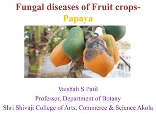
- 1. Fungal diseases of Fruit crops- Papaya Vaishali S.Patil Professor, Department of Botany Shri Shivaji College of Arts, Commerce & Science Akola
- 2. 1.Alternaria fruit spot- caused by Alternaria alternata Symptoms-depressed, circular to oval, lesions that eventually become black as a result of mass sporulation. Lesons are restricted to the surface of the fruit and do not cause extensive rotting of the flesh. However, lesions from multiple infection sites can coalesce as they expand and eventually cover the entire fruit surface. Control-A single postharvest water dip of 20 min, fungicides.
- 3. 2.Angular leaf spot caused by Leveillula taurica Symptoms- Petioles, stalks and flowers are rarely affected and fruits are occasionally infected. It penetrates the interior leaf tissues. On the ventral side powdery, whitish spots appear that gradually expand. On the dorsal side, yellow spots of varying intensity develop. On the dorsal side, powdery spots may also develop. Spots may later become necrotic. Control- Crop Resistance, proper irrigation, chemical control.
- 4. 3.Anthracnose caused by Colletotrichum gloeosporioides Symptoms -The spots on fruits first appear as brown superficial discolouration of the skin which develops into circular, slightly sunken areas. Gradually the lesions coalesce and sparse mycelia growth appears on the margins of the spots. Infection at early stages of fruit results in mummification and deformation. Control-removing the fruit as soon as it matures, removing all dead leaves and fruit from the vicinity of the plants, and removing infected fruits from the trees.
- 5. 4.Black spot caused by Asperisporium caricae, Cercospora papayae, Phomopsis caricae-papayae Symptoms- Water-soaked spots appear on the upper leaf surface. Later, small, black spots are visible on the underside of the leaf. Black spots may also be found on the fruit. The tissue beneath them becomes corky, but the fruit does not rot. Control-Fungicide
- 6. 5.Blossom spot caused by Choanephora cucurbitarum Symptoms- Ringspot disease are exhibited on the foliage fruit and main stem of the plant. The main stem growing at the time of infection frequently develops dark-green spots and streaks of an oily appearance. These are usually most common on the middle two-thirds of the stem. Control- Refrigerated storage and careful handling will minimize decay postharvest.
- 7. 6.Black rot caused by Mycosphaerella caricae Symptoms- Spots appear on leaves, flowers and young fruit.Fruit surface lesions are slightly sunken, circular, black. Margins of lesions are light brown and translucent. The lesion surface dries and wrinkles with age and turns black as it becomes covered with hyphae and pycnidia of the imperfect stage. Under humid conditions it is common to see light- colored tendrils of pycnidiospores oozing from pycnidia. The infected tissue is dry, firm and is initially tan in color and eventually turns black. Infection of flowers and young fruit are initially brown then become dark and sunken. Lesions often extend through the peduncle to the succulent stem tissue where it too takes on the characteristic dark, sunken appearance. Control- Hot water treatment, fungicides
- 8. 7.Brown spot caused by Corynespora cassiicola, Cercospora melonis,Cercospora vignicola, Helminthosporium cassiicola, Helminthosporium vignae,Helminthosporium vignicola Symptoms- On the lower leaves, small, angular brown spots, often with white centres that fall out . The spots have a characteristic well-defined yellow halos. Oval, dark brown spots also occur on the leaf stalks. However, in wet conditions, the spots grow much larger, join together, and develop into spots that are zoned, or have target-like rings. Spots on fruits are not common, but occasionally occur as dark, sunken spots on the fruits. Control- Remove infected papaya trees from the field, fungicide.
- 9. 8.Chocolate spot caused by Colletotrichum gloeosporioide Symptoms-superficial lesions, seldom slightly sunken, irregular to rounded, well defined, with characteristic reddish- brown colour. As the fruit ripens the lesions can either remain superficial or grow and become sunken, resembling anthracnose. Control-Remove and destroy infected plants or plant parts and do not compost them, use resistant/tolerant varieties, plant certified, disease free/healthy planting materials, plant trees in well-drained soil, ensure rotations with non-host plants, ensure proper weed control, to prevent post-harvest losses avoid fruit wounding, i.e. through bruising, scratching, or puncturing the fruits, fungicides.
- 10. 9.Collar rot caused by Cylindrocladium crotalariae, Calonectria crotalariae Symptoms-The stem of the tree will become water soaked and weak, usually right at ground level. This water-soaked area will develop into a brown or black lesion and begin to rot. Sometimes a white, fluffy growth of fungus is visible. The leaves may turn yellow and droop, and eventually the entire tree will fail and collapse. Control-plant papaya saplings in well-draining soil. Dig up infected plants and destroy them, and do not plant more trees in the same spot.
- 11. 10.Damping off caused by Colletotrichum gloeosporioides,Phytophthora palmivora,Phytophthora nicotianae,Phytophthora parasitica,Pythium aphanidermatum,Pythium debaryanum,Pythium ultimum,Pythium sp.,Rhizoctonia solani,Thanatephorus cucumeris Symptoms- seeds fail to germinate & slowly kills young plants. The fungus eventually rots out the stem. Control-resistant varieties, fungicide, Prepare the soil well and ensure that it drains quickly, crop rotation, Sanitize all containers and tools.
- 12. 11.Dry rot caused by Phoma caricae-papayae,Ascochyta caricae,Ascochyta caricae-papayae,Mycosphaerella caricae Symptoms-Fruit surface lesions are slightly sunken, circular, black . Margins of lesions are light brown and translucent. The lesion surface dries and wrinkles with age and turns black as it becomes covered with hyphae and pycnidia of the imperfect stage. Under humid conditions it is common to see light-colored tendrils of pycnidiospores oozing from pycnidia. The infected tissue is dry, firm and is initially tan in color and eventually turns black. Stem-end rot caused by this fungus takes on the characteristics similar to fruit surface lesions. Infection of flowers and young fruit are initially brown then become dark and sunken. Lesions often extend through the peduncle to the succulent stem tissue where it too takes on the characteristic dark, sunken appearance. Control-Hot water treatment, fungicide.
- 13. 12.Foot rot caused by Pythium aphanidermatum,Pythium ultimum Symptoms -Water-soaked patches on the stem near the ground level. These patches enlarge rapidly and girdle the stem, causing rotting of the tissues, which then turn dark brown or black. Such affected plants withstand strong wind and topple over and die. If the disease attack is mild, only one side of the stem rots and the plants remain stunted. Fruit if formed are shriveled and malformed. Gradually the plant dies. Control- well-draining soil, as well as irrigation that does not touch the trunk. Applications of copper solution shortly after planting and during the time of fruit formation will also help.
- 14. 13.Fruit rot caused by Monilia sp Symptoms-Initial fruit lesions are brown, circular, and firm. Eventually the whole fruit decays and turns brown. Tufts of mycelium and conidia (cream-white to buff colored) sprout from the skin of the infected fruit, often arranged in concentric rings. Rotted fruits may either fall to the ground or dry out on the tree, leaving a hard, shriveled ‘mummy’. Control-fungicides, emoval of mummified fruit and pruning of infected twigs, with subsequent burning or deep-burying, and the removal of wild host plants.
- 15. 14.Fruit spot caused by Cercospora mamaonis Symptoms-Fruit spots start as tiny black dots that eventually enlarge. The spots are superficial, slightly raised, a result of the tissue beneath the epidermis becoming corky, and do not develop into a fruit rot. The spots are somewhat obscure on green fruits but become readily visible when the skin color turns yellow as the fruit ripens. Actual damage to fruits is minor except its impact on their appearance and marketability. Control-fungicide
- 16. 15.Fusarium fruit rot caused by Fusarium solani, Fusarium spp. Symptoms-small, pitted, corky spots to large, sunken areas covered with a white or gray mold on fruit. Control-Crop rotations, fungicide , Avoidance of wounding during harvest and packing, storage in dry conditions, and proper handling during transit and marketing reduce post- harvest decay.
- 17. 16.Guignardia spot caused by Guignardia sp Symptoms- Numerous, circular to oval, sunken greenish-black lesions expand in diameter very slowly , the fungus may grow deep into the flesh causing a black, firm discoloration. Control-heat treatments.
- 18. 17.Greasy spot caused by Corynespora cassiicola,Cercospora melonis, Cercospora vignicola, Helminthosporium cassiicola, Helminthosporium vignae, Helminthosporium vignicola Symptoms-The leaf spots are grayish-white, subcircular to irregular, heavily infected leaves turn yellow and dry up. The fruit spots start as tiny spots which turn black and enlarge. The tissue just beneath the epidermis of the fruit becomes corky; the spot does not develop into a fruit rot. Control-Fungicides
- 19. 18.Internal blight caused by Cladosporium sp., Fusarium sp., Penicillium sp. Symptoms- Infected tissue is watersoaked and translucent in the early stages to black and firm in the later stages of infection. Infected fruits yellow prematurely and unevenly in the fruit column. Control-proper seed selection, fungicides.
- 20. 19.Lasiodiplodia fruit rot caused by Lasiodiplodia theobromae, Botryodiplodia theobromae, Botryodiplodia gossypii, Diplodia theobromae, Diplodia gossypina, Diplodia natalensis, Lasiodiplodia triflorae Symptoms- A wider, more extensive translucent margin on fruit ,mummify the entire fruit. Gray mycelium forms over the infected area and later turns black from the masses of pycnidia that form. The infected flesh takes on a bluish-black discoloration in the soft water-soaked tissue. Air pockets often form in the infected area, presumably caused by the flesh shrinking, and later become filled with gray mycelium Control- hot water treatment.
- 21. 20.Leaf spot caused by Alternaria sp., Asperisporium caricae, Cercospora mamaonis, Cercospora papayae, Choanephora cucurbitarum, Curvularia caricae-papayae, Gloeosporium sp., Phoma caricae-papayae, Mycosphaerella caricae, Phyllosticta sp. Symptoms- irregular dark brown to black spots appear on the lower leaf surface of leaves. On the upper leaf surface, the infection causes slightly sunken tan spots to occur. Black spots have also been observed on the surface of fruits. Control- fungicides.
- 22. 21.Petiole spot caused by Didymella sp. Symptoms- It causes moist dark brown stem cankers that often develop from de-leafing and other wounds. These lesions gradually spread and eventually surround the stem and/or petioles, thus disrupting the sap flow. Ultimately, yellowing and wilting of the leaflets and plant parts below the lesions.Brownish tiny globular structures (pycnidia) dot the damaged tissues. Control- Long rotations with crops, Wounding fruit during harvest and in storage must be avoided, resistant varieties,fungicides.
- 23. 22.Phytophthora blight caused by Phytophthora palmivora, Phytophthora nicotianae var. Parasitica, Phytophthora parasitica Symptoms- Water-soaked lesions exude milky latex. Fruits may eventually mummify and fall. Large lesions, often forming first where the fruit contacts the stem of the plant, are covered with whitish mycelium and masses. Fruits can rot, turn soft, and fall prematurely. As lesions enlarge, infected areas of the stems way weaken, causing stem damage or breaking. Foliage on affected stems may collapse. Roots may become dark and rotten, causing stunting of plant growth and yellow, collapsed leaves. Severely infected plants may die. Control- well drained soil, steamed or fumigated prior to planting, hot water treatment, Avoid damage or injury to papaya stems during cultivation, Variety selection, fungicides.
- 24. 23.Powdery mildew caused by Erysiphe cichoracearum, Erysiphe sp., Oidium caricae, Oidium indicum, Ovulariopsis papayae, Sphaerotheca fuliginea, Sphaerotheca humuli Symptoms- On the undersurface of disease leaves are found patches of whitish powder growth. On upper surfaces, leaves at the infection site show blotches of yellow or pale green usually near vein, surrounded by normally colored tissue. Fungus may attack the stem of young seedling. The spots enlarge and cover the entire leaf area. Severely infected leaves may become chlorotic and distorted before falling. Affected fruits are small in size and malformed. Control-Fungicide
- 25. 24.Phytophthora fruit rot caused by Phytophthora capsici, Phytophthora nicotianae var., parasitica, Phytophthora parasitica, Phytophthora palmivora Symptoms-Water-soaked lesions exude milky latex. Fruits may eventually mummify and fall, fruit rot initially appears as small, circular, water-soaked lesions. Large lesions, often forming first where the fruit contacts the stem of the plant, are covered with whitish mycelium and masses. Fruits can rot, turn soft, and fall prematurely. Control- well drained soil, pick up, remove and destroy fallen fruits, Intercrop, steamed or fumigated, hot water, Avoid damage or injury,fungicides.
- 26. 25.Rhizopus soft rot caused by Rhizopus stolonifer,Rhizopus nigricans Symptoms-soft and watery rot that quickly collapses the entire fruit but leaves the cuticle intact. The fungus can grow out through any break in the cuticle and spread rapidly to adjacent fruits, often destroying the entire contents of a box within a few days. The infected fruit is often covered by coarse, gray, hairy mycelia that form a mass of black sporangia at their tips. Control-sanitation, use of chlorinated water tanks, wounds should be minimized, Heat treatment, fungicide.
- 27. 26.Root rot caused by Phytophthora palmivora, Pythium aphanidermatum, Pythium ultimum, Pythium spp., Rhizoctonia solani, Thanatephorus cucumeris Symptoms-Ripe and unripe fruit are infected while still attached to the stem; lower fruit are infected first. Rots develop quickly and become covered in a white growth containing spores of the water mould, causing the fruit to shrivel and fall. Stems, also become infected through leaf and fruit scars. Rots develop, and these cause the tops of the trees to wilt and die. Rots also form at the base of the stem near soil level; in this case the decaying roots are the source of infection. Roots become infected with a soft, wet rot that affects the taproot and surface roots as well as the base of the stem. Leaves wilt, turn yellow and collapse, with only a few small leaves remaining at the top of the tree. Control-Soil drainage, crop rotation, hygiene, fungicides.
- 28. 27.Sclerotium blight caused by Sclerotium rolfsii, Athelia rolfsii Symptoms - development of coarse white strands of mycelium growing in a fan-shaped pattern on lower stems, leaf litter, and soil . After 7 to 14 days, tan-to-brown, mustard-seed-sized sclerotia form on the mycelial mat. Early symptoms consist of water-soaked lesions on crown and lower stem tissue. The disease usually is recognized by the yellowing and wilting of foliage, followed by a complete collapse of the plant. Soft, water-soaked, sunken, slightly yellowish lesions develop on fruit. These lesions quickly spread throughout most or all of the fruit, which will eventually become soft and collapse within 3 to 4 days of infection. The skin of the fruit often crack open and fine white mycelium and developing sclerotia spreads over the surface and quickly fills lesion cavities Control-Crop rotations, selecting fields that are free of S. Rolfsii, Close plant spacing & over-irrigation, Injury during cultivation should be Avoided deep plowing, fungicide, fumigants, Biological Control.
- 29. 28.Seedling blight caused by Colletotrichum gloeosporioides Symptoms- causing cotyledon or leaf spots, defoliation, stem lesions, collar rots, and damping-off. Over 80% of the seedlings were diseased in 3 weeks. The fungus spread from leaf or cotyledon lesions to stems and eventually caused stem or collar rot and damping-off. Control- The affected fruits should be remove and destroyed. The fruits should be harvested as soon as they mature, hot water treatment or a fungicidal wax treatment.
- 30. 29.Stem-end rot caused by Alternaria alterneta, Colletotrichum gloeosporioides, Fusarium sp.Lasiodiplodia theobromae, Botryodiplodia theobromae, Botryodiplodia gossypii, Diplodia theobromae, Diplodia gossypina, Diplodia natalensis, Lasiodiplodia triflorae, Phoma caricae-papayae, Ascochyta caricae, Ascochyta caricae-papayae, Mycosphaerella caricae, Phomopsis sp.Rhizopus stolonifer Symptoms- A dark rot develops from the stem end as fruit ripen after harvest. A dark brown to black rot begins at the stem end as a dark brown ring and the rot proceeds towards the other end. The rot produces dark streaking of the water-conducting tissues Control- fungicide, Avoid harvesting immature fruit. Cool fruit immediately after harvest and store in well-ventilated containers.
- 31. 30.Stemphylium fruit spot caused by Stemphylium lycopersici, Thyrospora lycopersici, Stemphylium floridanum Symptoms- fruit spot are small, round, dark brown lesions. The lesions become sunken and develop reddish-brown to purple margins as they enlarge. A velvety, dark green spore mass forms in the lesion center. White to gray mycelia grow over the lesion in the advanced stages. Internally, the infected tissue is discolored from a reddish brown to a dark brown color, appears dry, and small air pockets may develop. Control- hot and cold treatments, fungicides.
- 32. 31.Stem rot caused by Fusarium solani, Nectria haematococca, Fusarium sp., Phytophthora palmivora, Pythium aphanidermatum, Pythium ultimum Symptoms-The stem of the tree will become water soaked and weak, usually right at ground level. This water-soaked area will develop into a brown or black lesion and begin to rot. Sometimes a white, fluffy growth of fungus is visible. The leaves may turn yellow and droop, and eventually the entire tree will fail and collapse. Control-plant papaya saplings in well-draining soil. When planting saplings, handle them with care. Dig up infected plants and destroy them
- 33. 32.Target spot caused by Phyllosticta caricae-papayae Symptoms-The spots, on the leaves, vary considerably in size, some being small and rounded or irregular, oval, or elongated, almost white in the centre, they are often bounded by a yellowish or brownish margin which merges into the normal green of the leaf. The central portion of the spots is thin and papery, and finally it becomes brittle and drops out. When numerous spots are present, the leaf withers and falls. Control-Removing and destroying leaves from trees infected with eye spot, wetting foliage during irrigation should be avoided, Air circulation may be improved by pruning and by preventing overcrowded plantings. avoid susceptible species and cultivars , fungicide .
- 34. 33.Verticillium wilt caused by Verticillium dahliae Symptoms-Sudden wilting of leaves on one or more branches, rapid browning. Can start at base and move upwards. Young trees may be killed. Older trees may become stunted and lose productivity. Vascular tissue may be stained. Control-Avoid planting in known infected soils or fumigate soil.
- 35. 34.Wet fruit rot caused by Phomopsis sp. Symptoms- The infected area is slightly depressed, soft and translucent. The fungus is rapid growing causing lesions to expand very quickly with the infected area extending to the seed cavity. The cuticle over the infected area remains intact and develops a delicate, wrinkled pattern that is parallel to the leading edge of the lesion. Black, scattered pycnidia usually form in the infected area after about seven days. The infected tissue is soft, mushy and wet. The infected area can become covered by white to gray mycelium. Control-Removing dead and senescing leaves and petioles from the tree, hot water treatment for 20 minutes at 48C, fungicides.