Downloaded 160 times


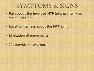


This document provides information on metatarsalgia and related conditions. It discusses the classification of metatarsalgia as primary, secondary, or unrelated to weight distribution disorders. Primary metatarsalgia is caused by an imbalance in weight distribution between the toes and metatarsal heads and can be due to functional or structural factors. Secondary metatarsalgia has other underlying causes such as rheumatoid arthritis. The document also covers Freiberg's disease, Morton's neuroma, and their symptoms, causes, imaging, classifications, and treatment options including conservative and surgical management.


