Patellofemoral Pain Syndrome
Represented by
Dr.Farha PT
Sports Physiotherapist
• Introduction
• Clinically relevant anatomy
• Function of Patella
• Biomechanical faults
• Clinical Presentation
• Difference diagnosis
• Examination
• Rehabilitaion
Introduction
• Patellofemoral Pain Syndrome (PFPS) is an umbrella term used for pain
arising from patellofemoral joint itself, or adjacent soft tissues.
• Historically it has been referred to as anterior knee pain but sometime pain
can be felt in all aspect of the knee.
• The main cause of PFPS is overuse injuries around in the patellofemoral
region because the PF joint is essentially a ‘Soft tissue’ joint.
• It is sometimes called "runner's knee" . it is common in people who participate
in sports—particularly females and young adults—but PFPS can occur in
nonathletic, as well. The pain and stiffness caused by PFPS can make it difficult
to climb stairs, kneel down, and perform other everyday activities.
Clinically relevant Anatomy
• Your knee is the largest joint in your body and one of the most complex. It
is made up of the lower end of the femur (thighbone), the upper end of the
tibia (shinbone), and the patella (kneecap).
• Ligaments and tendons connect the femur to the bones
of the lower leg. The four main ligaments in the knee
attach to the bones and act like strong ropes to hold
the bones together.
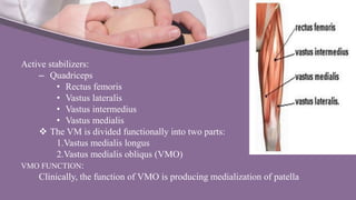
Active stabilizers:
– Quadriceps
• Rectus femoris
• Vastus lateralis
• Vastus intermedius
• Vastus medialis
 The VM is divided functionally into two parts:
1.Vastus medialis longus
2.Vastus medialis obliqus (VMO)
VMO FUNCTION:
Clinically, the function of VMO is producing medialization of patella
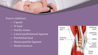
Passive stabilizers:
– Capsule
– IT band
– Patellar tendon
– Lateral patellofemoral ligament
– Patellotibial band
– Meniscopatellar ligament
– Medial meniscus
Patella
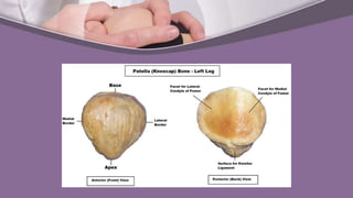
• Largest sesamoid bone in body, developed in tendon of quadriceps femoris.
– Features-
• Base
• An apex- Covered by expansion of rectus femoris
Separated from skin by prepatellar bursa
• 3 borders- Superior, Lateral, Medial
• 2 surfaces- Anterior,
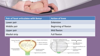
Posterior- Divided by 2 vertical and 2 transverse lines into 3
pairs of medial and lateral articular facets and one medial strip.
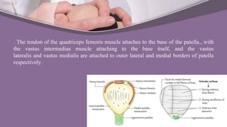
. The tendon of the quadriceps femoris muscle attaches to the base of the patella., with
the vastus intermedius muscle attaching to the base itself, and the vastus
lateralis and vastus medialis are attached to outer lateral and medial borders of patella
respectively.
• The posterior surface of the patella articulates with the femur, and is
marked by two facets:
– Medial facet – articulates with the medial condyle of the femur.
– Lateral facet – articulates with the lateral condyle of the femur.
• Articular areas:
– Divided by one vertical ridges : Larger lateral and smaller medial portion.
– Another vertical ridge separates a medial strip from medial portion. This strip
articulates with medial side of the intercondylar notch of the femur during full
flexion.
– The rest of the medial and lateral portion of the articular surface are divided by
two transverse lines into three pairs of facets.
– During different phases of movements of the knee, different portions of the
patella articulate with femur.
Pair of facet articulates with femur Action of knee
Lower pair Extension
Middle pair Beginning of flexion
Upper pair Mid flexion
Medial strip Full flexion
Function of Patella
• Increase the extensor moment of the Quadriceps
• Protect the anterior aspect of knee joint from physical trauma
• Patella work as a frictionless pulley, in which the force of the patellar
tendon equals the force of the quadriceps tendon
Biomechanical faults
• Patella maltracking
• Weakness of the quadriceps muscles - It may adversely affect the PF
mechanism.
• Weakness of the VMO- It allows the patella to track too far laterally.
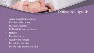
• Tight IT Band- it places excessive lateral force on the patella and can also
externally rotate the tibia, upsetting the balance of the PF mechanism.
• Tight Hamstring- It places more posterior force on the knee, causing pressure
between the patella and the femur to increase.
• Tight calf muscle- It can lead to compensatory foot pronation and can
increase the posterior force on the knee.
• Hip stability and hip abductor strength
– Recent research has shown that functional malalignment does not arise in
the knee joint but rather by internal rotation of the femur due to weakness
of hip external rotators and abductors (M. gluteus medius and minimus)
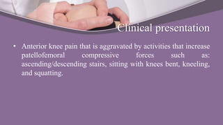
• Increase Q angle
– Due to increase Q angle knee goes to valgus position. At this time femur
and tibia rotate internally.
– Internal rotation of the femur might be the result of weakness of hip
abductors, internal rotation of tibia might arise from rear foot eversion or
pes-pronatus.
– Functional valgus may lead to lateral patellar maltracking.
Clinical presentation
• Anterior knee pain that is aggravated by activities that increase
patellofemoral compressive forces such as:
ascending/descending stairs, sitting with knees bent, kneeling,
and squatting.
Difference diagnosis
• Acute patellar dislocation
• Patellar subluxation
• Patellar tendinitis
• IT Band friction syndrome
• Bursitis
• Patellar fracture
• Quadriceps rupture
• Prepatellar bursitis
• Patella baja and Patella alta
Examination
• Subjective examination:
– History
– Behavior of symptoms
– Contributing factors
• Insidious onset- PFPS
• Overload event (excessive stairs climbing)-PFPS
• Traumatic events
– Pattern of Pain
• Pain only during activity-Think biomechanics
• Pain only after activity-Think inflammatory
• Pain that improve with exercise- Think about muscles
• Physical examination:
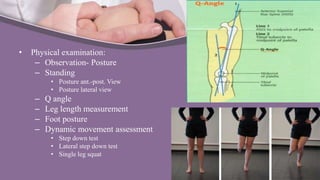
– Observation- Posture
– Standing
• Posture ant.-post. View
• Posture lateral view
– Q angle
– Leg length measurement
– Foot posture
– Dynamic movement assessment
• Step down test
• Lateral step down test
• Single leg squat
– Gait
– Hypermobility
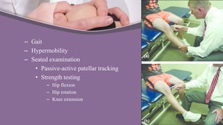
– Seated examination
• Passive-active patellar tracking
• Strength testing
– Hip flexion
– Hip rotation
– Knee extension
• Supine examination
– Palpation
– Flexibility test
– ROM of knee
– Special test
• Screening for knee lig.
• Screening for meniscus
• Neurological exam
– Reflexes
– Balance
• Side lying examination
– Flexibility test- IT Band
– Strength test
• Hip abd.-add.
• Prone examination
– Flexibility test - Quadriceps
– Strength test- Hip extensors, Knee flexor
Rehabilitation
Surgical versus conservative treatment:
Kettunen et. al. have demonstrated in a prospective randomized study with
PFPS patients that an arthroscopy, in combination with physiotherapy, had no
positive effect compared to physiotherapy alone. Therefore, the treatment of
PFPS is primarily non-operative.
• Pharmacological treatment
– NSAID’s
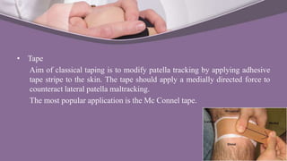
• Tape
Aim of classical taping is to modify patella tracking by applying adhesive
tape stripe to the skin. The tape should apply a medially directed force to
counteract lateral patella maltracking.
The most popular application is the Mc Connel tape.
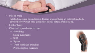
• Patella brace
Patella braces are non adhesive devices also applying an external medially
directed force which may counteract lateral patella maltracking.
• Foot orthosis
• Close and open chain exercises
– Stretching
– Static quadriceps
– SLR
– Squats
– Trunk stabilizer exercises
– Proprioceptive exercises
Patellofemoral Pain Syndrome