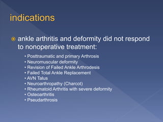
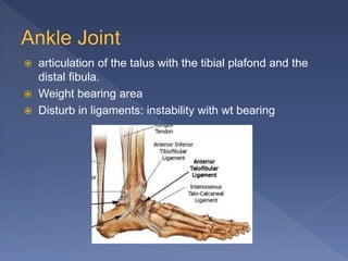
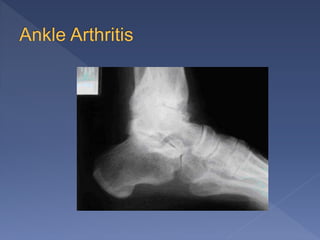
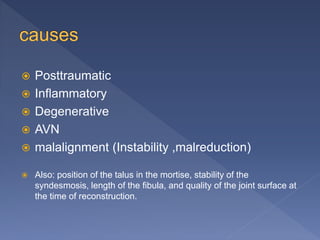
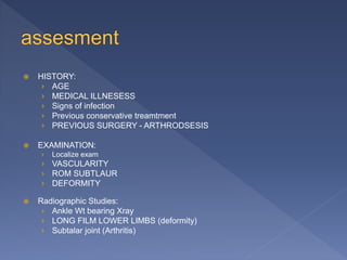
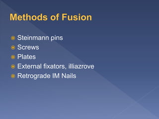
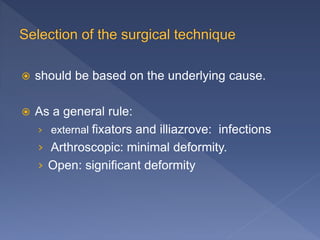
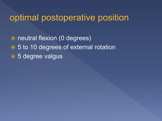
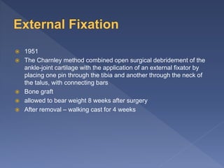
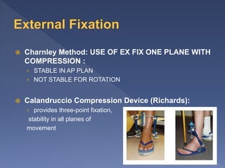
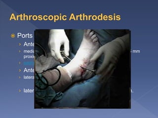
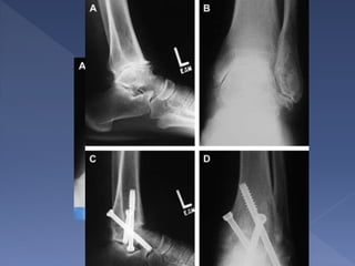
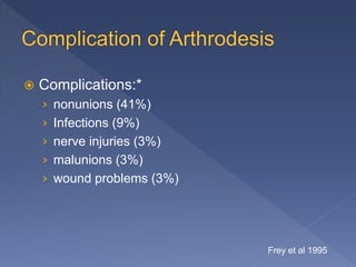
2012@
 articulation of the talus with the tibial plafond and the
distal fibula.
 Weight bearing area
 Disturb in ligaments: instability with wt bearing
 Posttraumatic
 Inflammatory
 Degenerative
 AVN
 malalignment (Instability ,malreduction)
 Also: position of the talus in the mortise, stability of the
syndesmosis, length of the fibula, and quality of the joint surface at
the time of reconstruction.
 Mechanical Pain
 ↓ROM
 Limitation of activity
 Limping
 P/E:
 Deformity, swelling, tenderness, ROM,
crepitation
 Xary
 CT
 MRI
 shoe inserts: cushioned heel and a stiff, rockerbottom sole
 ankle-foot orthosis
 walking cast for 6 weeks
 NSAID
 Wt reduction
 Activity modification
 Intra-articular injections
 Arthroscopic ankle-joint debridement
Ankle Arthrodesis
 Ankle Replacement
 ankle arthritis and deformity did not respond
to nonoperative treatment:
• Posttraumatic and primary Arthrosis
• Neuromuscular deformity
• Revision of Failed Ankle Arthrodesis
• Failed Total Ankle Replacement
• AVN Talus
• Neuroarthropathy (Charcot)
• Rheumatoid Arthritis with severe deformity
• Osteoarthritis
• Pseudarthrosis
 HISTORY:
› AGE
› MEDICAL ILLNESESS
› Signs of infection
› Previous conservative treamtment
› PREVIOUS SURGERY - ARTHRODSESIS
 EXAMINATION:
› Localize exam
› VASCULARITY
› ROM SUBTLAUR
› DEFORMITY
 Radiographic Studies:
› Ankle Wt bearing Xray
› LONG FILM LOWER LIMBS (deformity)
› Subtalar joint (Arthritis)
 Steinmann pins
 Screws
 Plates
 External fixators, illiazrove
 Retrograde IM Nails
 should be based on the underlying cause.
 As a general rule:
› external fixators and illiazrove: infections
› Arthroscopic: minimal deformity.
› Open: significant deformity
 neutral flexion (0 degrees)
 5 to 10 degrees of external rotation
 5 degree valgus
 1951
 The Charnley method combined open surgical debridement of the
ankle-joint cartilage with the application of an external fixator by
placing one pin through the tibia and another through the neck of
the talus, with connecting bars
 Bone graft
 allowed to bear weight 8 weeks after surgery
 After removal – walking cast for 4 weeks
 Charnley Method: USE OF EX FIX ONE PLANE WITH
COMPRESSION :
› STABLE IN AP PLAN
› NOT STABLE FOR ROTATION
 Calandruccio Compression Device (Richards):
› provides three-point fixation,
stability in all planes of
movement
 Proper in case of active infection.
 20 years old male
 Fracture Rt Ankel
 ORIF done
 C/o pain , with activity , even at rest
Jan 2012 Bone scan:
Hyperemia with increased uptake
suspicion for infection
Ct scan:
distal syndesmotic screw loosening,
associated with irregularities at the
ankle joint with surrounding fluid collection.
Feb 2012
Removal at end of August 2012
On last visit December 2012
 Ports 2 or 3
› Anteromedial:
› medial to the tibialis nterior tendon, and located about 5 mm
proximal to the medial malleolus
› saphenous nerve and vein
› Anterolateral:
› lateral to peroneus tertius tendon
› lateral to the extensor digitorum longus tendon.
 Distraction:
› 25 IB will give 1 to 1.5 mm of distraction
› 30 ib more than 1 hr , reversable nerve injury
 A 4.5-mm bur and curettes -remove articular surfaces
 Compression: Interanal or external
 Prevered: 2 cannulated screw
› One medial mallulus to lateral talus
› One lateral tibia to neck of talus
 NWB 5 weeks
 Progress gradual till radiological healing
 Advantage:
› faster rates of union, decreased complications,
reduced postoperative pain, shorter hospital
stays, minimal loss of length of the lower limb.
 Disadvantage:
› Only for minimal deformity, because it is difficult to
correct ankle deformity arthroscopically.
 with severe ankle-joint deformity
 better visualization
 two-incision
› Direct over fibula
› Along anterior third of medial malluls
 maintain full-thickness flaps and protect
tendons and N,V
 Lateral: osteotomy 10 cm from the fibular tip,
The remaining fibular fragment can then be
excised.
 preserve the medial malleolus so as to
provide an area of solid fixation for the screw
and to preserve the medial blood supply to
the talus
 Cut of talus 5mm paralel to tibial cut
 drilled or curetted until bleeding bone
 two guide pins for large (7.0- to 7.3-mm)
cannulated screws
 Care must be taken that the pins do not violate
the subtalar joint
 short threaded cannulated screws with washers
 bulky cast padding 2 weeks
 NWB short leg cast
 Start WTB after radiological evidence of healing
 usually occurs 8 to 12 weeks postoperatively
 (TTCA)
 Minimal invasive
 Better mechanical Proparties.
 Some types allow compression
 The starting point is of the most importance
Indication:
AVN talus
Failed Total Ankle Arthroplasty
Pseudoarthrosis
Neuropathic arthropathy
Contraindications:
 Infection
 Sever vascular disease
 Sever tibial malalignment
 Normal subtalar joint (relative)
 Plantur pad inssuficency
 Entry point:
› 3 cm longitudinal plantur incision
› Anterior to subcalcaneak fat pad
› Lateral to mid line
 Nail length:
› Nail end should extend at least 1 ½ to 2 tibial
diameter above any potential stress risers
 NWB for 6 weeks
 12 weeks in case of nuropathy or use of
bonegraft
Complications
 nonunion
 fracture at or above the fixation construct
 Nerve and artery damage
 Complications:*
› nonunions (41%)
› Infections (9%)
› nerve injuries (3%)
› malunions (3%)
› wound problems (3%)
Frey et al 1995
 Risk factors associated with nonunion:
› severe fracture, an open injury, local infection,
evidence of osteonecrosis of the talus, and
coexisting major medical problems
› past history of undergoing a subtalar or triple
arthrodesis
› Smoking risk increase 16 times
Ankle arthodesis

Ankle arthodesis

  • 1.
  • 2.
     articulation ofthe talus with the tibial plafond and the distal fibula.  Weight bearing area  Disturb in ligaments: instability with wt bearing
  • 4.
     Posttraumatic  Inflammatory Degenerative  AVN  malalignment (Instability ,malreduction)  Also: position of the talus in the mortise, stability of the syndesmosis, length of the fibula, and quality of the joint surface at the time of reconstruction.
  • 5.
     Mechanical Pain ↓ROM  Limitation of activity  Limping  P/E:  Deformity, swelling, tenderness, ROM, crepitation
  • 6.
  • 7.
     shoe inserts:cushioned heel and a stiff, rockerbottom sole  ankle-foot orthosis  walking cast for 6 weeks  NSAID  Wt reduction  Activity modification  Intra-articular injections
  • 8.
     Arthroscopic ankle-jointdebridement Ankle Arthrodesis  Ankle Replacement
  • 10.
     ankle arthritisand deformity did not respond to nonoperative treatment: • Posttraumatic and primary Arthrosis • Neuromuscular deformity • Revision of Failed Ankle Arthrodesis • Failed Total Ankle Replacement • AVN Talus • Neuroarthropathy (Charcot) • Rheumatoid Arthritis with severe deformity • Osteoarthritis • Pseudarthrosis
  • 11.
     HISTORY: › AGE ›MEDICAL ILLNESESS › Signs of infection › Previous conservative treamtment › PREVIOUS SURGERY - ARTHRODSESIS  EXAMINATION: › Localize exam › VASCULARITY › ROM SUBTLAUR › DEFORMITY  Radiographic Studies: › Ankle Wt bearing Xray › LONG FILM LOWER LIMBS (deformity) › Subtalar joint (Arthritis)
  • 12.
     Steinmann pins Screws  Plates  External fixators, illiazrove  Retrograde IM Nails
  • 13.
     should bebased on the underlying cause.  As a general rule: › external fixators and illiazrove: infections › Arthroscopic: minimal deformity. › Open: significant deformity
  • 14.
     neutral flexion(0 degrees)  5 to 10 degrees of external rotation  5 degree valgus
  • 15.
     1951  TheCharnley method combined open surgical debridement of the ankle-joint cartilage with the application of an external fixator by placing one pin through the tibia and another through the neck of the talus, with connecting bars  Bone graft  allowed to bear weight 8 weeks after surgery  After removal – walking cast for 4 weeks
  • 16.
     Charnley Method:USE OF EX FIX ONE PLANE WITH COMPRESSION : › STABLE IN AP PLAN › NOT STABLE FOR ROTATION  Calandruccio Compression Device (Richards): › provides three-point fixation, stability in all planes of movement
  • 18.
     Proper incase of active infection.
  • 19.
     20 yearsold male  Fracture Rt Ankel  ORIF done  C/o pain , with activity , even at rest
  • 20.
    Jan 2012 Bonescan: Hyperemia with increased uptake suspicion for infection Ct scan: distal syndesmotic screw loosening, associated with irregularities at the ankle joint with surrounding fluid collection.
  • 21.
  • 22.
    Removal at endof August 2012
  • 24.
    On last visitDecember 2012
  • 26.
     Ports 2or 3 › Anteromedial: › medial to the tibialis nterior tendon, and located about 5 mm proximal to the medial malleolus › saphenous nerve and vein › Anterolateral: › lateral to peroneus tertius tendon › lateral to the extensor digitorum longus tendon.
  • 27.
     Distraction: › 25IB will give 1 to 1.5 mm of distraction › 30 ib more than 1 hr , reversable nerve injury  A 4.5-mm bur and curettes -remove articular surfaces  Compression: Interanal or external  Prevered: 2 cannulated screw › One medial mallulus to lateral talus › One lateral tibia to neck of talus  NWB 5 weeks  Progress gradual till radiological healing
  • 29.
     Advantage: › fasterrates of union, decreased complications, reduced postoperative pain, shorter hospital stays, minimal loss of length of the lower limb.  Disadvantage: › Only for minimal deformity, because it is difficult to correct ankle deformity arthroscopically.
  • 30.
     with severeankle-joint deformity  better visualization  two-incision › Direct over fibula › Along anterior third of medial malluls  maintain full-thickness flaps and protect tendons and N,V
  • 31.
     Lateral: osteotomy10 cm from the fibular tip, The remaining fibular fragment can then be excised.  preserve the medial malleolus so as to provide an area of solid fixation for the screw and to preserve the medial blood supply to the talus
  • 32.
     Cut oftalus 5mm paralel to tibial cut  drilled or curetted until bleeding bone
  • 34.
     two guidepins for large (7.0- to 7.3-mm) cannulated screws  Care must be taken that the pins do not violate the subtalar joint  short threaded cannulated screws with washers  bulky cast padding 2 weeks  NWB short leg cast  Start WTB after radiological evidence of healing  usually occurs 8 to 12 weeks postoperatively
  • 35.
     (TTCA)  Minimalinvasive  Better mechanical Proparties.  Some types allow compression  The starting point is of the most importance
  • 37.
    Indication: AVN talus Failed TotalAnkle Arthroplasty Pseudoarthrosis Neuropathic arthropathy Contraindications:  Infection  Sever vascular disease  Sever tibial malalignment  Normal subtalar joint (relative)  Plantur pad inssuficency
  • 38.
     Entry point: ›3 cm longitudinal plantur incision › Anterior to subcalcaneak fat pad › Lateral to mid line
  • 39.
     Nail length: ›Nail end should extend at least 1 ½ to 2 tibial diameter above any potential stress risers
  • 40.
     NWB for6 weeks  12 weeks in case of nuropathy or use of bonegraft
  • 41.
    Complications  nonunion  fractureat or above the fixation construct  Nerve and artery damage
  • 42.
     Complications:* › nonunions(41%) › Infections (9%) › nerve injuries (3%) › malunions (3%) › wound problems (3%) Frey et al 1995
  • 43.
     Risk factorsassociated with nonunion: › severe fracture, an open injury, local infection, evidence of osteonecrosis of the talus, and coexisting major medical problems › past history of undergoing a subtalar or triple arthrodesis › Smoking risk increase 16 times

Editor's Notes

  • #5 Damage to the ankle joint from trauma or disease can result in progressive loss of the tibiotalar articular cartilage surface, with resulting inflammation, synovitis, osteophyte formation, progressive loss of anklejoint motion, weight-bearing pain, and functional disability.
  • #31 After the distal 10 cm of the fibula has been exposed, the superior peroneal retinaculum is incised posteriorly, and the peroneal tendons are mobilized while protecting the sural and superficial peroneal nerves.
  • #34 1- 2- take care to subcutanous nerves 3- expose distal fibula 4- cutting 5-expose ankle 6 tibial cut 7 A medial exposure medial to the bed of Tibialis anterior is required in every case.  8 cut talus parallel
  • #35 The first pin is started at the posterolateral corner of the tibia and is placed across the joint and into the neck of the talus. The second guide pin is placed from the medial malleolus into the lateral aspect of the talus. Alternatively, the second pin may be placed from the lateral process of the talus into the medial cortex of the tibia
  • #36 Tibio talo calcaneal Arthrodesis
  • #43 Frey et al 1995 - Malaligned Fusion: - genu recurvatum, or backknee, will result from a plantar-flexed foot; - heel lift may compensate for moderate plantar flexion (5-10 deg), but significant problems arise with excessive plantar flexion;    - to avoid vaulting over plantar-flexed foot, patient must turn leg out, & secondary medial collateral laxity can occur;    - if foot is translated medially or laterally, other subtalar and gait problems can occur; - when subtalar joint is inverted or in varus position, transverse tarsal    joint is locked, making rigid foot that needs to be vaulted over;