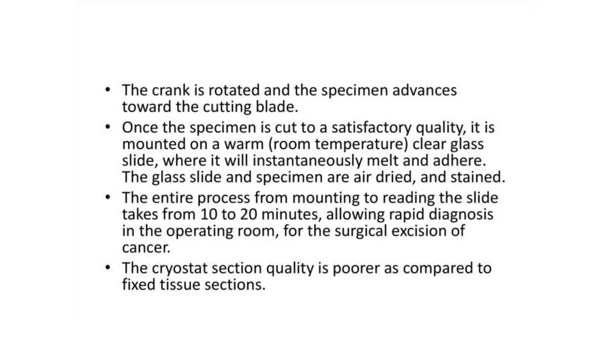
1. To avoid drying, the tissue should be kept in OCT compound or freezing medium.
2. Tissues can be fixed with formalin.
3. Paraffin or celloidin is used as embedding media
4. Carbon dioxide gas is most commonly used with freezing microtome.
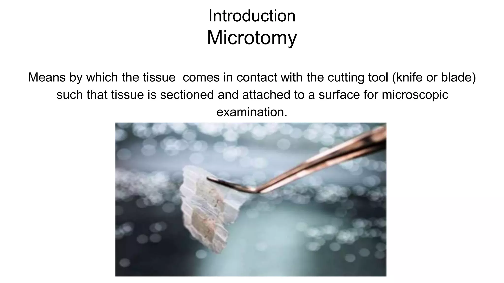
Introduction
Microtomy
Means by whichthe tissue comes in contact with the cutting tool (knife or blade)
such that tissue is sectioned and attached to a surface for microscopic
examination.
Types of Microtome
Thereare several types of microtome, each designed for a specific purpose,
although many have multifunctional roles.
6.
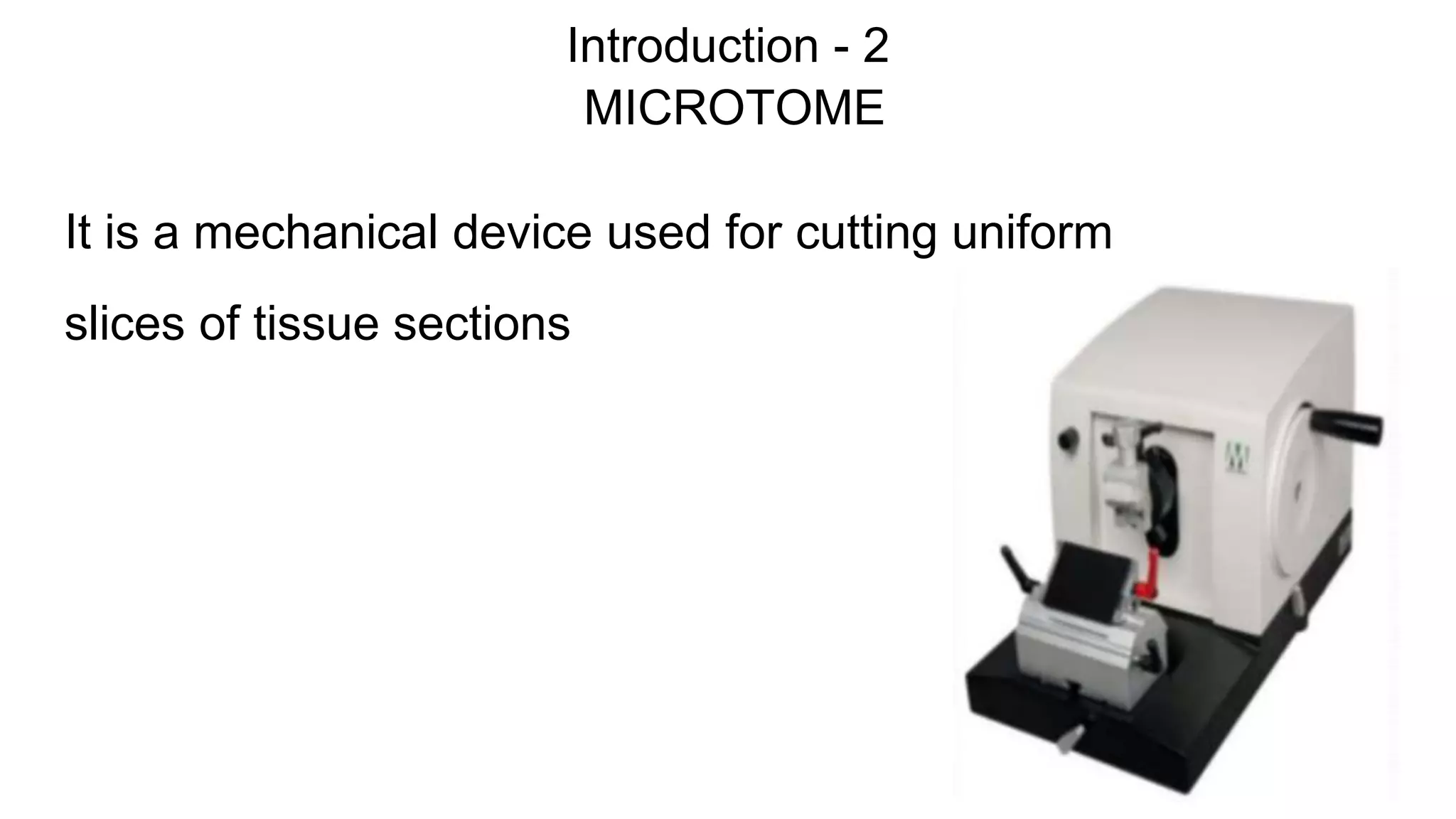
Rotary microtome
Rotation ofa fine advancing hand-wheel by 360° degrees, moving the specimen
vertically past the cutting surface and returning it to the starting position.
Most common type
7.
Types of rotarymicrotome
Manual rotary microtome
Completely manipulated by the
operator
8.
Types of rotarymicrotome
Semi automated rotary microtome
One motor to advance either fine or course hand wheel
9.
Types of rotarymicrotome
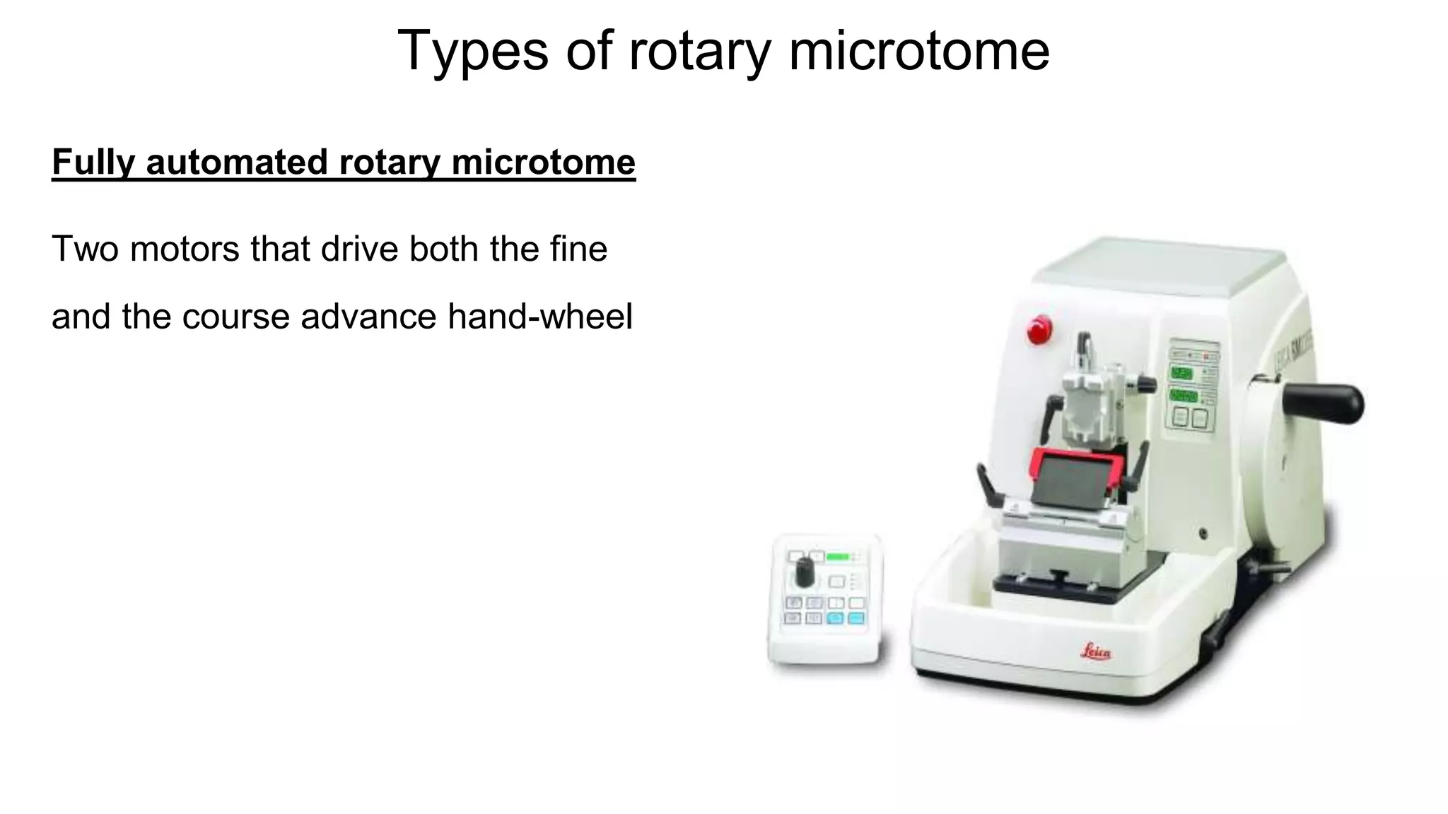
Fully automated rotary microtome
Two motors that drive both the fine
and the course advance hand-wheel
10.
Types of Microtome
Basesledge microtome
- Specimen is stationary
- Knife slides across the top of the
specimen during sectioning
- Used for large blocks, hard
tissues, or whole mounts,
- Neuro and ophthalmic pathology.
11.
Types of Microtome
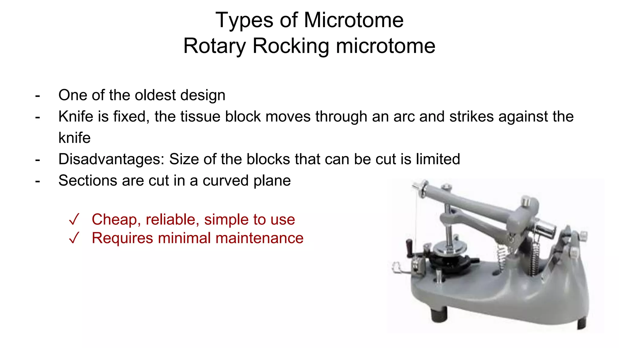
RotaryRocking microtome
- One of the oldest design
- Knife is fixed, the tissue block moves through an arc and strikes against the
knife
- Disadvantages: Size of the blocks that can be cut is limited
- Sections are cut in a curved plane
✓ Cheap, reliable, simple to use
✓ Requires minimal maintenance
12.
Types of Microtome
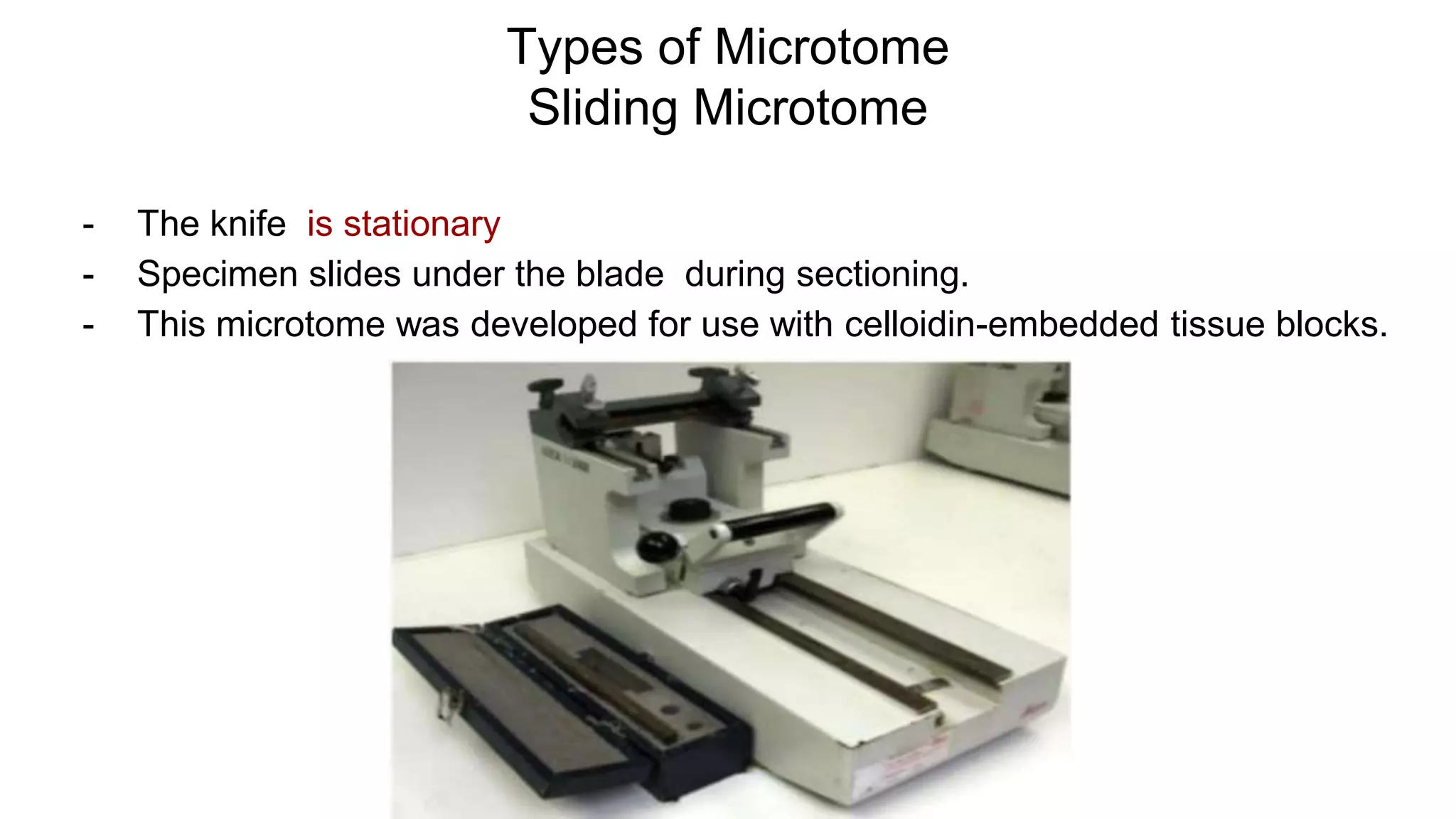
SlidingMicrotome
- The knife is stationary
- Specimen slides under the blade during sectioning.
- This microtome was developed for use with celloidin-embedded tissue blocks.
13.
Types of Microtome
UltraMicrotome
- Exclusively for electron microscopy
- Ultrathin sections upto 10nm
- Glass/ diamond/sapphire knives
- Block is brought close to the knife
edge under a microscope
- As each section is cut, it is floated
on a water bath adjacent to the
knife
14.
Types of Microtome
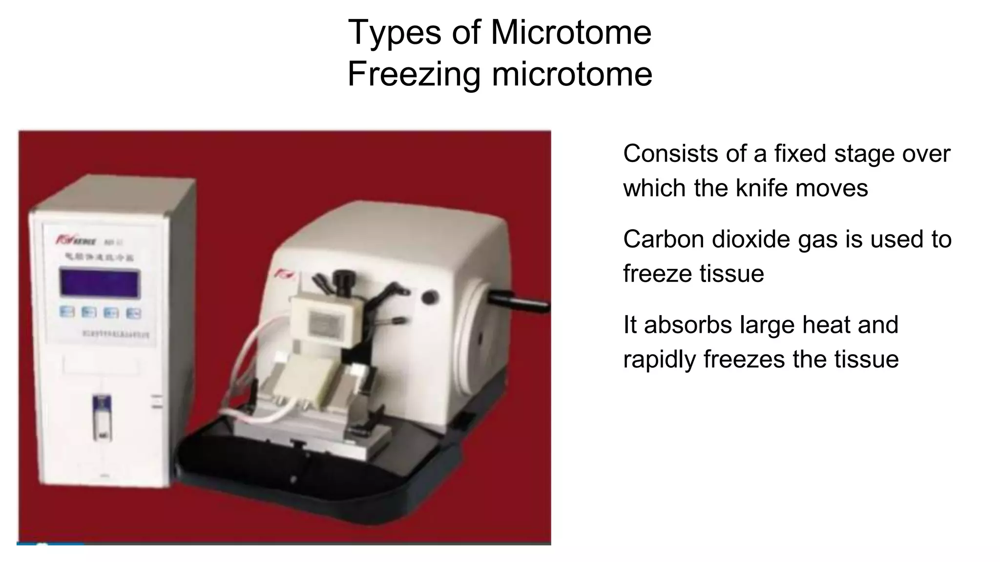
Freezingmicrotome
Consists of a fixed stage over
which the knife moves
Carbon dioxide gas is used to
freeze tissue
It absorbs large heat and
rapidly freezes the tissue
15.
Types of Microtome
Vibratingmicrotome
- High speed vibration is used
- Designed to cut tissues which have
not been fixed or frozen
- Sections are thicker
- Tissue is immersed in water
- Enzyme histochemistry and
ultrastructure histochemistry
16.
Types of Microtome
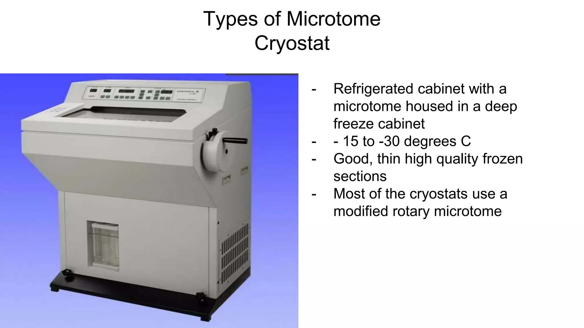
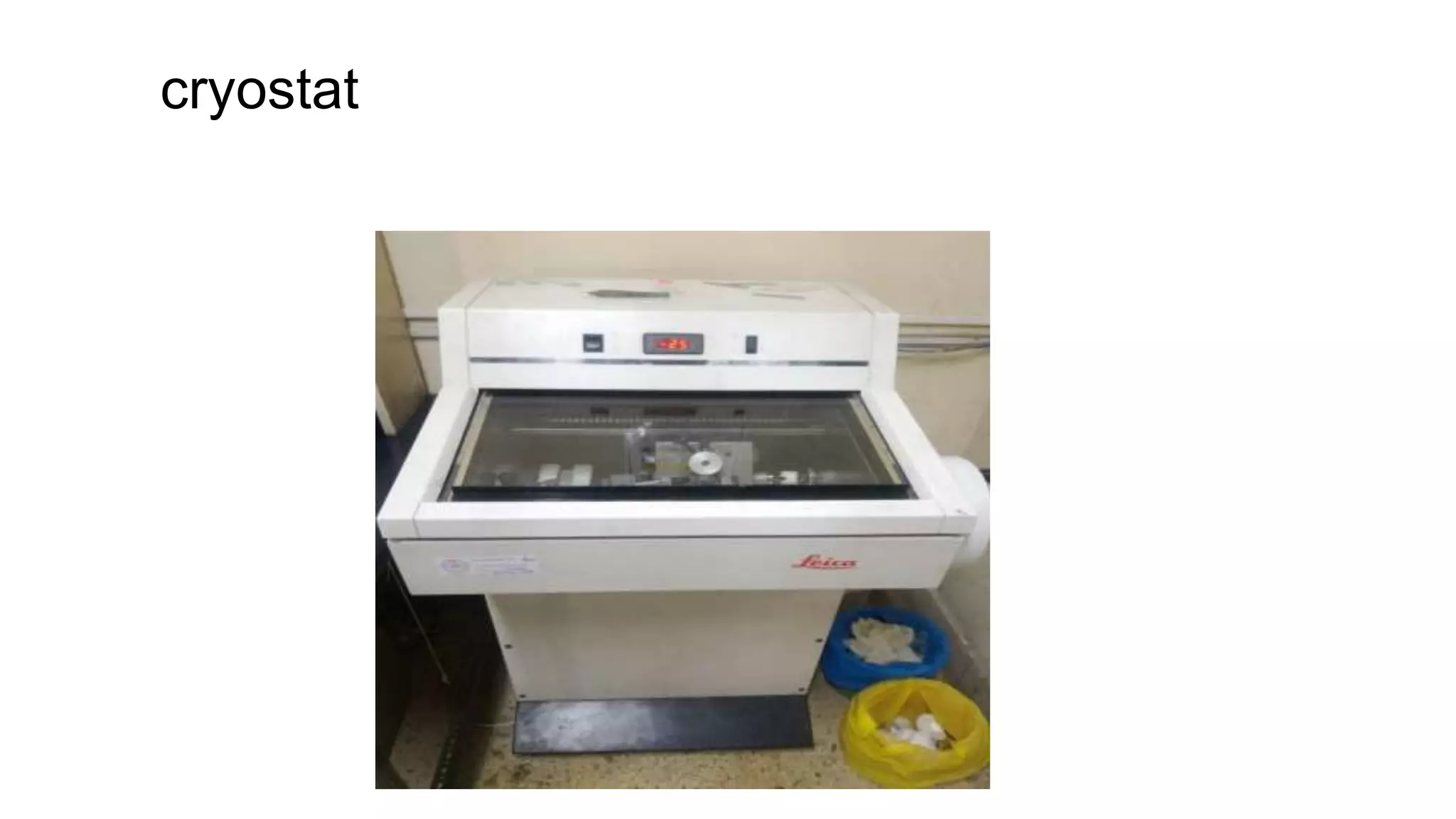
Cryostat
-Refrigerated cabinet with a
microtome housed in a deep
freeze cabinet
- - 15 to -30 degrees C
- Good, thin high quality frozen
sections
- Most of the cryostats use a
modified rotary microtome
17.
PRINCIPLE of FROZENSECTION
● When a tissue is frozen, the water within the tissue turns to ice and in
this state the tissue is firm, with the ice acting as the embedding
medium.
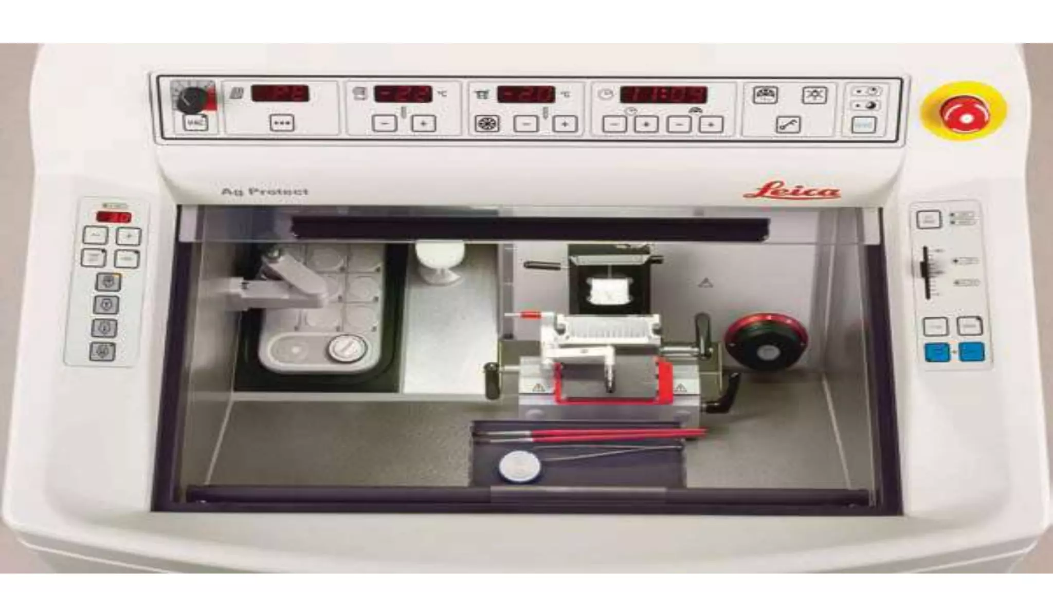
● Access tothe chamber is via a sliding window.
● Working temperature : 0 to -35 degree Celsius
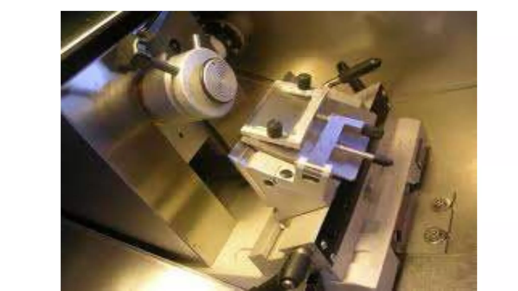
● Rotary microtome controlled by an external hand wheel.
● Freezing shelf
● knife holder
ANTI ROLL PLATE
❖Prevent rolling or curling of tissue
❖ Glass plate supported on an aluminium frame
❖ Provides gap between underside of glass and upper surface of knife.
TEMPERATURE SETTINGS
● Digitaldisplay
● For most tissues: -15 to -23 degree Celsius is used
● For tissues with more fat: Colder temperature
● Temperature log maintained
● Defrosting done daily
● Rapid freezing to be done to reduce freeze artefacts
1. To avoiddrying, the tissue should be kept in ......................
2. Tissues can be fixed with ......................
3. ...................... or ...................... is used as embedding media
4. ...................... gas is most commonly used with freezing microtome