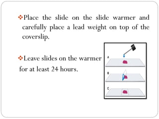
HISTOLOGY TECHNIQUES
PERMANENT MOUNTING
BY
SYEDA SADAFWAJAHAT
introduction
❖Histology also known as “Microanatomy” is
the study of cells, tissues and organs under
the microscope (Light microscope or
Electron microscope).
❖It is related to Cell biology (Cytology),
Anatomy, Physiology and Pathology.
Types of tissueS
Types of tissueS
SOURCES OF SPECIMEN
❖Specimens received for histological
examination may come from a number of
different sources.
1. Excision specimens (surgical biopsies),
where whole organs or affected areas are
removed at operation.
2. Incisional biopsy specimens, where tissue is
removed for diagnosis from within an affected
area.
3. Punch biopsies, where punches are used to
remove a small piece of suspicious tissue for
examination (often from the skin).
4. Shave biopsies, where small fragments of
tissue are “shaved” from a surface (usually skin)
5. Curetting, where tissue is removed in small
pieces from the lining of the uterus or cervix .
6. Core biopsies, where a small tissue sample is
removed using a special needle sometimes
through the skin (percutaneously).
PERMANENT MOUNTING
❖In histology or a pathology, mounting is the
last procedure in the series that ends with a
permanent histological preparation on the
table, well after the tissue processing and
staining.
PROCEDURE
PROPERTIES OF AN IDEAL
MOUNTING MEDIA
❖Refractive index (RI) should be as close as
possible to that of glass, i.e., 1.5.
❖ It should be colorless and transparent.
❖It should be dry to a non-stick consistency
and harden relatively quickly.
❖It should be able to completely permeate
and fill tissue interstices.
❖It should have no adverse effect on tissue
components.
❖It should be resistant to contamination.
CLASSIFICATION OF
MOUNTING MEDIA
❖There are two types of mounting medium.
1. Resinous media (hydrophobic/adhesive).
2. Aqueous media (hydrophilic/non-adhesive).
1. Resinous MEDIA
(hydrophobic/adhesive)
❖These are natural or synthetic resins
dissolved in benzene, toluene or xylene.
❖Used when a permanent mount is required.
❖Adhesives harden through solvent
evaporation and thereby fix the
accompanying coverslip to the slide.
❖Resinous mountants are the type most
frequently used.
❖It is further divided into
1. Natural resinous media like Candida
Balsam, Phenol Balsam and Dammar
Balsam.
2. Synthetic resinous media like DPX
(dibutyl phthalate and xylene), Permount
and Histamount.
2.AQUEOUS MEDIA
(HYDROPHILIC/NON-
ADHESIVE)
❖Aqueous media are used for mounting
sections from distilled water when the stains
would be removed by alcohol and xylene.
❖These media are of three types: The syrups,
Gelatin media and Gum Arabic media.
❖Aqueous mounting media require the
addition of bacteriostatic agents such as
phenol or crystal of thymol to prevent the
growth of fungi.
❖Examples are Water, Glycerine jelly,
Glycerine-Glycerol and Polyvinyl alcohol.
❖There are many commercial mounting media
which are available for mounting sections from
water.
PREPARING PERMANENTLY
MOUNTED SECTIONS
❖Place 2-3 drops of resin (mounting media)
over the section.
❖To avoid entrapping air bubbles, lower the
cover slip slowly from one side of the droplet.
❖Place the slide on the slide warmer and
carefully place a lead weight on top of the
coverslip.
❖Leave slides on the warmer
for at least 24 hours.
❖Observe the tissue section under the
microscope.
CONCLUSION
❖To conclude various commercial
manufacturers have various mounting
media. You should select a mounting media
that suits the viewing and preserving the
required sections for further research.
Histology

Histology

  • 1.
  • 2.
    introduction ❖Histology also knownas “Microanatomy” is the study of cells, tissues and organs under the microscope (Light microscope or Electron microscope). ❖It is related to Cell biology (Cytology), Anatomy, Physiology and Pathology.
  • 4.
  • 5.
  • 6.
    SOURCES OF SPECIMEN ❖Specimensreceived for histological examination may come from a number of different sources. 1. Excision specimens (surgical biopsies), where whole organs or affected areas are removed at operation.
  • 7.
    2. Incisional biopsyspecimens, where tissue is removed for diagnosis from within an affected area. 3. Punch biopsies, where punches are used to remove a small piece of suspicious tissue for examination (often from the skin).
  • 8.
    4. Shave biopsies,where small fragments of tissue are “shaved” from a surface (usually skin) 5. Curetting, where tissue is removed in small pieces from the lining of the uterus or cervix . 6. Core biopsies, where a small tissue sample is removed using a special needle sometimes through the skin (percutaneously).
  • 9.
    PERMANENT MOUNTING ❖In histologyor a pathology, mounting is the last procedure in the series that ends with a permanent histological preparation on the table, well after the tissue processing and staining.
  • 10.
  • 12.
    PROPERTIES OF ANIDEAL MOUNTING MEDIA ❖Refractive index (RI) should be as close as possible to that of glass, i.e., 1.5. ❖ It should be colorless and transparent. ❖It should be dry to a non-stick consistency and harden relatively quickly.
  • 13.
    ❖It should beable to completely permeate and fill tissue interstices. ❖It should have no adverse effect on tissue components. ❖It should be resistant to contamination.
  • 14.
    CLASSIFICATION OF MOUNTING MEDIA ❖Thereare two types of mounting medium. 1. Resinous media (hydrophobic/adhesive). 2. Aqueous media (hydrophilic/non-adhesive).
  • 15.
    1. Resinous MEDIA (hydrophobic/adhesive) ❖Theseare natural or synthetic resins dissolved in benzene, toluene or xylene. ❖Used when a permanent mount is required.
  • 16.
    ❖Adhesives harden throughsolvent evaporation and thereby fix the accompanying coverslip to the slide. ❖Resinous mountants are the type most frequently used. ❖It is further divided into
  • 17.
    1. Natural resinousmedia like Candida Balsam, Phenol Balsam and Dammar Balsam. 2. Synthetic resinous media like DPX (dibutyl phthalate and xylene), Permount and Histamount.
  • 19.
    2.AQUEOUS MEDIA (HYDROPHILIC/NON- ADHESIVE) ❖Aqueous mediaare used for mounting sections from distilled water when the stains would be removed by alcohol and xylene. ❖These media are of three types: The syrups, Gelatin media and Gum Arabic media.
  • 20.
    ❖Aqueous mounting mediarequire the addition of bacteriostatic agents such as phenol or crystal of thymol to prevent the growth of fungi. ❖Examples are Water, Glycerine jelly, Glycerine-Glycerol and Polyvinyl alcohol.
  • 21.
    ❖There are manycommercial mounting media which are available for mounting sections from water.
  • 22.
    PREPARING PERMANENTLY MOUNTED SECTIONS ❖Place2-3 drops of resin (mounting media) over the section. ❖To avoid entrapping air bubbles, lower the cover slip slowly from one side of the droplet.
  • 23.
    ❖Place the slideon the slide warmer and carefully place a lead weight on top of the coverslip. ❖Leave slides on the warmer for at least 24 hours.
  • 24.
    ❖Observe the tissuesection under the microscope.
  • 25.
    CONCLUSION ❖To conclude variouscommercial manufacturers have various mounting media. You should select a mounting media that suits the viewing and preserving the required sections for further research.