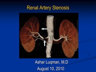
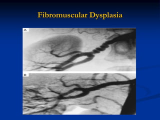
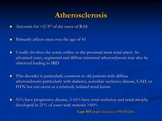
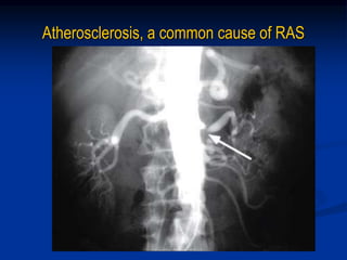
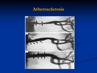
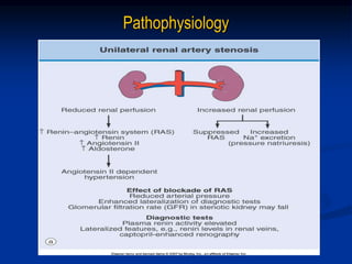
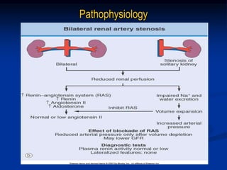
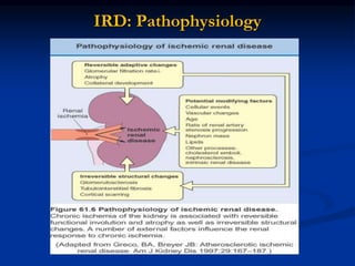
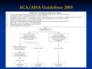
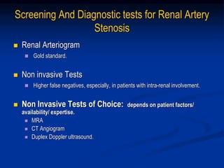
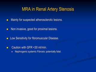
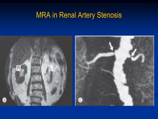
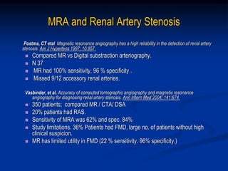
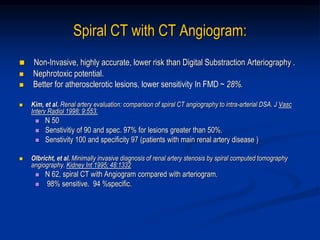
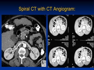
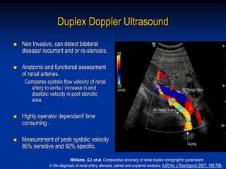
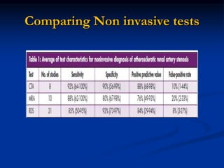
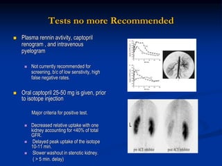
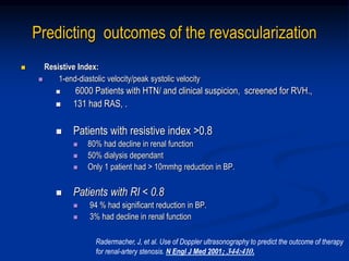
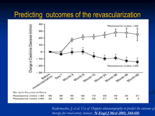
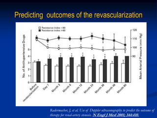
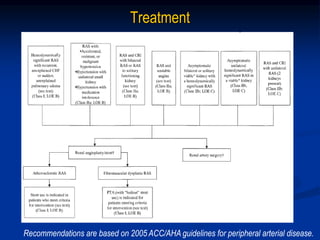
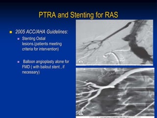
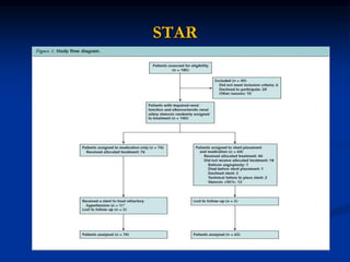
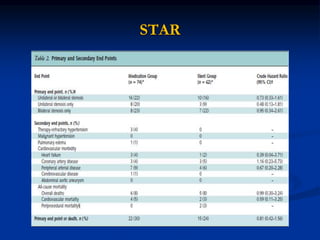
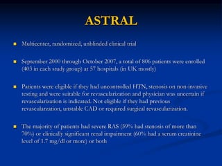
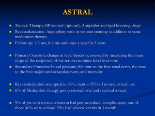
Renal Artery Stenosis
Ashar Luqman, M.D
August 10, 2010
Definition
 Renal artery stenosis is the narrowing of the renal artery, most often caused
by atherosclerosis or fibromuscular dysplasia. This narrowing of the renal
artery can impede blood flow to the target kidney. Hypertension and atrophy
of the affected kidney may result from renal artery stenosis, ultimately leading
to renal failure if not treated
 Pressure gradient of 20 mmHg across stenotic area during angiography is
usually considered to be of hemodynamic significance
Epidemiology
Common Disease: Incidence
General Population 0.1%
HTN 4.0%
HTN and suspected CAD 10-20%
Malignant HTN 20-30%
Malignant HTN and Renal Insufficiency 30-40%
Epidemiology
 Ultrasound screening in patients (mean age of 77): 6.8%
 Male : female 2:1
 No racial difference
(Hansen. J Vasc Surg. 2002;36:443–451)
 Autopsies in 221 patients > 50 years: 27%
 H/o diastolic HTN: 53%
(Holley. Am J Med. 1964;37:14 –22)
 Diagnostic cath for suspected CAD: up to 20%
 Third most common reason of ESRD (after DM and HTN), seen in 10-20%
of ESRD patients > 50 yrs old
(Rihal. Mayo Clin Proc. 2002;77:309 –316)
Causes
 Common causes
 Atherosclerosis
 Fibromuscular dysplasia
 Less Common causes
 Vasculitis (Takayasu’s arteritis)
 Dissection of the renal artery.
 Thromboembolic disease
 Renal artery aneurysm
 Renal artery coarctation
 Extrinsic compression
 Radiation injury
Fibromuscular Dysplasia
 Accounts for 10% of all cases of RAS. 90% cases involve media
 Affect females, age 15-50
 Rarely leads to renal artery occlusion
 Genetic predisposition, smoking, hormonal factors, and disorders of the vasa
vasorum are possible risk factors
 Angiography:
Site: distal renal artery
Beaded, aneurysmal appearance
 Treatment:
Ballon Angioplasty :
Successful in 82-100%
Restenosis in 10%
Cure HTN in 60%
Safian. NEJM. 2001;344: 341-442
Fibromuscular Dysplasia
Atherosclerosis
 Accounts for >2/3rd of the cases of RAS
 Primarily affects men over the age of 45
 Usually involves the aortic orifice or the proximal main renal artery. In
advanced cases, segmental and diffuse intrarenal atherosclerosis may also be
observed leading to IRD
 This disorder is particularly common in old patients with diffuse
atherosclerosis particularly with diabetes, aortoiliac occlusive disease, CAD, or
HTN but can occur as a relatively isolated renal lesion
 51% have progressive disease, 3-16% have total occlusion and renal atrophy
developed in 21% of cases with stenosis >60%
Caps MT et al Circulation 1998;98:2866
Atherosclerosis, a common cause of RAS
Atherosclerosis
Pathophysiology
Pathophysiology
Clinical Findings
 HTN:
 Difficult-to-control hypertension despite adequate medical
treatment
 Accelerated or malignant hypertension
 Severe hypertension (diastolic blood pressure >120 mm Hg) or
resistant hypertension
 Paradoxical worsening of hypertension with diuretic therapy
 Onset of hypertension occurring in patients younger than 30
years or older than 50 years
 Hypertension with an asymmetric kidney
Clinical Findings
 Renal abnormalities
 Unexplained azotemia (suggestive of atherosclerotic
renal-artery stenosis)
 Azotemia induced by treatment with an angiotensin-
converting–enzyme inhibitor
 Unilateral small kidney
 Unexplained hypokalemia
Clinical Findings
 Other findings:
 Abdominal bruit, flank bruit, or both
 Severe retinopathy
 Carotid, coronary, or peripheral vascular disease
 Unexplained congestive heart failure or acute
pulmonary edema
Renovascular HTN and Pulmonary Edema
 Recurrent Pumonary Edema :
 HTN induced increase in after-load.
 Diastolic dysfunction ( LVH )
 Sodium retention, with bilateral disease.
Pickering, TG, et al. Recurrent pulmonary oedema in hypertension due to bilateral renal artery stenosis:
Treatment by angioplasty or surgical revascularization. Lancet 1988; 2:551.
RVH and Hyponatremia
 Renovascular hypertension and hyponatremia.
 Retrospective study of 32 patients with renovascular
hypertension
 mean plasma concentration of sodium was 129.7 meq/L,
correlated inversely with plasma renin levels.
 ? sec. to dipsogenic effects of angiotensin II.
Agarwal, M,etal. Hyponatremic-hypertensive syndrome with renal ischemia:
An underrecognized disorder. Hypertension 1999; 33:1020
Protienuria with RVH
 Nephrotic range protienuria !.
 Regression of protienuria with correction of vascular lesion.
 Presence of protienuria, DOES NOT exlcude renovascular HTN.
Chen R etal, Reversible renin mediated massive protienuria
successfully treated by nephrectomy. J Urol 1995;153:133-134.
Clinical Syndromes in RAS
Renovascular HTN
 RVH: The most common correctable cause of secondary HTN.
 A syndrome of elevated blood pressure, and decreased renal
perfusion.
 Relatively Less common in blacks.
 Incidence
 ~ < 1% in patients with mild to moderate HTN.
 10-45% in patients with acute ( superimposed on essential ), severe, and or resistant
HTN.
Davis etal, Prevalence of renovascular hypertension in patients with
grade III or IV retinopathy. N Engl J Med 1979; 301:1273
RAS and Hypertension;
Possible Clinical Presentations
 True reno-vascular HTN.
 Essential HTN with presence of RAS, but not contributing to
hypertension.
 Essential HTN with superimposed HTN secondary to RAS.
 RAS with presence of renal parenchymal disease. (Ischemic CKD)
Ischemic Renal Disease
 Critical reduction in renal blood flow to functioning renal parenchyma leading
to decreased GFR, which overtime causes progressive renal injury and
atrophyand thus, permanent loss of renal structural and functional integrity
 Ischemic renal disease has three anatomic variations:
1. Unilateral stenosis and occlusion in a single kidney
2. Bilateral critical renal artery stenosis or occlusion
3. Unilateral stenosis or occluison with contralateral atrophy/nonfunction
IRD Causing ESRD: 11-22%
Ischemic Renal Disease
IRD: Pathophysiology
Who should we screen?
Why we need to screen?
ACA/AHA Guidelines 2005
Screening And Diagnostic tests for Renal Artery
Stenosis
 Renal Arteriogram
 Gold standard.
 Non invasive Tests
 Higher false negatives, especially, in patients with intra-renal involvement.
 Non Invasive Tests of Choice: depends on patient factors/
availability/ expertise.
 MRA
 CT Angiogram
 Duplex Doppler ultrasound.
MRA in Renal Artery Stenosis
 Mainly for suspected atherosclerotic lesions.
 Non invasive, good for proximal lesions.
 Low Sensitivity for fibromuscular Disease.
 Caution with GFR <30 ml/min.
 Nephrogenic systemic Fibrosis: potentially fatal.
MRA in Renal Artery Stenosis
MRA and Renal Artery Stenosis
Postma, CT etal Magnetic resonance angiography has a high reliability in the detection of renal artery
stenosis. Am J Hypertens 1997; 10:957.
 Compared MR vs Digital substraction arteriography.
 N 37
 MR had 100% sensitivity, 96 % specificity .
 Missed 9/12 accessory renal arteries.
Vasbinder, et al. Accuracy of computed tomographic angiography and magnetic resonance
angiography for diagnosing renal artery stenosis. Ann Intern Med 2004; 141:674.
 350 patients; compared MR / CTA/ DSA
 20% patients had RAS.
 Sensitivity of MRA was 62% and spec. 84%
 Study limitations. 36% Patients had FMD, large no. of patients without high
clinical suspicion.
 MR has limited utility in FMD (22 % sensitivity. 96% specificity.)
Spiral CT with CT Angiogram:
 Non-Invasive, highly accurate, lower risk than Digital Substraction Arteriography .
 Nephrotoxic potential.
 Better for atherosclerotic lesions, lower sensitivity In FMD ~ 28%.
 Kim, et al. Renal artery evaluation: comparison of spiral CT angiography to intra-arterial DSA. J Vasc
Interv Radiol 1998; 9:553.
 N 50
 Senstivitiy of 90 and spec. 97% for lesions greater than 50%.
 Senstivity 100 and specificity 97 (patients with main renal artery disease )
 Olbricht, et al. Minimally invasive diagnosis of renal artery stenosis by spiral computed tomography
angiography. Kidney Int 1995; 48:1332
 N 62, spiral CT with Angiogram compared with arteriogram.
 98% sensitive. 94 %specific.
Spiral CT with CT Angiogram:
Duplex Doppler Ultrasound
 Non Invasive, can detect bilateral
disease/ recurrent and or re-stenosis.
 Anatomic and functional assessment
of renal arteries.
Compares systolic flow velocity of renal
artery to aorta./ increase in end
diastolic velocity in post stenotic
area.
 Highly operator dependant/ time
consuming .
 Measurement of peak systolic velocity
85% sensitive and 92% specific.
Williams, GJ, et al. Comparative accuracy of renal duplex sonographic parameters
in the diagnosis of renal artery stenosis: paired and unpaired analysis. AJR Am J Roentgenol 2007; 188:798.
Comparing Non invasive tests
Tests no more Recommended
 Plasma rennin avtivity, captopril
renogram , and intravenous
pyelogram
 Not currently recommended for
screening, b/c of low senstivity, high
false negative rates.
 Oral captopril 25-50 mg is given, prior
to isotope injection
Major criteria for positive test.
 Decreased relative uptake with one
kidney accounting for <40% of total
GFR.
 Delayed peak uptake of the isotope
10-11 min.
 Slower washout in stenotic kidney.
( > 5 min. delay)
CLINICAL SIGNIFICANCE OF STENOTIC
LESIONS.
Clinical history is important.
>75% Occlusion of 1 or both arteries on
arteriogram is diagnostic.
50% stenosis with post-stenotic dilatation is
suggestive of RVH.
Predicting outcomes of the revascularization
 Resistive Index:
 1-end-diastolic velocity/peak systolic velocity
 6000 Patients with HTN/ and clinical suspicion, screened for RVH.,
 131 had RAS, .
 Patients with resistive index >0.8
 80% had decline in renal function
 50% dialysis dependant
 Only 1 patient had > 10mmhg reduction in BP.
 Patients with RI < 0.8
 94 % had significant reduction in BP.
 3% had decline in renal function
Radermacher, J, et al. Use of Doppler ultrasonography to predict the outcome of therapy
for renal-artery stenosis. N Engl J Med 2001; 344:410.
Predicting outcomes of the revascularization
Radermacher, J, et al. Use of Doppler ultrasonography to predict the outcome of
therapy for renal-artery stenosis. N Engl J Med 2001; 344:410.
Predicting outcomes of the revascularization
Radermacher, J, et al. Use of Doppler ultrasonography to predict the outcome of
therapy for renal-artery stenosis. N Engl J Med 2001; 344:410.
Treatment
Recommendations are based on 2005 ACC/AHA guidelines for peripheral arterial disease.
Medical Therapy
 Optimal medical therapy in recent
randomized trials has included
aggressive blood pressure (BP)
control with multiple agents, lipid
lowering therapy with a statin,
antiplatelet agents and smoking
cessation
 ACE-I, CCB, BB (Level of evidence A)
 ARB ( Level of Evidence B)
 Concern with ACE-I
 Reduction In GFR.
 Usually minimal elevation in SCr.,
only 5-10% have larger increase
 Severe bilateral disease:
Marked reduction in GFR with ANY
HTN agent. -- intervention
recommended
PTRA and Stenting for RAS
 2005 ACC/AHA Guidelines:
 Stenting Ostial
lesions.(patients meeting
criteria for intervention)
 Balloon angioplasty alone for
FMD ( with bailout stent , if
necessary)
PTRA and Stenting for RAS
 Angioplasty alone is less successful.
 Meta-analysis of 24 studies comapring
Angioplasty vs stent placenment at 6-29
months
Restenosis: 26% VS 17%
Tech. success: 77% vs 98%
 Small non randomized studies shown
benefit for
 BP control and improvement in renal
function
 However those studies were not well
powered
Highly Effective in Ostial lesions.
 65-80% success rate. 11-17% re-
stenosis.
Surgical Revascularization
 Surgical Revascularization :
 Bilateral Repair, unilateral repair
with contralateral nephrectomy of
non functioning , atrophic kidney.
 70-90% cure rate reported.
 3-6% mortality.
 Indications:
Aortic Reconstruction
( Aneurysm, aorto-iliac occlusive
disease )
Multiple small renal arteries.
Outcome
%
FMD
N 575
ASRD
N 631
Cured 62 37
Improved 26 46
Cured/+
Improved
89 84
Failure 10 15
Stanley JC, Surgical Treatment of renovascular hypertension. Am J Surg 1997;174:102-110)
Surgical Revascularization Vs Other Therapies
 42 prospective or retrospective
cohort studies of either
endovascular or surgical treatment
of ARAS documented 30-day
mortality rates that were 3.1%
higher(1.8%–4.4%) among
surgically treated patients, despite
the fact that these patients who
underwent surgical repair were
younger (62 versus 68 years), less
likely to have IHD (53% versus
60%), and less likely to have DM
(14% versus 26%)
Abela R, et. An analysis comparing open surgical
and endovascular treatment of atherosclerotic
renal artery stenosis. Eur J Vasc Endovasc Surg.
2009;38:666-675
 Uzzo et al compared medical
therapy with surgery:
 52 pts, with ckd, uncontrolled
HTN and >75% RAS in any one
of renal artery
 No difference in prim outcome
(HTN, worsening Cr, CV
endpoint) after 6 yr f/u
 Balzer et al compared 50 ARAS
complications with angioplasty vs
surgery
 Surgery was associated with non
significant (25% vs 18%) in 4 yr
f/u
Revascularization VS Medical Therapy
 Randomized controlled trials
 DRASTIC ( Dutch renal artery stenosis Intervention co-op trial)
Apr 2000 NEJM
 STAR ( Stent placement Atherosclerotic renal artery trial) : June
2009 Ann Int Med
 ASTRAL ( Angioplasty and stent in Renal artery Lesions): Nov
2009 NEJM
 CORAL ( CV outcomes in Renal Atherosclerotic Lesions):
Enrollment finished in Jan 2010, follow up will cont till 2014
DRASTIC
 Prospective, randomized study was conducted at 26 centers in the
Netherlands between January 1993 and November 1998
 Renal Arteriography done in 543 patients because their diastolic blood
pressure, measured at three consecutive visits was at least 95 mm Hg despite
treatment with a standardized regimen of two antihypertensive drugs or
because their serum Cr on the second or third visit had risen by at least 0.2
mg/dl during treatment with an ACE
DRASTIC
 169 were found to have ostial or nonostial renal-artery stenosis (defined as a
decrease in luminal diameter of more than50 percent) and thus were
considered candidates for the treatment phase
 Out of those 63 were excluded because of
 Single functioning kidney and a serum Cr greater than 1.7 mg per deciliter
 Affected kidney that was less than 8.0 cm long, as determined by USG
 Total occlusion of the renal artery
 Aortic aneurysm necessitating surgery;
 Renal-artery stenosis due to fibromuscular dysplasia
 106 were eligible for the treatment phase and were randomly assigned to
undergo balloon angioplasty or to receive antihypertensive-drug therapy
DRASTIC
DRASTIC
DRASTIC
DRASTIC
 Limitations:
High Cross over rate (44%)
Low number of stents used
 Conclusion:
It is prudent to use balloon angioplasty in those pts whose BP is difficult to
control with three meds or more or who have worsening renal function but
not in any other subset of patient
STAR
 Randomized trial involving 10 centers (9 in the Netherlands and 1 in France).
 Patients were randomly assigned to undergo renal artery stent placement
combined with medical treatment or medical treatment only.
 Patients were monitored for 2 years
 Inclusion Criteria:
Impaired renal Function ( CrCl <80 by CG on 2 separate occasions)
Unilateral or Bilateral ostial stenosis ( 50% or more by CTA, MRA or digital
subtratction angio)
Stable BP( <140/90) with a stable regimen without ACE or ARB prefrably
 Exclusion Criteria:
Renal size less than 8 cm
Renal artery diameter less than 4 mm
CrCl less than 15 mL/min per 1.73 m2
DM and Malignant HTN
STAR
 Medication Group: Target BP of 140/90 with CCB, diuretics, BB, alpha
blockers followed by ACE or ARB, plus 10-20 mg atorvastatin, ASA and
smoking cessation counselling
 Stent Group: Same Medication regimen plus Palmaz-Corinthian IQ/Palmaz-
Genesis stent was placed in every ostial stenosis
 Follow up at 1 and 3 mo and then every 3 mo for 2 yrs
 End points:
 Primary: Worsening renal function (20% or more decrease in CrCl)
 Secondary: CV morbidity and mortality, complications, Pulmonary edema and
Malignant HTN
STAR
STAR
STAR
 Limitations:
Non invasive techniques used for
diagnosis (False positive)
Low power( Lower than expected
event rate and short follow up)
Findings compatible with harm and
efficacy and are inconclusive
 Conclusion:
No statistically significant diff. in
worsening renal fx and mortality
b/w stent placemnet vs medical
therapy
ASTRAL
 Multicenter, randomized, unblinded clinical trial
 September 2000 through October 2007, a total of 806 patients were enrolled
(403 in each study group) at 57 hospitals (in UK mostly)
 Patients were eligible if they had uncontrolled HTN, stenosis on non-invasive
testing and were suitable for revascularization and physician was uncertain if
revascularization is indicated. Not eligible if they had previous
revascularzation, unstable CAD or required surgical revascularization.
 The majority of patients had severe RAS (59% had stenosis of more than
70%) or clinically significant renal impairment (60% had a serum creatinine
level of 1.7 mg/dl or more) or both
ASTRAL
 Medical Therapy: BP control (optimal), Antiplatlet and lipid lowering drugs
 Revascularization: Angioplasty with or without stenting in addition to same
medication therapy
 Follow up: 1-3 mo, 6-8 mo and once a year for 5 years
 Primary Outcome: change in renal function, assessed by measuring the mean
slope of the reciprocal of the serum creatinine level over time
 Secondary Outcome: blood pressure, the time to the first renal event, the time
to the first major cardiovascular event, and mortality
 Revascularization attempted in 83%, stent in 95% of revascularized pts.
 6% of Medication therapy group crossed over and received a stent
 9% of pts with revascularization had periprocedural complications, out of
those 40% were serious. 20% had adverse events in 1 month
ASTRAL
ASTRAL
 Sub-group Analysis:
 No significant differences in the
primary outcome in any of the
subroups defined according to the
serum Cr level, e-GFR, severity of
renal-artery stenosis, kidney length,
and previous rate of progression of
renal impairment
 Limitation:
 Inclusion criteria based on
uncertainty of treating physician
 Conclusion:
 Revascularization carried
substantial risk but was not
associated with any benefit with
respect to renal function, blood
pressure, renal or cardiovascular
events, or mortality when
compared head to head in a
randomized large trial with medical
therapy alone
Summary
 Renovascular disease is an important correctable cause of
secondary HTN.
 Decision to screen for RVH should be based on strong clinical
suspicion, pt’s renal function, and ability to tolerate intervention.
 MRA and CT Angiogram provide accurate non-invasive assessment.
 Medical Therapy have been proven to be of equal efficacy in
management of RAS as compared to revascularization in recently
published RCT
 Surgical Intervention no longer recommended sec. to increase
mortality
Questions?

ras (1).ppt

  • 1.
    Renal Artery Stenosis AsharLuqman, M.D August 10, 2010
  • 2.
    Definition  Renal arterystenosis is the narrowing of the renal artery, most often caused by atherosclerosis or fibromuscular dysplasia. This narrowing of the renal artery can impede blood flow to the target kidney. Hypertension and atrophy of the affected kidney may result from renal artery stenosis, ultimately leading to renal failure if not treated  Pressure gradient of 20 mmHg across stenotic area during angiography is usually considered to be of hemodynamic significance
  • 3.
    Epidemiology Common Disease: Incidence GeneralPopulation 0.1% HTN 4.0% HTN and suspected CAD 10-20% Malignant HTN 20-30% Malignant HTN and Renal Insufficiency 30-40%
  • 4.
    Epidemiology  Ultrasound screeningin patients (mean age of 77): 6.8%  Male : female 2:1  No racial difference (Hansen. J Vasc Surg. 2002;36:443–451)  Autopsies in 221 patients > 50 years: 27%  H/o diastolic HTN: 53% (Holley. Am J Med. 1964;37:14 –22)  Diagnostic cath for suspected CAD: up to 20%  Third most common reason of ESRD (after DM and HTN), seen in 10-20% of ESRD patients > 50 yrs old (Rihal. Mayo Clin Proc. 2002;77:309 –316)
  • 5.
    Causes  Common causes Atherosclerosis  Fibromuscular dysplasia  Less Common causes  Vasculitis (Takayasu’s arteritis)  Dissection of the renal artery.  Thromboembolic disease  Renal artery aneurysm  Renal artery coarctation  Extrinsic compression  Radiation injury
  • 6.
    Fibromuscular Dysplasia  Accountsfor 10% of all cases of RAS. 90% cases involve media  Affect females, age 15-50  Rarely leads to renal artery occlusion  Genetic predisposition, smoking, hormonal factors, and disorders of the vasa vasorum are possible risk factors  Angiography: Site: distal renal artery Beaded, aneurysmal appearance  Treatment: Ballon Angioplasty : Successful in 82-100% Restenosis in 10% Cure HTN in 60% Safian. NEJM. 2001;344: 341-442
  • 7.
  • 8.
    Atherosclerosis  Accounts for>2/3rd of the cases of RAS  Primarily affects men over the age of 45  Usually involves the aortic orifice or the proximal main renal artery. In advanced cases, segmental and diffuse intrarenal atherosclerosis may also be observed leading to IRD  This disorder is particularly common in old patients with diffuse atherosclerosis particularly with diabetes, aortoiliac occlusive disease, CAD, or HTN but can occur as a relatively isolated renal lesion  51% have progressive disease, 3-16% have total occlusion and renal atrophy developed in 21% of cases with stenosis >60% Caps MT et al Circulation 1998;98:2866
  • 9.
  • 10.
  • 11.
  • 12.
  • 13.
    Clinical Findings  HTN: Difficult-to-control hypertension despite adequate medical treatment  Accelerated or malignant hypertension  Severe hypertension (diastolic blood pressure >120 mm Hg) or resistant hypertension  Paradoxical worsening of hypertension with diuretic therapy  Onset of hypertension occurring in patients younger than 30 years or older than 50 years  Hypertension with an asymmetric kidney
  • 14.
    Clinical Findings  Renalabnormalities  Unexplained azotemia (suggestive of atherosclerotic renal-artery stenosis)  Azotemia induced by treatment with an angiotensin- converting–enzyme inhibitor  Unilateral small kidney  Unexplained hypokalemia
  • 15.
    Clinical Findings  Otherfindings:  Abdominal bruit, flank bruit, or both  Severe retinopathy  Carotid, coronary, or peripheral vascular disease  Unexplained congestive heart failure or acute pulmonary edema
  • 16.
    Renovascular HTN andPulmonary Edema  Recurrent Pumonary Edema :  HTN induced increase in after-load.  Diastolic dysfunction ( LVH )  Sodium retention, with bilateral disease. Pickering, TG, et al. Recurrent pulmonary oedema in hypertension due to bilateral renal artery stenosis: Treatment by angioplasty or surgical revascularization. Lancet 1988; 2:551.
  • 17.
    RVH and Hyponatremia Renovascular hypertension and hyponatremia.  Retrospective study of 32 patients with renovascular hypertension  mean plasma concentration of sodium was 129.7 meq/L, correlated inversely with plasma renin levels.  ? sec. to dipsogenic effects of angiotensin II. Agarwal, M,etal. Hyponatremic-hypertensive syndrome with renal ischemia: An underrecognized disorder. Hypertension 1999; 33:1020
  • 18.
    Protienuria with RVH Nephrotic range protienuria !.  Regression of protienuria with correction of vascular lesion.  Presence of protienuria, DOES NOT exlcude renovascular HTN. Chen R etal, Reversible renin mediated massive protienuria successfully treated by nephrectomy. J Urol 1995;153:133-134.
  • 19.
  • 20.
    Renovascular HTN  RVH:The most common correctable cause of secondary HTN.  A syndrome of elevated blood pressure, and decreased renal perfusion.  Relatively Less common in blacks.  Incidence  ~ < 1% in patients with mild to moderate HTN.  10-45% in patients with acute ( superimposed on essential ), severe, and or resistant HTN. Davis etal, Prevalence of renovascular hypertension in patients with grade III or IV retinopathy. N Engl J Med 1979; 301:1273
  • 21.
    RAS and Hypertension; PossibleClinical Presentations  True reno-vascular HTN.  Essential HTN with presence of RAS, but not contributing to hypertension.  Essential HTN with superimposed HTN secondary to RAS.  RAS with presence of renal parenchymal disease. (Ischemic CKD)
  • 22.
    Ischemic Renal Disease Critical reduction in renal blood flow to functioning renal parenchyma leading to decreased GFR, which overtime causes progressive renal injury and atrophyand thus, permanent loss of renal structural and functional integrity  Ischemic renal disease has three anatomic variations: 1. Unilateral stenosis and occlusion in a single kidney 2. Bilateral critical renal artery stenosis or occlusion 3. Unilateral stenosis or occluison with contralateral atrophy/nonfunction IRD Causing ESRD: 11-22%
  • 23.
  • 24.
  • 25.
    Who should wescreen? Why we need to screen?
  • 26.
  • 27.
    Screening And Diagnostictests for Renal Artery Stenosis  Renal Arteriogram  Gold standard.  Non invasive Tests  Higher false negatives, especially, in patients with intra-renal involvement.  Non Invasive Tests of Choice: depends on patient factors/ availability/ expertise.  MRA  CT Angiogram  Duplex Doppler ultrasound.
  • 28.
    MRA in RenalArtery Stenosis  Mainly for suspected atherosclerotic lesions.  Non invasive, good for proximal lesions.  Low Sensitivity for fibromuscular Disease.  Caution with GFR <30 ml/min.  Nephrogenic systemic Fibrosis: potentially fatal.
  • 29.
    MRA in RenalArtery Stenosis
  • 30.
    MRA and RenalArtery Stenosis Postma, CT etal Magnetic resonance angiography has a high reliability in the detection of renal artery stenosis. Am J Hypertens 1997; 10:957.  Compared MR vs Digital substraction arteriography.  N 37  MR had 100% sensitivity, 96 % specificity .  Missed 9/12 accessory renal arteries. Vasbinder, et al. Accuracy of computed tomographic angiography and magnetic resonance angiography for diagnosing renal artery stenosis. Ann Intern Med 2004; 141:674.  350 patients; compared MR / CTA/ DSA  20% patients had RAS.  Sensitivity of MRA was 62% and spec. 84%  Study limitations. 36% Patients had FMD, large no. of patients without high clinical suspicion.  MR has limited utility in FMD (22 % sensitivity. 96% specificity.)
  • 31.
    Spiral CT withCT Angiogram:  Non-Invasive, highly accurate, lower risk than Digital Substraction Arteriography .  Nephrotoxic potential.  Better for atherosclerotic lesions, lower sensitivity In FMD ~ 28%.  Kim, et al. Renal artery evaluation: comparison of spiral CT angiography to intra-arterial DSA. J Vasc Interv Radiol 1998; 9:553.  N 50  Senstivitiy of 90 and spec. 97% for lesions greater than 50%.  Senstivity 100 and specificity 97 (patients with main renal artery disease )  Olbricht, et al. Minimally invasive diagnosis of renal artery stenosis by spiral computed tomography angiography. Kidney Int 1995; 48:1332  N 62, spiral CT with Angiogram compared with arteriogram.  98% sensitive. 94 %specific.
  • 32.
    Spiral CT withCT Angiogram:
  • 33.
    Duplex Doppler Ultrasound Non Invasive, can detect bilateral disease/ recurrent and or re-stenosis.  Anatomic and functional assessment of renal arteries. Compares systolic flow velocity of renal artery to aorta./ increase in end diastolic velocity in post stenotic area.  Highly operator dependant/ time consuming .  Measurement of peak systolic velocity 85% sensitive and 92% specific. Williams, GJ, et al. Comparative accuracy of renal duplex sonographic parameters in the diagnosis of renal artery stenosis: paired and unpaired analysis. AJR Am J Roentgenol 2007; 188:798.
  • 34.
  • 35.
    Tests no moreRecommended  Plasma rennin avtivity, captopril renogram , and intravenous pyelogram  Not currently recommended for screening, b/c of low senstivity, high false negative rates.  Oral captopril 25-50 mg is given, prior to isotope injection Major criteria for positive test.  Decreased relative uptake with one kidney accounting for <40% of total GFR.  Delayed peak uptake of the isotope 10-11 min.  Slower washout in stenotic kidney. ( > 5 min. delay)
  • 36.
    CLINICAL SIGNIFICANCE OFSTENOTIC LESIONS. Clinical history is important. >75% Occlusion of 1 or both arteries on arteriogram is diagnostic. 50% stenosis with post-stenotic dilatation is suggestive of RVH.
  • 37.
    Predicting outcomes ofthe revascularization  Resistive Index:  1-end-diastolic velocity/peak systolic velocity  6000 Patients with HTN/ and clinical suspicion, screened for RVH.,  131 had RAS, .  Patients with resistive index >0.8  80% had decline in renal function  50% dialysis dependant  Only 1 patient had > 10mmhg reduction in BP.  Patients with RI < 0.8  94 % had significant reduction in BP.  3% had decline in renal function Radermacher, J, et al. Use of Doppler ultrasonography to predict the outcome of therapy for renal-artery stenosis. N Engl J Med 2001; 344:410.
  • 38.
    Predicting outcomes ofthe revascularization Radermacher, J, et al. Use of Doppler ultrasonography to predict the outcome of therapy for renal-artery stenosis. N Engl J Med 2001; 344:410.
  • 39.
    Predicting outcomes ofthe revascularization Radermacher, J, et al. Use of Doppler ultrasonography to predict the outcome of therapy for renal-artery stenosis. N Engl J Med 2001; 344:410.
  • 40.
    Treatment Recommendations are basedon 2005 ACC/AHA guidelines for peripheral arterial disease.
  • 41.
    Medical Therapy  Optimalmedical therapy in recent randomized trials has included aggressive blood pressure (BP) control with multiple agents, lipid lowering therapy with a statin, antiplatelet agents and smoking cessation  ACE-I, CCB, BB (Level of evidence A)  ARB ( Level of Evidence B)  Concern with ACE-I  Reduction In GFR.  Usually minimal elevation in SCr., only 5-10% have larger increase  Severe bilateral disease: Marked reduction in GFR with ANY HTN agent. -- intervention recommended
  • 42.
    PTRA and Stentingfor RAS  2005 ACC/AHA Guidelines:  Stenting Ostial lesions.(patients meeting criteria for intervention)  Balloon angioplasty alone for FMD ( with bailout stent , if necessary)
  • 43.
    PTRA and Stentingfor RAS  Angioplasty alone is less successful.  Meta-analysis of 24 studies comapring Angioplasty vs stent placenment at 6-29 months Restenosis: 26% VS 17% Tech. success: 77% vs 98%  Small non randomized studies shown benefit for  BP control and improvement in renal function  However those studies were not well powered Highly Effective in Ostial lesions.  65-80% success rate. 11-17% re- stenosis.
  • 44.
    Surgical Revascularization  SurgicalRevascularization :  Bilateral Repair, unilateral repair with contralateral nephrectomy of non functioning , atrophic kidney.  70-90% cure rate reported.  3-6% mortality.  Indications: Aortic Reconstruction ( Aneurysm, aorto-iliac occlusive disease ) Multiple small renal arteries. Outcome % FMD N 575 ASRD N 631 Cured 62 37 Improved 26 46 Cured/+ Improved 89 84 Failure 10 15 Stanley JC, Surgical Treatment of renovascular hypertension. Am J Surg 1997;174:102-110)
  • 45.
    Surgical Revascularization VsOther Therapies  42 prospective or retrospective cohort studies of either endovascular or surgical treatment of ARAS documented 30-day mortality rates that were 3.1% higher(1.8%–4.4%) among surgically treated patients, despite the fact that these patients who underwent surgical repair were younger (62 versus 68 years), less likely to have IHD (53% versus 60%), and less likely to have DM (14% versus 26%) Abela R, et. An analysis comparing open surgical and endovascular treatment of atherosclerotic renal artery stenosis. Eur J Vasc Endovasc Surg. 2009;38:666-675  Uzzo et al compared medical therapy with surgery:  52 pts, with ckd, uncontrolled HTN and >75% RAS in any one of renal artery  No difference in prim outcome (HTN, worsening Cr, CV endpoint) after 6 yr f/u  Balzer et al compared 50 ARAS complications with angioplasty vs surgery  Surgery was associated with non significant (25% vs 18%) in 4 yr f/u
  • 46.
    Revascularization VS MedicalTherapy  Randomized controlled trials  DRASTIC ( Dutch renal artery stenosis Intervention co-op trial) Apr 2000 NEJM  STAR ( Stent placement Atherosclerotic renal artery trial) : June 2009 Ann Int Med  ASTRAL ( Angioplasty and stent in Renal artery Lesions): Nov 2009 NEJM  CORAL ( CV outcomes in Renal Atherosclerotic Lesions): Enrollment finished in Jan 2010, follow up will cont till 2014
  • 47.
    DRASTIC  Prospective, randomizedstudy was conducted at 26 centers in the Netherlands between January 1993 and November 1998  Renal Arteriography done in 543 patients because their diastolic blood pressure, measured at three consecutive visits was at least 95 mm Hg despite treatment with a standardized regimen of two antihypertensive drugs or because their serum Cr on the second or third visit had risen by at least 0.2 mg/dl during treatment with an ACE
  • 48.
    DRASTIC  169 werefound to have ostial or nonostial renal-artery stenosis (defined as a decrease in luminal diameter of more than50 percent) and thus were considered candidates for the treatment phase  Out of those 63 were excluded because of  Single functioning kidney and a serum Cr greater than 1.7 mg per deciliter  Affected kidney that was less than 8.0 cm long, as determined by USG  Total occlusion of the renal artery  Aortic aneurysm necessitating surgery;  Renal-artery stenosis due to fibromuscular dysplasia  106 were eligible for the treatment phase and were randomly assigned to undergo balloon angioplasty or to receive antihypertensive-drug therapy
  • 49.
  • 50.
  • 51.
  • 52.
    DRASTIC  Limitations: High Crossover rate (44%) Low number of stents used  Conclusion: It is prudent to use balloon angioplasty in those pts whose BP is difficult to control with three meds or more or who have worsening renal function but not in any other subset of patient
  • 53.
    STAR  Randomized trialinvolving 10 centers (9 in the Netherlands and 1 in France).  Patients were randomly assigned to undergo renal artery stent placement combined with medical treatment or medical treatment only.  Patients were monitored for 2 years  Inclusion Criteria: Impaired renal Function ( CrCl <80 by CG on 2 separate occasions) Unilateral or Bilateral ostial stenosis ( 50% or more by CTA, MRA or digital subtratction angio) Stable BP( <140/90) with a stable regimen without ACE or ARB prefrably  Exclusion Criteria: Renal size less than 8 cm Renal artery diameter less than 4 mm CrCl less than 15 mL/min per 1.73 m2 DM and Malignant HTN
  • 54.
    STAR  Medication Group:Target BP of 140/90 with CCB, diuretics, BB, alpha blockers followed by ACE or ARB, plus 10-20 mg atorvastatin, ASA and smoking cessation counselling  Stent Group: Same Medication regimen plus Palmaz-Corinthian IQ/Palmaz- Genesis stent was placed in every ostial stenosis  Follow up at 1 and 3 mo and then every 3 mo for 2 yrs  End points:  Primary: Worsening renal function (20% or more decrease in CrCl)  Secondary: CV morbidity and mortality, complications, Pulmonary edema and Malignant HTN
  • 55.
  • 56.
  • 57.
    STAR  Limitations: Non invasivetechniques used for diagnosis (False positive) Low power( Lower than expected event rate and short follow up) Findings compatible with harm and efficacy and are inconclusive  Conclusion: No statistically significant diff. in worsening renal fx and mortality b/w stent placemnet vs medical therapy
  • 58.
    ASTRAL  Multicenter, randomized,unblinded clinical trial  September 2000 through October 2007, a total of 806 patients were enrolled (403 in each study group) at 57 hospitals (in UK mostly)  Patients were eligible if they had uncontrolled HTN, stenosis on non-invasive testing and were suitable for revascularization and physician was uncertain if revascularization is indicated. Not eligible if they had previous revascularzation, unstable CAD or required surgical revascularization.  The majority of patients had severe RAS (59% had stenosis of more than 70%) or clinically significant renal impairment (60% had a serum creatinine level of 1.7 mg/dl or more) or both
  • 59.
    ASTRAL  Medical Therapy:BP control (optimal), Antiplatlet and lipid lowering drugs  Revascularization: Angioplasty with or without stenting in addition to same medication therapy  Follow up: 1-3 mo, 6-8 mo and once a year for 5 years  Primary Outcome: change in renal function, assessed by measuring the mean slope of the reciprocal of the serum creatinine level over time  Secondary Outcome: blood pressure, the time to the first renal event, the time to the first major cardiovascular event, and mortality  Revascularization attempted in 83%, stent in 95% of revascularized pts.  6% of Medication therapy group crossed over and received a stent  9% of pts with revascularization had periprocedural complications, out of those 40% were serious. 20% had adverse events in 1 month
  • 62.
  • 63.
    ASTRAL  Sub-group Analysis: No significant differences in the primary outcome in any of the subroups defined according to the serum Cr level, e-GFR, severity of renal-artery stenosis, kidney length, and previous rate of progression of renal impairment  Limitation:  Inclusion criteria based on uncertainty of treating physician  Conclusion:  Revascularization carried substantial risk but was not associated with any benefit with respect to renal function, blood pressure, renal or cardiovascular events, or mortality when compared head to head in a randomized large trial with medical therapy alone
  • 64.
    Summary  Renovascular diseaseis an important correctable cause of secondary HTN.  Decision to screen for RVH should be based on strong clinical suspicion, pt’s renal function, and ability to tolerate intervention.  MRA and CT Angiogram provide accurate non-invasive assessment.  Medical Therapy have been proven to be of equal efficacy in management of RAS as compared to revascularization in recently published RCT  Surgical Intervention no longer recommended sec. to increase mortality
  • 65.