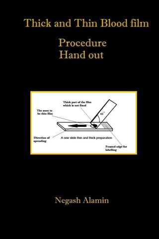
Title: Thick and thin blood films
Name: Negash Alamin
Profession: Clinical Laboratory Scientist
Email: drnegash@gmail.com
Date: 4/11/17
Principles of the procedure
Thick and thin blood films are a routine practice in the hematological
department to diagnose various pathogens that cause ailments in the
human organism. There are two portions in this routinely used method of
pathogen diagnosis: one in called the thick part where the general load of
the parasite infecting the RBC or just situated in the blood tissue; but, the
thin film is a part of the stained smear which more likely to be widely
ramified thus enabling us to see what type of pathogen species specifically
has infected the individual. Thus, both sections of the film are important;
but, the thin part is useful to us in specific diagnosis.
Materials required
1- Gloves
2- Test tube with patient blood.
3- Glass slides: one as a spreader and one as the receiver with frost
end for labeling.
4- The slides need to be dust, scratch, detergent, and grease free.
5- Pasteur pipette used to draw blood out of the test tube on to the
frosted slide.
Procedure
1- Open the vial containing the blood of patient carefully.
2- Insert the Pasteur pipette and withdraw a sufficient amount of
blood.
3- Displace the small amount of blood of the ¾ section of the frosted
slide.
4- Prepare your spreader by chipping of its angles.
5- Position the spreader 45° in relation to your frosted slide.
6- Place it directly over the drop of blood and slide.
7- If both thick and thin are performed on the same slide in case of
experienced lab technicians; then, the thin part of the film will only
be fixed leaving out the bulged drop of blood on the frosted side.
8- A good smear is a one with parabolic tongue symmetry.
9- It should not be short with small blood; it should not be very thick
or jagged with lots of blood; it just needs to be right.
Clinical significance
The blood film is an invaluable method for diagnosing several maladies in
the clinical laboratory. A blood film can be stained by different and various
methods as in Wright stain, Gram stain, Giemsa stain and other several
combinational methods. After blood film is performed it can be stained by
Wright stain for cell morphology analysis, Gram stain for bacteriology
inquiry and Giemsa stain for parasitological diagnosis. But, nowadays
methods of diagnosis are becoming extremely simple due to advancement in
technology diseases that needed blood film like malaria are now diagnosed
immunologically by modern strip tests; saving capital spent on reagents,
time and work load. But in general we can find these parasites in a typical
blood film.
Table 1 This table is not conclusive of all vermin that we can find in a blood film. The
entire field of bacteriological pathogens has not been mentioned here.
Pathogens one can find in a
blood film and their associated
disease
Stages they are discovered
(diagnostic stage)
Microfilariae
Brugia malayi (lymphatic
filariasis)
Microfilaria
Loa loa (subcutaneous filariasis) Microfilaria
Wuchereria bancrofti
(elephantiasis)
Microfilaria
Mansonella perstans (serous
cavity filariasis)
Microfilaria
Malaria species
Plasomdium vivax Trophozoite or schizont
Plasmodium falciparium Trophozoite or schizont
Plasmodium malariae Trophozoite or schizont
Plasmodium ovale (has two
subspecies)
Trophozoite or schizont
Plasmodium knowlesi (rare & Trophozoite or schizont
zoonotic)
Trypanosoma species
Trypanosoma brucei which has
two sub species and causes
sleeping sickness
Trypomastigote
Trypanosoma cruzi (Chagas
disease)
Trypomastigote
Additional vermins
Leshmania species
(Leshmaniasis)
Amastigotes

Thick and thin blood smear Procedure

  • 2.
    Title: Thick andthin blood films Name: Negash Alamin Profession: Clinical Laboratory Scientist Email: drnegash@gmail.com Date: 4/11/17
  • 3.
    Principles of theprocedure Thick and thin blood films are a routine practice in the hematological department to diagnose various pathogens that cause ailments in the human organism. There are two portions in this routinely used method of pathogen diagnosis: one in called the thick part where the general load of the parasite infecting the RBC or just situated in the blood tissue; but, the thin film is a part of the stained smear which more likely to be widely ramified thus enabling us to see what type of pathogen species specifically has infected the individual. Thus, both sections of the film are important; but, the thin part is useful to us in specific diagnosis. Materials required 1- Gloves 2- Test tube with patient blood. 3- Glass slides: one as a spreader and one as the receiver with frost end for labeling. 4- The slides need to be dust, scratch, detergent, and grease free. 5- Pasteur pipette used to draw blood out of the test tube on to the frosted slide. Procedure 1- Open the vial containing the blood of patient carefully. 2- Insert the Pasteur pipette and withdraw a sufficient amount of blood. 3- Displace the small amount of blood of the ¾ section of the frosted slide. 4- Prepare your spreader by chipping of its angles. 5- Position the spreader 45° in relation to your frosted slide. 6- Place it directly over the drop of blood and slide.
  • 4.
    7- If boththick and thin are performed on the same slide in case of experienced lab technicians; then, the thin part of the film will only be fixed leaving out the bulged drop of blood on the frosted side. 8- A good smear is a one with parabolic tongue symmetry. 9- It should not be short with small blood; it should not be very thick or jagged with lots of blood; it just needs to be right. Clinical significance The blood film is an invaluable method for diagnosing several maladies in the clinical laboratory. A blood film can be stained by different and various methods as in Wright stain, Gram stain, Giemsa stain and other several combinational methods. After blood film is performed it can be stained by
  • 5.
    Wright stain forcell morphology analysis, Gram stain for bacteriology inquiry and Giemsa stain for parasitological diagnosis. But, nowadays methods of diagnosis are becoming extremely simple due to advancement in technology diseases that needed blood film like malaria are now diagnosed immunologically by modern strip tests; saving capital spent on reagents, time and work load. But in general we can find these parasites in a typical blood film. Table 1 This table is not conclusive of all vermin that we can find in a blood film. The entire field of bacteriological pathogens has not been mentioned here. Pathogens one can find in a blood film and their associated disease Stages they are discovered (diagnostic stage) Microfilariae Brugia malayi (lymphatic filariasis) Microfilaria Loa loa (subcutaneous filariasis) Microfilaria Wuchereria bancrofti (elephantiasis) Microfilaria Mansonella perstans (serous cavity filariasis) Microfilaria Malaria species Plasomdium vivax Trophozoite or schizont Plasmodium falciparium Trophozoite or schizont Plasmodium malariae Trophozoite or schizont Plasmodium ovale (has two subspecies) Trophozoite or schizont Plasmodium knowlesi (rare & Trophozoite or schizont
  • 6.
    zoonotic) Trypanosoma species Trypanosoma bruceiwhich has two sub species and causes sleeping sickness Trypomastigote Trypanosoma cruzi (Chagas disease) Trypomastigote Additional vermins Leshmania species (Leshmaniasis) Amastigotes