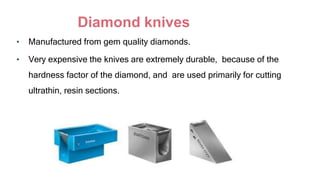
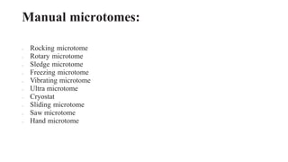
1. Acharya N.G. RangaAgricultural University
S. V.Agricultural College, Tirupati
COURSE NO : PL PATH-502
COURSE TITLE : Plant virology
TOPIC : Microtomy
SUBMITTED BY:
M. Lokeswaridevi
TAM -2020-030,
Dept of Entomology
2. Microtomy
• The means by which tissue can be sectioned and attached to a
surface for further microscopic examination.
Derived from the Greek mikros, meaning “small”, and temnein,
meaning “to cut”)
It is a mechanical device for cutting thin uniform slices of tissue
sections.
3. History
• The earliest form of microtomy
was the freehand sectioning of
fresh or fixed material using a
sharp razor.
4. • This allowed for the observation of samples using light microscopes in
a transmission mode.
• One of the first devices for the preparation of such cuts was invented
in 1770 by George Adams, Jr. (1750–1795) and further developed by
Alexander Cummings.
• Invention of the microtome is given to the anatomist Wilhelm His.
5. Based on material of the knife:
• Steel knives
• Glass knives
• Diamond knives
• Sapphire knives
6. Steel Knives
• Steel microtome knives are manufactured from high quality carbon or
tool grade steel which is heat treated to harden the edge.
• The steel should be rust resistant, free from impurities and contain
anti-corrosives.
8. Tungsten carbide knives
• Noncorrosive
• Practically nonmagnetic
• 100 times harder than hardened tool
steel.
9. Non-Corrosive Knives For Cryostats
• These are manufactured from hardened, heat treated stainless steel free
from all impurities.
• They contain 12 to 15% chromium.
10. Disposable Blades
• These are essentially refined, thickened razor blades.
• All disposable blades are manufactured from high quality stainless
steel.
• The edge of disposable blades can be coated with platinum or
chromium to enhance strength and prolong cutting life
12. Hard but brittle
Care is required while handling
These knives deteriorate with storage due to changes in the
'flow' or 'strain' of the glass after fracture and from oxidation
impurities remaining in the hardened glass after manufacture.
13. Diamond knives
• Manufactured from gem quality diamonds.
• Very expensive the knives are extremely durable, because of the
hardness factor of the diamond, and are used primarily for cutting
ultrathin, resin sections.
14. Sapphire knives
Manufactured from one piece of solid sapphire artificially
produced from an alumina monocrystal under computer
controlled thermal conditions.
15. The knife edge can be classified according to its
shape / profile.
• PROFILE – A: Strongly Plano concave/biconcave
• PROFILE – B: Plano concave
• PROFILE – C: Wedge Shaped
• PROFILE – D: Plane Shaped/Tool edge shaped
24. Rocking Microtome
• This instrument is one of the
oldest in design, relatively
cheap, and is exclusively
designed for sectioning
paraffin blocks.
• This microtome comprise of
three moving parts, which is
extremely reliable and
requires minimum
maintenance.
25. Mechanism of action
⚫Knife is fixed, the block of the tissue moves through an arc to strike the knife.
⚫Between strokes the block is moved towards the knife for the required
thickness of sections by means of a ratchet operated micrometer thread.
⚫Steady backward and forward movement of the handle gives ribbons of
good sections.
26. Rotary Microtome
• Derives its name from the
rotary action of the hand-
wheel which actuates the
cuttingmovements.
• Section thickness settings
range from 0.5μm to 60μm
on most machines.
• Machines of this sort are
general purpose microtomes
for cutting semi-thin to thin
sections for light microscopy
28. Sledge microtome
• Become a popular machine for
routine use since the world war II
• Its action is much slower when
compared to rocking or rotary
type.
• A large knife is used (24cm in
length) and the knife is usually
wedge-shaped which reduces the
possibility of vibration and
requires less honing.
29. Sliding Microtome
• In a sliding microtome, the
knife is moved horizontally
against a fixed block which
progresses against it in an
inclined plane.
• The sliding microtome can be
used for paraffin-wax embedded
sections although it was designed
for cutting celloidin- embedded
sections
30. Freezing Microtome
• The machine is clamped to
the edge of a bench and is
connected to a cylinder of
CO2 by means of a specially
strengthened flexible metal
tube.
• Consistent, high quality, thin
sections are very difficult to
obtain with this type of
microtome
31. Vibrating Microtome
• The vibrating microtome was
conceived as a microtome
which could produce high
quality sections of fresh,
unfixed material from animal
or botanical sources.
• The name of the instrument
was derived from the high
speed vibration produced by
a safety razor blade which
provided the cutting power.
32. Ultra Microtome
• Is used to prepare ultra-thin
sections for light and
electron microscopy .
• Are usually embedded in
hard resin before cutting.
• That sections can be cut as
thin as 10 nanometers.
• Two forms of advance
mechanism have been
developed in this style of
microtome.
33. Saw Microtome
• Saw microtome's cut sections from very
hard material such as undecalcified bone,
glass or ceramics.
• The samples, commonly embedded in
resins, are moved extremely slowly against
a diamond coated saw rotating at
approximately 600 rpm.
• It is possible to produce sections of 20 μm
or greater, provided the saw blade is in
perfect condition.
• The saw microtome is not capable for
producing very thin sections.
34. Hand Microtome
• The hand microtome is limited to
sectioning intrinsically rigid
botanical material, and it is
difficult to obtain thin sections
from animal tissues
35. Cryostat Microtome
• The introduction of fluorescent antibody staining techniques
by coons, creech and jones in 1941 led to a need for thin
sections (3-5 μm) of fresh frozen tissue free of ice crystal
defects .
• To satisfy these criteria the tissue must be snap frozen at a
very low temperature.
• Linderstrom-lang and mogensen designed the first cryostat in
1938. Coons and his colleagues redesigned it in 1951.
36. • microtome of any type but preferably rustproof, which is enclosed and
operated within a deep freeze cabinet.
• The temperature may be regulated between-10ºC to -40ºC.
• Any crystostat can be used as an alternative to a freezing microtome
for rapid sectioning.
• The microtome may be adjusted to cut sections from 2- 16μm.
37. Laser Microtome
• Laser microtome is used for precise, non-
contact sectioning and was designed to slice
samples with high precision.
• It’s equipped with state- of-the-art
femtosecond laser technology.
• It enables non-contact cutting inside
biological tissues and various materials
without causing thermal damage.
• Depending on the material being processed,
slice thicknesses of about 5 to 100 μm are
feasible.
38.
39. Principle:
The device operates using a cutting action of an infra-red laser.As the
laser emits a radiation in the near infra-red, in this wavelength regime
the laser can interact with biological materials.
40. Computerized Microtome
• It is equipped with the advanced
rapid thermostatic switch,
semiconductor freezing, cryo
scalped and cryoplate.
• The computerized microtome can
carry out the rapid freezing
section or routine paraffin section
(dual-purpose).
41. • This microtome attains slice thickness in the range of 1- 25μm with
least slice adjusting graduation of 1μm and a maximal slice section
of 32x32mm.
• The temperature of cryo scalpel and cryoplate range between 0ºC ~ -
18ºC and -10ºC ~ -40ºC respectively
42. Embedding and Sectioning
Embedment
Light microscopy
Tissue infiltrated with molten paraffin wax - which is allowed to cool and
harden.
Requires dehydration and infiltration with a paraffin solvent - aromatic
hydrocarbon (xylene, toluene, benzene)
Provides sufficient support to section to about 3 micrometers minimum with a
steel knife.
Paraffin can infiltrate deeply into tissue, allowing large blocks and ultimately
large sections to be obtained.
43. Precautions to be taken before section cutting
• Fix Specimens Properly
• Process Tissue Properly
• Embed Specimens Carefully
• Locate Microtome Appropriately
• Set Blade Clearance Angle Optimally
• Maximize Blade Life
• Ensure Blocks are Cold
44. Faults
• Section too thick
• Holes from rough trimming
• Knife lines (vertical striations)
• Disruption
• Fine cracks or micro-chatter
• Coarse chatter
45. SOLUTION
⚫ Tighten blade and block holders
⚫ Reduce clearance angle
⚫ Use softening fluid
⚫ Rehydrate and surface decalcify
⚫ Re-embed in fresh paraffin
⚫ Replace or use new area of blade
⚫ Clean blade edge to clean excess paraffin
47. REFERENCES
• MICROTOMES AND MICROTOME KNIVES – A REVIEW AND
PROPOSED CLASSIFICATION
• Ellis RC. The microtome: function and design. Woods and Ellis 2000
• Du pont. Sorvall JB-4 Microtome. The light microtome for super thin sections.
Newtown, USA; Du Pont Instruments. 1991.
• http://users.adam.com. au/royellis/microt/microt.ht.
• Walter F. The microtome manual of the technique of preparation and of
section cutting. Germany; Ernst Leitz Wetzlar GMBH. 1980