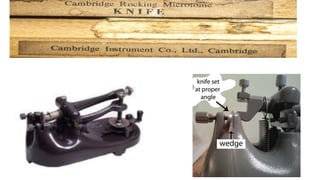
1) A microtome is a tool used to cut extremely thin slices of material for microscopic examination. It was developed to allow for the precise cutting of tissue sections thin enough for light to pass through.
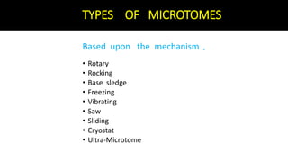
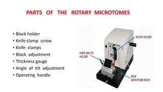
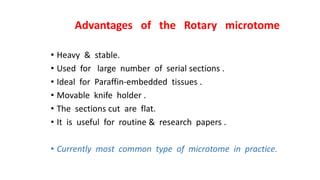
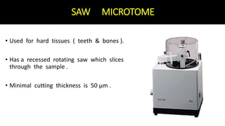
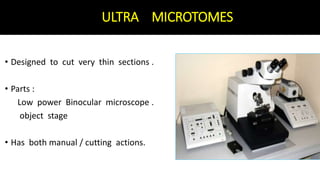
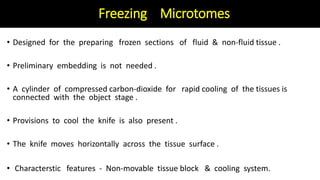
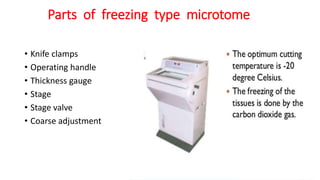
2) There are several types of microtomes including rotary, rocking, base sledge, freezing, vibrating, saw, sliding, cryostat, and ultramicrotome. The rotary microtome is the most common type used for routine research due to its ability to cut flat serial sections.
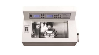
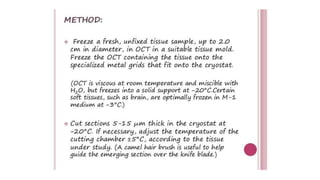
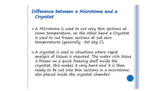
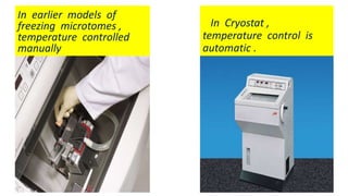
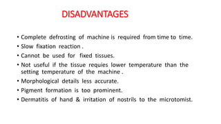
3) A cryostat allows cutting of unfixed fresh or frozen tissue sections and maintains the tissue at low temperatures during sectioning to preserve cellular structures, though it cannot be used for fixed