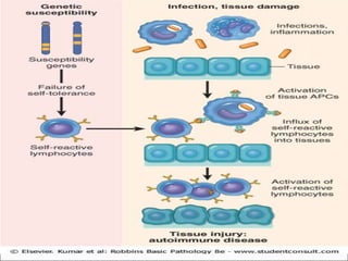
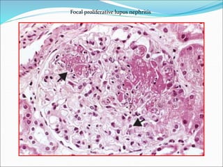
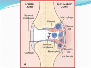
AT WAR WITHIN : The double edged sword
of IMMUNITY
Normally
protective
Immunodeficiency
states
Deregulated immune
response against self
antigens :
AUTOIMMUNE
DISEASES
Exaggerated immune
response :
HYPERSENSITIVITY
REACTIONS
 Immune reaction to self-antigens is autoimmunity
 A disorder is categorized as truly autoimmune when:
(1) an immune reaction is specific for some self-
antigen or self-tissue
(2) evidence that such a reaction is not secondary to
tissue damage but is of primary pathogenic significance
(3) the absence of another well-defined cause of the
disease
 Organ specific autoimmunity - one particular organ or
cell type (Hashimoto’s thyroiditis)
 Systemic autoimmunity - multiple autoantibodies or
cell-mediated reactions against numerous self-
antigens (SLE)
 Immunological tolerance is the phenomenon of
unresponsiveness to an antigen as a result of exposure
of lymphocytes to that antigen.
 Self-tolerance refers to lack of responsiveness to an
individual's own antigens
 Lack of the tolerance leads to autoimmunity
 Self-tolerance can be classified into two groups:
central tolerance and peripheral tolerance
 Central Tolerance-immature self-reactive T- and B-
lymphocyte clones that recognize self-antigens during
their maturation are killed
 Negative selection or deletion in T cells
 Receptor editing in B cells
 Peripheral Tolerance – silencing of autoreactive B
and T cells in the peripheral tissue
 Anergy: This refers to prolonged or irreversible
functional inactivation of lymphocytes
 Suppression by regulatory T cells
 Deletion by activation-induced cell death
Mechanisms of Autoimmunity
 Inheritance of susceptibility genes that may disrupt
different tolerance pathways
 Infections and tissue alterations that may expose self-
antigens and activate APCs and self reactive
lymphocytes in the tissues.
Role of Infections and Tissue Injury
 Molecular mimicry - Viruses and certain bacteria
such as streptococci and Klebsiella organisms share
cross-reacting epitopes with self-antigens, such that
responses to the microbial antigen may attack self-
tissues.
Features of Autoimmunity
 An autoimmune response itself promote further
autoimmune attack by a process that has been called
epitope spreading.
 Clinicopathologic features depend on the type of T cell
response.
 Precise categorization is difficult.
Systemic Lupus Erythematosus
 Multisystem autoimmune disease
 Acute or insidious in its onset
 It is a chronic, remitting and relapsing, often febrile
illness characterized principally by injury to the skin,
joints, kidney and serosal membranes
 The fundamental defect in SLE is a failure to maintain
self-tolerance.
Spectrum of Autoantibodies in SLE
 ANAs - directed against nuclear antigens
(1) antibodies to DNA,
(2) antibodies to histones,
(3) antibodies to nonhistone proteins bound to RNA,
and
(4) antibodies to nucleolar antigens.
 Antibodies against blood cells
 Antiphospholipid antibodies are present in 40% to 50% of
lupus patients and react with a wide variety of proteins in
complex with phospholipids.
Morphology
 An acute necrotizing vasculitis involving capillaries,
small arteries and arterioles can be present in any
tissue.
 The arteritis is characterized by fibrinoid deposits in
the vessel walls.
 In chronic stages, vessels undergo fibrous thickening
with luminal narrowing.
Morphology
 The most characteristic lesions result from immune
complex deposition in:
 blood vessels
 kidneys
 connective tissue
 skin
 Kidney-Lupus nephritis affects 50% of SLE patients.
 The principal mechanism of injury is immune
complex deposition in the glomeruli, tubular or
peritubular capillary basement membranes, or larger
blood vessels.
 Six patterns are recognized: minimal mesangial (class
I); mesangial proliferative (class II); focal lupus
nephritis (class III); diffuse lupus nephritis (class IV);
membranous lupus nephritis (class V) and Advanced
sclerosing lupus nephritis (classVI).
 Minimal mesangial : immune complex deposition in
the mesangium, identified by immunofluorescence
and by EM but without structural changes by light
microscopy
 Mesangial proliferative lupus nephritis: mesangial cell
and matrix proliferation. and granular mesangial
deposits of immunoglobulin and complement without
involvement of glomerular capillaries
 Focal Lupus nephritis: involvement of fewer than 50%
of glomeruli. Glomeruli exhibit swelling and
proliferation of endothelial and mesangial cells,
leukocyte accumulation and hyaline thrombi. There is
also often extracapillary proliferation with crescent
formation
 Diffuse Lupus nephritis: Involvement of more than
50% of gloumeruli. Most common and severe form of
lupus nephritis. Subendothelial immune complex
deposits cause circumferential thickening of the
capillary wall, forming “wire loop” structures on light
microscopy
Focal proliferative lupus nephritis
Diffuse proliferative lupus nephritis
Wire loop lesions
Deposition of IgG antibody in a granular pattern, detected by
immunofluorescence
 Membranous Lupus nephritis: diffuse thickening of
the capillary walls due to deposition of subepithelial
immune complexes
 Advanced sclerosing lupus nephritis: characterized by
sclerosis >90% of the glomeruli and represents end-
stage renal disease.
 The skin is involved in 50% of patients
 Erythematous or maculopapular eruption over the
malar eminences and bridge of the nose ("butterfly
pattern“).
 Exposure to sunlight (UV light) exacerbates the
erythema (so-called photosensitivity)
 Joint involvement is typically a nonerosive synovitis
with little deformity.
 Central nervous system (CNS) involvement
ascribed to vascular lesions causing ischemia or
multifocal cerebral microinfarcts.
 Pericardium and pleura, show a variety of
inflammatory changes ranging (in the acute phase)
from serous effusions to fibrinous exudates and
progressing to fibrous opacification in the chronic
stage.
 Involvement of the heart is manifested primarily in
the form of pericarditis.
 Myocarditis, in the form of a nonspecific mononuclear
cell infiltrate, and valvular lesions, called Libman-
Sacks endocarditis, also occur but are less common
in the current era of aggressive corticosteroid therapy.
 The valvular nonbacterial verrucous endocarditis
takes the form of irregular, 1- to 3-mm warty deposits,
distinctively on either surface of the leaflets
Rheumatoid Arthritis
 Rheumatoid arthritis (RA) is a systemic, chronic
inflammatory disease affecting many tissues but
principally attacking the joints.
 Characterised by nonsuppurative proliferative synovitis
that frequently progresses to destroy articular cartilage
and underlying bone with resulting disabling arthritis
 RA typically presents as symmetric arthritis,
principally affecting the small joints of the hands
and feet, ankles, knees, wrists, elbows, and shoulders.
 Typically, the proximal interphalangeal and
metacarpophalangeal joints are affected, but distal
interphalangeal joints are spared.
Morphology
(1) Synovial cell hyperplasia and proliferation
(2) Dense perivascular inflammatory cell infiltrates
(frequently forming lymphoid follicles) in the
synovium composed of CD4+ T cells, plasma cells, and
macrophages
(3) Increased vascularity due to angiogenesis
(4) Neutrophils and aggregates of organizing fibrin on
the synovial surface and in the joint space; and
(5) Increased osteoclast activity in the underlying bone,
leading to synovial penetration and bone erosion
 The articular cartilage subjacent to the pannus is
eroded and, in time, virtually destroyed.
 The subarticular bone may also be attacked and
eroded.
 Eventually the pannus fills the joint space, and
subsequent fibrosis and calcification may cause
permanent ankylosis.
 Rheumatoid subcutaneous nodules along the
extensor surface of the forearm or other areas
subjected to mechanical pressure; rarely they can form
in the lungs, spleen, heart, aorta, and other viscera.
 Rheumatoid nodules are firm, nontender, oval or
rounded masses as large as 2 cm in diameter.
 Microscopically, they are characterized by a central
focus of fibrinoid necrosis surrounded by a palisade of
macrophages, which in turn is rimmed by granulation
tissue
Sjögren Syndrome
 Clinicopathologic entity characterized by dry eyes
(keratoconjunctivitis sicca) and dry mouth
(xerostomia), resulting from immune-mediated
destruction of the lacrimal and salivary glands.
 It occurs as an isolated disorder (primary form), also
known as the sicca syndrome, or more often in
association with another autoimmune disease
(secondary form).
 90% of Sjögren syndrome cases occur in women
between the ages of 35 and 45 years.
 Antibodies directed against two ribonucleoprotein
antigens, SS-A (Ro) and SS-B (la) , can be detected in
as many as 90% of patients
 Patients present with dry mouth, lack of tears
Morphology
 Intense lymphocyte and plasma-cell infiltrate, forming
lymphoid follicles with germinal centers with
associated destruction of the native architecture
 Keratoconjunctivitis and Xerostomia
 Dryness and crusting of the nose
 Secondary laryngitis, bronchitis, and pneumonitis
 Approximately 25% of the patients (especially those
with anti-SS-A antibodies) develop extraglandular
disease affecting the CNS, skin, kidneys, and muscles.
Systemic Sclerosis (Scleroderma)
 Systemic sclerosis is a chronic disease characterized
by:
(1) chronic inflammation thought to be the result of
autoimmunity,
(2) widespread damage to small blood vessels, and
(3) progressive interstitial and perivascular fibrosis in
the skin and multiple organs
 Diffuse scleroderma - characterized by widespread
skin involvement at onset, with rapid progression and
early visceral involvement.
 Limited scleroderma - the skin involvement is often
confined to fingers, forearms, and face. Visceral
involvement occurs late
The progressive fibrosis characteristic of the disease is the culmination
of multiple abnormalities, including the actions of fibrogenic cytokines
produced by infiltrating leukocytes, hyperresponsiveness of fibroblasts
to these cytokines, and scarring following upon ischemic damage
caused by the vascular lesions
Clinical features
 Female-to-male ratio of 3 : 1,
 50- to 60-year age group.
 Distinctive features are the striking cutaneous
changes, notably skin thickening.
 Raynaud's phenomenon, manifested as episodic
vasoconstriction of the arteries and arterioles of the
extremities,
 Dysphagia due to esophageal fibrosis and its resultant
hypomotility is present in more than 50% of patients
 CREST syndrome
Seen in some patients with limited systemic
sclerosis.
It is characterized by calcinosis, raynaud
phenomenon, esophageal dysfunction, sclerodactyly,
telangiectasia,
Presence of anticentromere antibodies.
Morphology - Skin
 Diffuse, sclerotic atrophy of the skin
 Edema and perivascular infiltrates containing CD4+ T
cells
 Capillaries and small arteries show thickening of the
basal lamina, endothelial cell damage, and partial
occlusion
 Increase of compact collagen in the dermis, with
thinning of the epidermis, loss of rete pegs, atrophy of
the dermal appendages, and hyaline thickening of the
vessels.
Alimentary Tract
 Affected in approximately 90% of patients
 Progressive atrophy and collagenous fibrous
replacement of the muscularis at any level of the gut
but are most severe in the esophagus
 Thinned mucosa ,excessive collagenization of the
lamina propria and submucosa. Loss of villi and
microvilli in the small bowel
MIXED CONNECTIVE TISSUE DISEASE
 A disease with clinical features that are a mixture of
the features of SLE, systemic sclerosis, and
polymyositis.
 The disease is characterized serologically by high titers
of antibodies to ribonucleoprotein particle–containing
U1 ribonucleoprotein.
AUTOIMMUNE DISEASES.ppt

AUTOIMMUNE DISEASES.ppt

  • 2.
    AT WAR WITHIN: The double edged sword of IMMUNITY Normally protective Immunodeficiency states Deregulated immune response against self antigens : AUTOIMMUNE DISEASES Exaggerated immune response : HYPERSENSITIVITY REACTIONS
  • 3.
     Immune reactionto self-antigens is autoimmunity  A disorder is categorized as truly autoimmune when: (1) an immune reaction is specific for some self- antigen or self-tissue (2) evidence that such a reaction is not secondary to tissue damage but is of primary pathogenic significance (3) the absence of another well-defined cause of the disease
  • 4.
     Organ specificautoimmunity - one particular organ or cell type (Hashimoto’s thyroiditis)  Systemic autoimmunity - multiple autoantibodies or cell-mediated reactions against numerous self- antigens (SLE)
  • 5.
     Immunological toleranceis the phenomenon of unresponsiveness to an antigen as a result of exposure of lymphocytes to that antigen.  Self-tolerance refers to lack of responsiveness to an individual's own antigens  Lack of the tolerance leads to autoimmunity  Self-tolerance can be classified into two groups: central tolerance and peripheral tolerance
  • 6.
     Central Tolerance-immatureself-reactive T- and B- lymphocyte clones that recognize self-antigens during their maturation are killed  Negative selection or deletion in T cells  Receptor editing in B cells
  • 7.
     Peripheral Tolerance– silencing of autoreactive B and T cells in the peripheral tissue  Anergy: This refers to prolonged or irreversible functional inactivation of lymphocytes  Suppression by regulatory T cells  Deletion by activation-induced cell death
  • 8.
    Mechanisms of Autoimmunity Inheritance of susceptibility genes that may disrupt different tolerance pathways  Infections and tissue alterations that may expose self- antigens and activate APCs and self reactive lymphocytes in the tissues.
  • 10.
    Role of Infectionsand Tissue Injury  Molecular mimicry - Viruses and certain bacteria such as streptococci and Klebsiella organisms share cross-reacting epitopes with self-antigens, such that responses to the microbial antigen may attack self- tissues.
  • 11.
    Features of Autoimmunity An autoimmune response itself promote further autoimmune attack by a process that has been called epitope spreading.  Clinicopathologic features depend on the type of T cell response.  Precise categorization is difficult.
  • 12.
    Systemic Lupus Erythematosus Multisystem autoimmune disease  Acute or insidious in its onset  It is a chronic, remitting and relapsing, often febrile illness characterized principally by injury to the skin, joints, kidney and serosal membranes  The fundamental defect in SLE is a failure to maintain self-tolerance.
  • 15.
    Spectrum of Autoantibodiesin SLE  ANAs - directed against nuclear antigens (1) antibodies to DNA, (2) antibodies to histones, (3) antibodies to nonhistone proteins bound to RNA, and (4) antibodies to nucleolar antigens.  Antibodies against blood cells  Antiphospholipid antibodies are present in 40% to 50% of lupus patients and react with a wide variety of proteins in complex with phospholipids.
  • 18.
    Morphology  An acutenecrotizing vasculitis involving capillaries, small arteries and arterioles can be present in any tissue.  The arteritis is characterized by fibrinoid deposits in the vessel walls.  In chronic stages, vessels undergo fibrous thickening with luminal narrowing.
  • 19.
    Morphology  The mostcharacteristic lesions result from immune complex deposition in:  blood vessels  kidneys  connective tissue  skin
  • 20.
     Kidney-Lupus nephritisaffects 50% of SLE patients.  The principal mechanism of injury is immune complex deposition in the glomeruli, tubular or peritubular capillary basement membranes, or larger blood vessels.  Six patterns are recognized: minimal mesangial (class I); mesangial proliferative (class II); focal lupus nephritis (class III); diffuse lupus nephritis (class IV); membranous lupus nephritis (class V) and Advanced sclerosing lupus nephritis (classVI).
  • 21.
     Minimal mesangial: immune complex deposition in the mesangium, identified by immunofluorescence and by EM but without structural changes by light microscopy  Mesangial proliferative lupus nephritis: mesangial cell and matrix proliferation. and granular mesangial deposits of immunoglobulin and complement without involvement of glomerular capillaries
  • 22.
     Focal Lupusnephritis: involvement of fewer than 50% of glomeruli. Glomeruli exhibit swelling and proliferation of endothelial and mesangial cells, leukocyte accumulation and hyaline thrombi. There is also often extracapillary proliferation with crescent formation  Diffuse Lupus nephritis: Involvement of more than 50% of gloumeruli. Most common and severe form of lupus nephritis. Subendothelial immune complex deposits cause circumferential thickening of the capillary wall, forming “wire loop” structures on light microscopy
  • 23.
  • 24.
  • 25.
  • 26.
    Deposition of IgGantibody in a granular pattern, detected by immunofluorescence
  • 27.
     Membranous Lupusnephritis: diffuse thickening of the capillary walls due to deposition of subepithelial immune complexes  Advanced sclerosing lupus nephritis: characterized by sclerosis >90% of the glomeruli and represents end- stage renal disease.
  • 28.
     The skinis involved in 50% of patients  Erythematous or maculopapular eruption over the malar eminences and bridge of the nose ("butterfly pattern“).  Exposure to sunlight (UV light) exacerbates the erythema (so-called photosensitivity)
  • 30.
     Joint involvementis typically a nonerosive synovitis with little deformity.  Central nervous system (CNS) involvement ascribed to vascular lesions causing ischemia or multifocal cerebral microinfarcts.  Pericardium and pleura, show a variety of inflammatory changes ranging (in the acute phase) from serous effusions to fibrinous exudates and progressing to fibrous opacification in the chronic stage.
  • 31.
     Involvement ofthe heart is manifested primarily in the form of pericarditis.  Myocarditis, in the form of a nonspecific mononuclear cell infiltrate, and valvular lesions, called Libman- Sacks endocarditis, also occur but are less common in the current era of aggressive corticosteroid therapy.  The valvular nonbacterial verrucous endocarditis takes the form of irregular, 1- to 3-mm warty deposits, distinctively on either surface of the leaflets
  • 34.
    Rheumatoid Arthritis  Rheumatoidarthritis (RA) is a systemic, chronic inflammatory disease affecting many tissues but principally attacking the joints.  Characterised by nonsuppurative proliferative synovitis that frequently progresses to destroy articular cartilage and underlying bone with resulting disabling arthritis
  • 35.
     RA typicallypresents as symmetric arthritis, principally affecting the small joints of the hands and feet, ankles, knees, wrists, elbows, and shoulders.  Typically, the proximal interphalangeal and metacarpophalangeal joints are affected, but distal interphalangeal joints are spared.
  • 36.
    Morphology (1) Synovial cellhyperplasia and proliferation (2) Dense perivascular inflammatory cell infiltrates (frequently forming lymphoid follicles) in the synovium composed of CD4+ T cells, plasma cells, and macrophages (3) Increased vascularity due to angiogenesis (4) Neutrophils and aggregates of organizing fibrin on the synovial surface and in the joint space; and (5) Increased osteoclast activity in the underlying bone, leading to synovial penetration and bone erosion
  • 37.
     The articularcartilage subjacent to the pannus is eroded and, in time, virtually destroyed.  The subarticular bone may also be attacked and eroded.  Eventually the pannus fills the joint space, and subsequent fibrosis and calcification may cause permanent ankylosis.
  • 40.
     Rheumatoid subcutaneousnodules along the extensor surface of the forearm or other areas subjected to mechanical pressure; rarely they can form in the lungs, spleen, heart, aorta, and other viscera.  Rheumatoid nodules are firm, nontender, oval or rounded masses as large as 2 cm in diameter.  Microscopically, they are characterized by a central focus of fibrinoid necrosis surrounded by a palisade of macrophages, which in turn is rimmed by granulation tissue
  • 42.
    Sjögren Syndrome  Clinicopathologicentity characterized by dry eyes (keratoconjunctivitis sicca) and dry mouth (xerostomia), resulting from immune-mediated destruction of the lacrimal and salivary glands.  It occurs as an isolated disorder (primary form), also known as the sicca syndrome, or more often in association with another autoimmune disease (secondary form).
  • 43.
     90% ofSjögren syndrome cases occur in women between the ages of 35 and 45 years.  Antibodies directed against two ribonucleoprotein antigens, SS-A (Ro) and SS-B (la) , can be detected in as many as 90% of patients  Patients present with dry mouth, lack of tears
  • 44.
    Morphology  Intense lymphocyteand plasma-cell infiltrate, forming lymphoid follicles with germinal centers with associated destruction of the native architecture  Keratoconjunctivitis and Xerostomia  Dryness and crusting of the nose  Secondary laryngitis, bronchitis, and pneumonitis  Approximately 25% of the patients (especially those with anti-SS-A antibodies) develop extraglandular disease affecting the CNS, skin, kidneys, and muscles.
  • 46.
    Systemic Sclerosis (Scleroderma) Systemic sclerosis is a chronic disease characterized by: (1) chronic inflammation thought to be the result of autoimmunity, (2) widespread damage to small blood vessels, and (3) progressive interstitial and perivascular fibrosis in the skin and multiple organs
  • 47.
     Diffuse scleroderma- characterized by widespread skin involvement at onset, with rapid progression and early visceral involvement.  Limited scleroderma - the skin involvement is often confined to fingers, forearms, and face. Visceral involvement occurs late
  • 48.
    The progressive fibrosischaracteristic of the disease is the culmination of multiple abnormalities, including the actions of fibrogenic cytokines produced by infiltrating leukocytes, hyperresponsiveness of fibroblasts to these cytokines, and scarring following upon ischemic damage caused by the vascular lesions
  • 49.
    Clinical features  Female-to-maleratio of 3 : 1,  50- to 60-year age group.  Distinctive features are the striking cutaneous changes, notably skin thickening.  Raynaud's phenomenon, manifested as episodic vasoconstriction of the arteries and arterioles of the extremities,  Dysphagia due to esophageal fibrosis and its resultant hypomotility is present in more than 50% of patients
  • 50.
     CREST syndrome Seenin some patients with limited systemic sclerosis. It is characterized by calcinosis, raynaud phenomenon, esophageal dysfunction, sclerodactyly, telangiectasia, Presence of anticentromere antibodies.
  • 51.
    Morphology - Skin Diffuse, sclerotic atrophy of the skin  Edema and perivascular infiltrates containing CD4+ T cells  Capillaries and small arteries show thickening of the basal lamina, endothelial cell damage, and partial occlusion  Increase of compact collagen in the dermis, with thinning of the epidermis, loss of rete pegs, atrophy of the dermal appendages, and hyaline thickening of the vessels.
  • 54.
    Alimentary Tract  Affectedin approximately 90% of patients  Progressive atrophy and collagenous fibrous replacement of the muscularis at any level of the gut but are most severe in the esophagus  Thinned mucosa ,excessive collagenization of the lamina propria and submucosa. Loss of villi and microvilli in the small bowel
  • 55.
    MIXED CONNECTIVE TISSUEDISEASE  A disease with clinical features that are a mixture of the features of SLE, systemic sclerosis, and polymyositis.  The disease is characterized serologically by high titers of antibodies to ribonucleoprotein particle–containing U1 ribonucleoprotein.