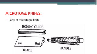
INTRODUCTION
• Microtomy :
Isthe means by which tissue can be sectioned and attached to a
surface for further microscopic examination.
• Microtome:
• Basic instrument used in microtomy.
• Mechanical device for cutting thin uniform slices of tissue -
sections.
4.
Types of microtomes
•These microtomes named according to the mechanism.
• Rocking microtome.
• Rotary microtome.
• Base sledge microtome.
• Sliding microtome.
• Freezing microtome.
• vibrating microtome.
• ultra microtome.
• laser microtome.
ROTATORY MICROTOMY :
•It is the most commonly used microtome in histopathology
laboratories
• Owing to the rotary action of handwheel that activates the
section cutting, this type of microtome is known as a rotary
microtome.
• This microtome is also known as 'Minot microtome' (invented
by Minot, 1886).
• This microtome is provided with fixed blade and tissue block
proceeds forwards during its up and down movement.
• Knife is fixed in holding clamps.
7.
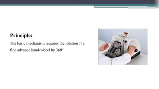
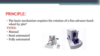
PRINCIPLE:
• The basicmechanism requires the rotation of a fine advance hand-
wheel by 360°
TYPES:
• Manual
• Semi automated
• Fully automated
8.
ADVANTAGES:
• It isa stable machine without any vibration during cutting.
• Most commonly used, suitable for paraffin and celloidin blocks.
• Serial sectioning is possible.
• Cutting angle can be adjusted.
• Ability to cut thin 2-3 um sections
SLEDGE MICROTOME
This microtomeis suitable
1. For cutting hard tissues like bone and teeth.
2. For large specimens that are not suitable for rotary microtome.
3. For celloidin sections (whole brain sections).
11.
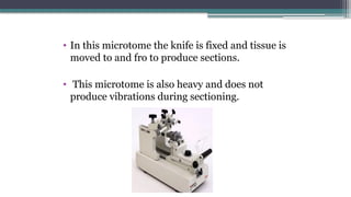
• In thismicrotome the knife is fixed and tissue is
moved to and fro to produce sections.
• This microtome is also heavy and does not
produce vibrations during sectioning.
12.
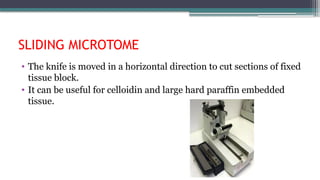
SLIDING MICROTOME
• Theknife is moved in a horizontal direction to cut sections of fixed
tissue block.
• It can be useful for celloidin and large hard paraffin embedded
tissue.
13.
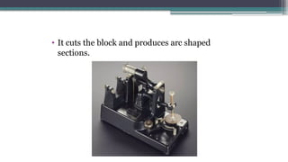
ROCKING MICROTOME
• Thismicrotome is an older version of the microtome.
• Tissue block moves with rocking action (hence the name rocking
microtome) over a fixed knife.
• Rocking microtome is useful for cutting tiny block.
14.
• It cutsthe block and produces arc shaped
sections.
15.
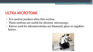
ULTRA MICROTOME
• Itis used to produce ultra thin section.
• These sections are useful for electron microscopy.
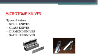
• Knives used for ultramicrotomy are diamond, glass or sapphire
knives.
16.
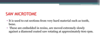
SAW MICROTOME
• Itis used to cut sections from very hard material such as tooth,
bone.
• These are embedded in resins, are moved extremely slowly
against a diamond coated saw rotating at approximately 600 rpm.
17.
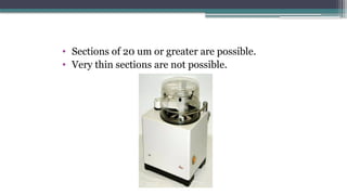
• Sections of20 um or greater are possible.
• Very thin sections are not possible.
18.
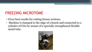
FREEZING MICROTOME
• Givesbest results for cutting frozen sections.
• Machine is clamped to the edge of a bench and connected to a
cylinder of CO2 by means of a specially strengthened flexible
metal tube.
19.
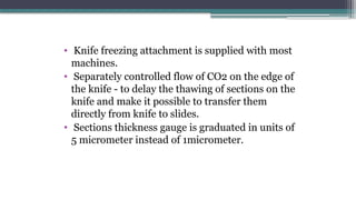
• Knife freezingattachment is supplied with most
machines.
• Separately controlled flow of CO2 on the edge of
the knife - to delay the thawing of sections on the
knife and make it possible to transfer them
directly from knife to slides.
• Sections thickness gauge is graduated in units of
5 micrometer instead of 1micrometer.
20.
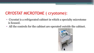
CRYOSTAT MICROTOME (cryotome):
• Cryostat is a refrigerated cabinet in which a specialty microtome
is housed.
• All the controls for the cabinet are operated outside the cabinet.
21.
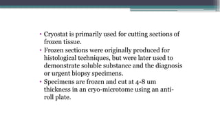
• Cryostat isprimarily used for cutting sections of
frozen tissue.
• Frozen sections were originally produced for
histological techniques, but were later used to
demonstrate soluble substance and the diagnosis
or urgent biopsy specimens.
• Specimens are frozen and cut at 4-8 um
thickness in an cryo-microtome using an anti-
roll plate.
22.
Principle
• When thetissue is frozen, the interstitial water turns into ice,
tissue becomes firm and acts as an embedding medium.
23.
USES -
• rapidproduction of sections for intra-operative diagnosis*
diagnostic and research enzyme histochemistry for labile enzymes.
• immunofluorescent methodology.
• immunohistochemistry techniques when heat and fixation may
inactivate or destroy the antigens.
• Diagnostic and research non-enzyme histochemistry, e.g. lipids
and some carbohydrates.
• silver demonstration methods, particularly neuropathology.
24.
Advantages
• Electronic temperaturecontrol.
• Electronically controlled advance and retraction of the block.
Specimen orientation facility.
• Digital visualization of chuck and cabinet temperature.
• Mechanical cutting speed control and section thickness.
• Automatic defrost mechanism.
• Automated decontamination and sterilization.
25.
LASER MICROTOME
• Contactfree slicing.
• Prior preparation of sample not required.
• Can also be used for very hard materials, such as bones or teeth as
well as some ceramics.
• Thickness: 10-100 um
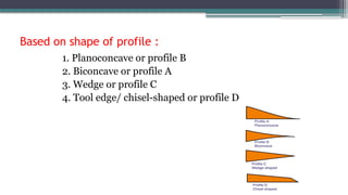
Based on shapeof profile :
1. Planoconcave or profile B
2. Biconcave or profile A
3. Wedge or profile C
4. Tool edge/ chisel-shaped or profile D
29.
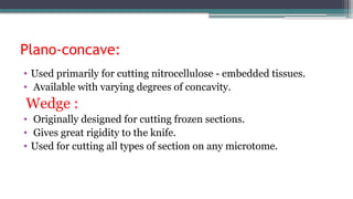
Plano-concave:
• Used primarilyfor cutting nitrocellulose - embedded tissues.
• Available with varying degrees of concavity.
Wedge :
• Originally designed for cutting frozen sections.
• Gives great rigidity to the knife.
• Used for cutting all types of section on any microtome.
30.
Biconcave :
• .Classicalknife shape introduced by Heiffor.
• Used with the rocking microtome.
• Relatively easy to sharpen.
• Less rigid, prone to more vibrations.
31.
Tool edge(D-profile):
• Tooledge(D-profile): Called 'chisel edge', similar to a
woodworker's chisel.
• Used primarily to section exceptionally hard tissue.
• Decalcified dense cortical bone.
• Undecalcified bone.
• Stouter than conventional knives to give added rigidity.
• Edge may be coated with tungsten-carbide for increased life.
32.
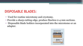
DISPOSABLE BLADES:
• Usedfor routine microtomy and cryotomy.
• Provide a sharp cutting edge, produce flawless 2-4 mm sections.
• Disposable blade holders incorporated into the microtome or an
adapter.
Bevel angle/Facet angle:
•The facet angle is the angle between the two facets that form the
cutting edge.
• Usually vary between 27-32. Smaller the bevel angle sharper is the
knife, however too small bevel angle permits elastic distortion of
the edge.
35.
Rake and clearanceangles/wedge angle:
• Standard wedge angle 15 degree.
• High rake angle and low clearance angle gives less compression to
the tissue block and produces a smooth plastic flow type during
sectioning.
• High rake angles suitable for soft tissues and need to be reduced
for harder tissues.
• The width of the two facet which makes the cutting edge of knife
has recommended from 0.1 to about 0.6mm.
36.
MICROTOME KNIFE SHARPENING:
• Manual procedure or automatic procedure.
1) Abrasive grinding of the facets [HONING]
2)Polishing [STROPPING]
37.
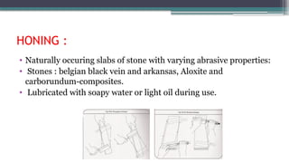
HONING :
• Naturallyoccuring slabs of stone with varying abrasive properties:
• Stones : belgian black vein and arkansas, Aloxite and
carborundum-composites.
• Lubricated with soapy water or light oil during use.
38.
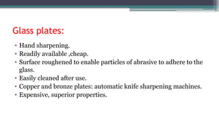
Glass plates:
• Handsharpening.
• Readily available ,cheap.
• Surface roughened to enable particles of abrasive to adhere to the
glass.
• Easily cleaned after use.
• Copper and bronze plates: automatic knife sharpening machines.
• Expensive, superior properties.
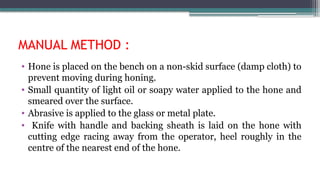
MANUAL METHOD :
•Hone is placed on the bench on a non-skid surface (damp cloth) to
prevent moving during honing.
• Small quantity of light oil or soapy water applied to the hone and
smeared over the surface.
• Abrasive is applied to the glass or metal plate.
• Knife with handle and backing sheath is laid on the hone with
cutting edge racing away from the operator, heel roughly in the
centre of the nearest end of the hone.
41.
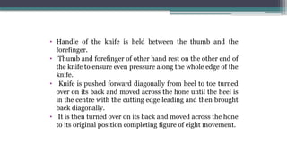
• Handle ofthe knife is held between the thumb and the
forefinger.
• Thumb and forefinger of other hand rest on the other end of
the knife to ensure even pressure along the whole edge of the
knife.
• Knife is pushed forward diagonally from heel to toe turned
over on its back and moved across the hone until the heel is
in the centre with the cutting edge leading and then brought
back diagonally.
• It is then turned over on its back and moved across the hone
to its original position completing figure of eight movement.
42.
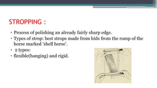
STROPPING :
• Processof polishing an already fairly sharp edge.
• Types of strop: best strops made from hide from the rump of the
horse marked 'shell horse'.
• 2 types:
• flexible(hanging) and rigid.
43.
Flexible type:
• Backof the strop is made of canvas and is intended to support the
leather during stropping.
• Strops should be kept soft by applying a small quantity of
vegetable oil into the back of the leather.
44.
Rigid type:
• Singleleather strop stretched over a wooden frame to give a
standard tension or a block of wood about 12x2x2 inches in size
having a handle at one end with four grades of leather or even a
soft stone cemented on each side.
• The sides of these strops are numbered and the knife is stropped
on No1, then No2 and so on finishing on the finest leather.
45.
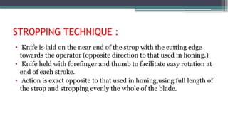
STROPPING TECHNIQUE :
•Knife is laid on the near end of the strop with the cutting edge
towards the operator (opposite direction to that used in honing.)
• Knife held with forefinger and thumb to facilitate easy rotation at
end of each stroke.
• Action is exact opposite to that used in honing,using full length of
the strop and stropping evenly the whole of the blade.
46.
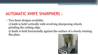
AUTOMATIC KNIFE SHARPNERS:
• Two basic designs available.
1) knife is held vertically with revolving sharpening wheels
grinding the cutting edge.
2) knife is held horizontally against the surface of a slowly rotating
flat plate.
47.
Microtomy- paraffin wax
•Factors involved in producing good paraffin-wax sections :
Temperature:
• Tissues are more easily sectioned at a lower temperature than that
of the atmosphere.
• Lowering temperature brings tissues of differing composition to a
more uniform consistency, degree of hardness-ensures a uniform
cutting process.
• Blocks are cooled by keeping, face down on ice-tray (2-3min).
48.
Knife angle
• Greaterthe rake angle(flatter the knife)more likely is a smooth
plastic flow type cutting action.
• Higher rake angles are more suitable to softer tissues.
• Lower rake angles for harder tissues.
49.
Speed of cutting:
•Soft tissues are cut more easily at a slow speed.
• Hard tissues are cut easily at a little fast rate.
• If sections are cut at too fast speed, compression will become
more marked.
• If cut too slowly, difficult to maintain the rhythmic action
required.
50.
PARAFFIN SECTION CUTTING
•Equipment required:
• Microtome.
• Flotation (water bath).
• Slide drying oven or hot plate.
• Fine pointed or curved forceps.
• Sable or camel haired brush.
• Scalpel.
• Slide rack.
• Clean slides.
• Teasing needle.
• Ice tray.
• Chemical resistant pencil or pen.
51.
CUTTING TECHNIQUE
• Insertappropriate knife in the knife-holder of the microtome and
screw it tightly in position.
• Correctly set the adjustable knife angles.
• Fix the block in the block holder of the microtome.
• Move the block holder forward or upward until the paraffin wax is
almost touching the knife edge.
• Ensure that the whole surface of the block will move parallel to the
edge of the knife.
52.
• Trim theexcess wax from the block surface and
expose the tissue, advance the block by setting
the thickness to about 15 micrometer.
• Care should be taken not to trim too coarsely as
A)Small biopsies may be lost.
B) tissue in the block may be torn giving rise to
considerable artefact.
C) unsuspected small foci of calcification may
cause tears in the tissue and nicks in the knife.
53.
• Once thesurface of tissue has been revealed
proceed to trim.
• Replace the trimming edge by a sharp one and
check it is tightly secured.
• Reset the thickness gauge to 4-5 micrometer.
• Insert the block to be cut and tighten securely.
• Bring the block face up until it nearly touches
the knife edge.
54.
• Paraffin-wax embeddedtissue, sections are
normally cut at a thickness of 4-5 micrometer.
• Thicker sections (10-20
micrometer) :demonstrate certain features of the
central nervous system.
• Thin sections(1-2 micrometer): for examining
highly cellular tissue such as lymph nodes.
55.
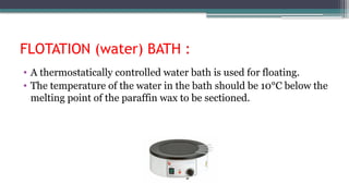
FLOTATION (water) BATH:
• A thermostatically controlled water bath is used for floating.
• The temperature of the water in the bath should be 10°C below the
melting point of the paraffin wax to be sectioned.
56.
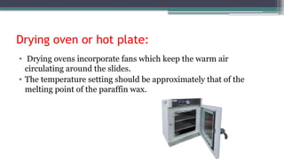
Drying oven orhot plate:
• Drying ovens incorporate fans which keep the warm air
circulating around the slides.
• The temperature setting should be approximately that of the
melting point of the paraffin wax.
57.
• If theoven is too hot there maybe distortion to
the cells causing dark pyknotic nuclei, nuclear
bubbling and loss of nuclear detail.
• Care should be taken when drying delicate or
central nervous system tissue, 37°Cfor 24 hours
is recommended.
58.
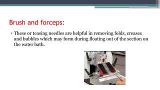
Brush and forceps:
•These or teasing needles are helpful in removing folds, creases
and bubbles which may form during floating out of the section on
the water bath.
59.
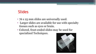
• 76 x25 mm slides are universally used.
• Larger slides are available for use with specialty
tissues such as eyes or brain.
• Colored, frost-ended slides may be used for
specialized Techniques.
Slides
60.
SECTION ADHESIVES
Poly-L-lysine (PLL):
•It is available as a 0.1% solution which is further diluted for use
1:10 with distilled water.
3-aminopropyltriethoxysilane (APES):
• Slides are dipped in a 2% solution of APES in acetone, drained,
dipped in acetone and drained again.
• These slides are useful for cytology and specimens which may be
bloody or contain proteinaceous material.
61.
Charged or plusslides:
• Laboratories often use slides which have been manufactured with
a permanent positive charge.
• These slides are superior in their resistance to cell and tissue loss
during staining or pre-treatments such as enzyme and antigen
retrieval.
62.
PRACTICAL MICROTOMY
Setup ofthe microtome:
• The water bath and the microtome should be ergonomically positioned to
reduce stress and tension.
• The water bath may be filled with distilled or tap water and adjusted to the
correct temperature for the paraffin wax.
• The blade should be sharp and defect free.
• The recommended clearance angle varies from 2-4° for paraffin and 5-7° for
frozen sections.
• The correct angle reduces friction as the blade passes through the block,
preventing compression of the section.
63.
SECTIONING
Trimming the tissueblocks :
• The paraffin block may be faced or "rough cut" by setting the
micrometer at15-30 um or by advancing the block using the coarse
feed mechanism.
Cutting sections
• Blocksshould be arranged in numerical order on an ice tray or
cooling mechanism, cooling both the tissue and the paraffin wax
to a consistent temperature.
• A small amount of water is absorbed into the tissue causing slight
swelling and making sectioning easier Over-soaking may cause
expansion and distortion of the tissue section
• Ideally, successive sections will stick edge to edge, forming a
ribbon. If the entire block is to be sectioned and retained, the
ribbons are stored.
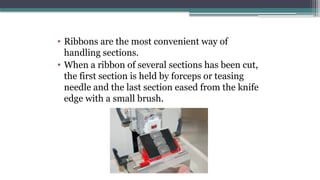
66.
• Ribbons arethe most convenient way of
handling sections.
• When a ribbon of several sections has been cut,
the first section is held by forceps or teasing
needle and the last section eased from the knife
edge with a small brush.
67.
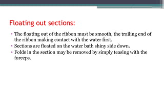
Floating out sections:
•The floating out of the ribbon must be smooth, the trailing end of
the ribbon making contact with the water first.
• Sections are floated on the water bath shiny side down.
• Folds in the section may be removed by simply teasing with the
forceps.
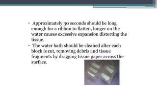
68.
• Approximately 30seconds should be long
enough for a ribbon to flatten, longer on the
water causes excessive expansion distorting the
tissue.
• The water bath should be cleaned after each
block is cut, removing debris and tissue
fragments by dragging tissue paper across the
surface.
69.
Drying sections:
• Thesmall amount of water held under the section will allow further
flattening to occur when heat is applied to dry the section.
• The temperature should be at the melting point of the paraffin wax.
• It is important to eliminate over-heating during the drying stage as
cellular details may be compromised.
• Less distortion will occur if the temperature is reduced and the time
prolonged.
• Overnight drying at 37°C or room temperature is recommended for
many tissues.
70.
Cutting hard tissues
•Cutting difficulties are more likely due to poor fixation or
overprocessing.
• Prolonged soaking of the block or exposing the block surface to
running tap water for 30 minutes overcomes many of the
problems associated with cutting hard tissues.
• A slight reduction in the knife angle may also yield results.
• Softening agents may be used on the surface of the block.
71.
Surface decalcification:
• Whensmall foci of calcium are present in the tissue. Section, the
block may be removed from the chuck after rough cutting the
tissue and placed face down in a dish which contains a small
amount of decalcifying solution.
• The exposure time will vary depending on the tissue.
• The block is rinsed well, blotted dry, chilled and returned to the
microtome.
• Diagnostic materials may be compromised if over decalcification
occurs.
REFERENCES
• Handbook ofHistopathological and Histochemical Techniques
(including museum techniques) THIRD EDITION - C. F. A.
CULLING.
• Bancroft’s THEORY and PRACTICE of HISTOLOGICAL
TECHNIQUESEIGHTH EDITION - S. Kim Suvarna, Christopher
Layton, John D. Bancroft.
• Sy J, Ang LC. Microtomy: Cutting Formalin-Fixed, Paraffin-
Embedded Sections. Methods Mol Biol. 2019;1897:269-278. doi:
10.1007/978-1-4939-8935-5_23. PMID: 30539451.
• A review of artifacts in histopathology. J Oral Maxillofac Pathol.
2018 May-Aug;22(2):279. doi: 10.4103/jomfp.JOMP
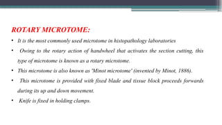
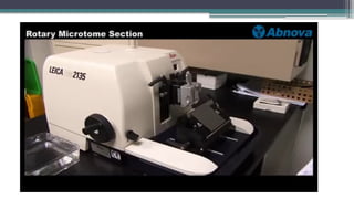
ROTARY MICROTOME:
• Itis the most commonly used microtome in histopathology laboratories
• Owing to the rotary action of handwheel that activates the section cutting, this
type of microtome is known as a rotary microtome.
• This microtome is also known as 'Minot microtome' (invented by Minot, 1886).
• This microtome is provided with fixed blade and tissue block proceeds forwards
during its up and down movement.
• Knife is fixed in holding clamps.
82.
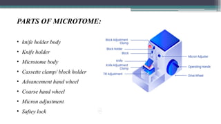
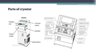
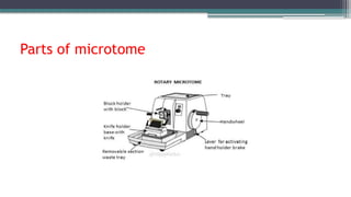
PARTS OF MICROTOME:
•knife holder body
• Knife holder
• Microtome body
• Cassette clamp/ block holder
• Advancement hand wheel
• Coarse hand wheel
• Micron adjustment
• Saftey lock
83.
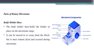
Parts of RotaryMicrotome
Knife Holder Base
• The knife holder base holds the holder in
place on the microtome stage.
• It can be moved to or away from the block,
but it must remain fixed and secured during
microtomy.
84.
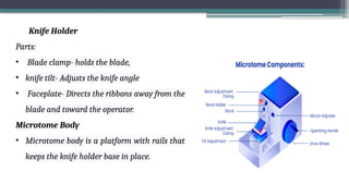
Knife Holder
Parts:
• Bladeclamp- holds the blade,
• knife tilt- Adjusts the knife angle
• Faceplate- Directs the ribbons away from the
blade and toward the operator.
Microtome Body
• Microtome body is a platform with rails that
keeps the knife holder base in place.
85.
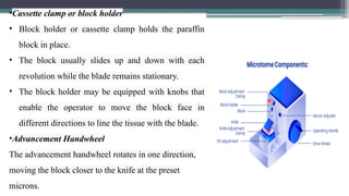
•Cassette clamp orblock holder
• Block holder or cassette clamp holds the paraffin
block in place.
• The block usually slides up and down with each
revolution while the blade remains stationary.
• The block holder may be equipped with knobs that
enable the operator to move the block face in
different directions to line the tissue with the blade.
•Advancement Handwheel
The advancement handwheel rotates in one direction,
moving the block closer to the knife at the preset
microns.
86.
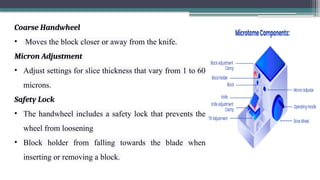
Coarse Handwheel
• Movesthe block closer or away from the knife.
Micron Adjustment
• Adjust settings for slice thickness that vary from 1 to 60
microns.
Safety Lock
• The handwheel includes a safety lock that prevents the
wheel from loosening
• Block holder from falling towards the blade when
inserting or removing a block.
87.
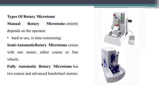
Types Of RotaryMicrotome
Manual Rotary Microtome entirely
depends on the operator.
• hard to use, is time-consuming.
Semi-AutomaticRotary Microtome comes
with one motor, either coarse or fine
wheels.
Fully Automatic Rotary Microtome has
two coarse and advanced handwheel motors.
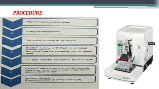
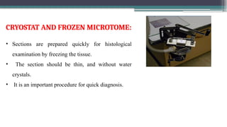
• Sections areprepared quickly for histological
examination by freezing the tissue.
• The section should be thin, and without water
crystals.
• It is an important procedure for quick diagnosis.
CRYOSTAT AND FROZEN MICROTOME:
92.
INDICATIONS:
• Quick diagnosis
•Study the margins of cancer
• Enzyme histochemistry
• Immunohistochemistry
• Detection of lipid
• Some molecular procedure
93.
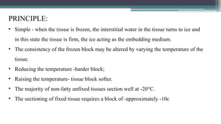
PRINCIPLE:
• Simple -when the tissue is frozen, the interstitial water in the tissue turns to ice and
in this state the tissue is firm, the ice acting as the embedding medium.
• The consistency of the frozen block may be altered by varying the temperature of the
tissue.
• Reducing the temperature -harder block;
• Raising the temperature- tissue block softer.
• The majority of non-fatty unfixed tissues section well at -20°C.
• The sectioning of fixed tissue requires a block of -approximately -10c
94.
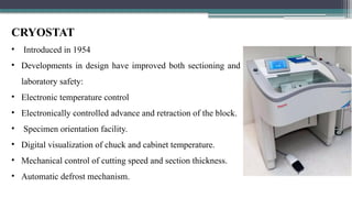
CRYOSTAT
• Introduced in1954
• Developments in design have improved both sectioning and
laboratory safety:
• Electronic temperature control
• Electronically controlled advance and retraction of the block.
• Specimen orientation facility.
• Digital visualization of chuck and cabinet temperature.
• Mechanical control of cutting speed and section thickness.
• Automatic defrost mechanism.
Freezing of freshunfixed tissue:
• The fresh tissue should be frozen as rapidly as possible without creating freeze
artifacts. Suitable techniques include
• Liquefied nitrogen (-190°C).
• Isopentane (2-methylbutane) cooled by liquid nitrogen (-150°C).
• Dry ice (-70°C).
• Carbon dioxide gas (-70°C)
• Aerosol sprays (-50°C).
• Internal freezing shelf or bar
97.
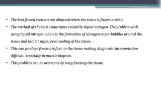
• The bestfrozen sections are obtained when the tissue is frozen quickly.
• The method of choice is isopentane cooled by liquid nitrogen. The problem with
using liquid nitrogen alone is the formation of nitrogen vapor bubbles around the
tissue and inhibit rapid, even cooling of the tissue.
• This can produce freeze artifact. in the tissue making diagnostic interpretation
difficult, especially in muscle biopsies.
• This problem can be overcome by snap freezing the tissue.
98.
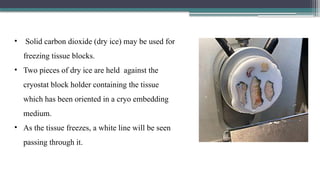
• Solid carbondioxide (dry ice) may be used for
freezing tissue blocks.
• Two pieces of dry ice are held against the
cryostat block holder containing the tissue
which has been oriented in a cryo embedding
medium.
• As the tissue freezes, a white line will be seen
passing through it.
99.
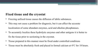
Fixed tissue andthe cryostat
• Freezing unfixed tissue causes the diffusion of labile substances.
• This may not cause a problem for diagnosis, but it can affect the accurate
localization of some abundant enzymes, acid and alkaline phosphatases.
• To accurately localize these hydrolytic enzymes and other antigens it is better to
fix the tissue prior to sectioning in the cryostat.
• Tissue prepared in this manner must be fixed under controlled conditions
• Tissue must be absolutely fresh and placed in formal calcium at 4°C for 18 hours.
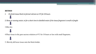
METHOD
1. Fix freshtissue block in formal calcium at 4°C for 18 hours.
2. Rinse in running water, or for a short time in distilled water if the tissue fragment is small or fragile
3.Blot dry.
4.Place tissue in the gum sucrose solution at 4°C for 18 hours or less with small fragments.
5. Blot dry &Freeze tissue onto the block holder.
102.
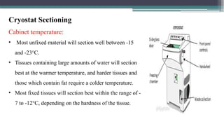
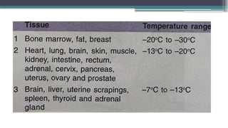
Cryostat Sectioning
Cabinet temperature:
•Most unfixed material will section well between -15
and -23°C.
• Tissues containing large amounts of water will section
best at the warmer temperature, and harder tissues and
those which contain fat require a colder temperature.
• Most fixed tissues will section best within the range of -
7 to -12°C, depending on the hardness of the tissue.
104.
Blade or knife:
•Disposable blades have become routine
in most clinical laboratories.
• They produce a perfect, sharp edge.
• They can be rapidly cooled because of
their size.
105.
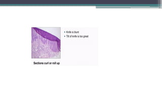
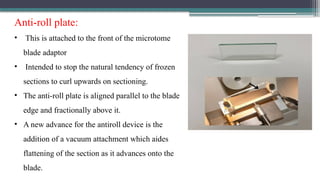
Anti-roll plate:
• Thisis attached to the front of the microtome
blade adaptor
• Intended to stop the natural tendency of frozen
sections to curl upwards on sectioning.
• The anti-roll plate is aligned parallel to the blade
edge and fractionally above it.
• A new advance for the antiroll device is the
addition of a vacuum attachment which aides
flattening of the section as it advances onto the
blade.
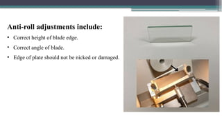
106.
Anti-roll adjustments include:
•Correct height of blade edge.
• Correct angle of blade.
• Edge of plate should not be nicked or damaged.
107.
Sectioning technique:
Factors:
• Speed,tissue type and temperature of the block and cabinet play important role in
frozen section.
• The cut section rests on the surface of the blade holder, a room temperature slide is held
above it and electrostatic attraction causes the tissue to adhere to the slide.
• Tissues which require harsh or lengthy staining procedures-positively charged or
coated slides should be used.
• These slides are usually coated with gelatin- formaldehyde (equal parts of 1% gelatin
and 2% formaldehyde) or poly-L-lysine (0.01% aqueous solution).
109.
Rapid biopsy forintraoperative diagnosis
• Frozen sections provide a valuable tool in the rapid diagnosis of tissues during surgery.
• The pathologist selects a piece of tissue and this is frozen using any of the techniques.
• slide is immediately submerged in cold acetone or 95% alcohol and the sections are
stained immediately by a rapid hematoxylin and eosin (H&E), methylene blue or
polychrome stain.
• With properly cut and stained slides a rapid diagnosis can be made for the surgeon.
110.
FREEZE DRYING :
Principle
•Freeze drying involves rapid freezing of the fresh tissue at -160°C followed by
removal of water (ice) by sublimation at -40°C. This tissue is then rapidly fixed by
vapours embedded in media.
111.
The technique minimizes:
•Loss of soluble substances.
• Displacement of cell constituents.
• Chemical alteration of reactive groups.
• Denaturation of proteins.
• Destruction or inactivation of enzymes.
112.
PROCEDURE:
1. Quenching
It israpid freezing at -160°C. It stops all chemical reactions.
2. Drying/sublimation
The water component of tissue is removed at -40°C by sublimation under a vacuum of
193 mPa or more.
3. Fixation
Dried tissue is brought to room temperature.
It is fixed by vapours of fixative (formaldehyde, glutaraldehyde or osmium tetraoxide).
113.
Applications for cryostat:
• Immunohistochemical methods.
• Demonstration of hydrolytic enzymes.
• Fluorescent antibody studies.
• Autoradiography.
• Microspectrofluorimetry of autofluorescent substances.
• Formaldehyde-induced fluorescence.
• Mucosubstances.
• Proteins* Scanning electron microscopy.
114.
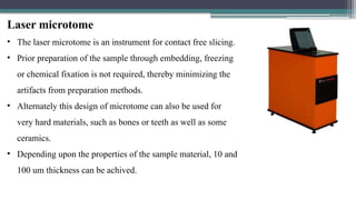
Laser microtome
• Thelaser microtome is an instrument for contact free slicing.
• Prior preparation of the sample through embedding, freezing
or chemical fixation is not required, thereby minimizing the
artifacts from preparation methods.
• Alternately this design of microtome can also be used for
very hard materials, such as bones or teeth as well as some
ceramics.
• Depending upon the properties of the sample material, 10 and
100 um thickness can be achived.
115.
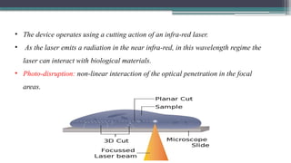
• The deviceoperates using a cutting action of an infra-red laser.
• As the laser emits a radiation in the near infra-red, in this wavelength regime the
laser can interact with biological materials.
• Photo-disruption: non-linear interaction of the optical penetration in the focal
areas.
116.
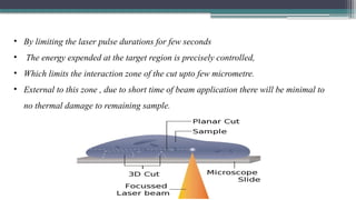
• By limitingthe laser pulse durations for few seconds
• The energy expended at the target region is precisely controlled,
• Which limits the interaction zone of the cut upto few micrometre.
• External to this zone , due to short time of beam application there will be minimal to
no thermal damage to remaining sample.


![MICROTOME KNIFE SHARPENING :
• Manual procedure or automatic procedure.
1) Abrasive grinding of the facets [HONING]
2)Polishing [STROPPING]](https://image.slidesharecdn.com/microtomykkk-250811020940-5ac0c4f9/85/microtomy-kkk-presenting-to-cryst-in-gl-36-320.jpg)