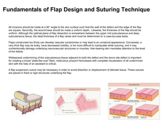
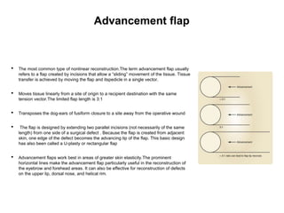
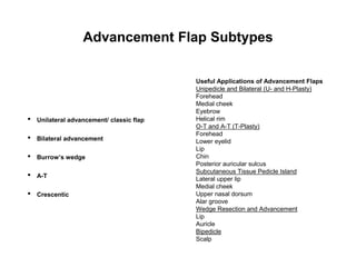
2. DEFINITIONS OF FLAP COMPONENTS
Body The skin being advanced, transposed, rotated, interpolated or imported into the primary defect
Pedicle Also known as the vascular base of the flap, it is the conduit of the vascular supply that will remain intact and maintain
the vascularization of the body of the flap intraoperatively and for the early postoperative period
Primary defect The area deficient in skin that will be reconstructed by the movement of the body of the flap
Secondary defect (donor site defect) The area devoid of skin created by the movement of the body of the flap
Primary flap movement The movement of the flap body into the defect
Secondary flap movement The tissue movement necessary to close the void or donor site defect created by the movement of
the body of the flap
Primary tension vector The direction of the force tending to counteract the movement of the body of the flap
Secondary tension vector The direction of the force created by the closure of the donor site defect
FLAPS CLASSIFICATION
3. All incisions should be made at a 90Ā° angle to the skin surface such that the wall of the defect and the edge of the flap
are square. Secondly, the wound base should be made a uniform depth.. Likewise, the thickness of the flap should be
uniform. Although the optimal plane of flap dissection is somewhere between the upper mid subcutaneous and deep
subcutaneous tissue, the ideal thickness of a flap varies and must be determined on a case-by-case basis.
Flaps constructed too thinly can develop vascular compromise or may lead to an unnatural appearance. Conversely, a
very thick flap may be bulky, have decreased mobility, or be more difficult to manipulate while suturing, and it may
unintentionally damage underlying neurovascular structures or muscles. Hair-bearing skin mandates attention to the level
of the follicle.
Widespread undermining of the subcutaneous tissue adjacent to both the defect and the donor site defect is important
for creating a broad āplate-like scarā.Next, meticulous pinpoint hemostasis with complete visualization of all undermined
skin with the help of an assistant is critical.
A flap suspension suture may be necessary in order to avoid distortion or displacement of delicate tissue. These sutures
are placed in fixed or rigid structures underlying the flap.
Fundamentals of Flap Design and Suturing Technique
4. ā¢ In part, the degree of wound closure tension is
related to skin extensibility. Skin extensibility is the
lengthening of skin under tension due to stretching of
elastic fibers.
ā¢ It is advantageous to recruit skin for repair of
a wound in areas of maximum skin extensibility. These
areas are identified by the lines of maximum extensibility
(LME).
ā¢ The orientation of skin excisions and repair of
wounds are usually made parallel to RSTLs when
possible. RSTLs result from orientation of collagen fibers
of the skin and are manifested in the aging face as skin
creases and wrinkles . RSTLs are perpendicular to LME.
ā¢ Orienting them this way places the maximum wound
closure tension parallel to LME and perpendicular
to RSTLs. This orientation results in wound repair that is
performed with the least amount of wound closure tension.
5. Linear closure
ā¢ Primary closures are most used in surgeries with smaller number of phases, non-aggressive
histological subtypes and lesions located outside the nose. Nasal tumors or with aggressive histology
are associated with the use of other closure modalities.
ā¢ The M-plasty is a tissue-sparing technique that permits primary wound closure while limiting incision
length. The technique can also be used to remove standing cutaneous dog ear deformities.
ā¢ The S-plasty is a common technique used by surgeons to repair defects that traverse convex surfaces.
The technique described provides a simple, and convenient, approach to removing dog-ears in a
curvilinear fashion
ā¢ Z-plasty is a plastic surgery technique used to improve the functional and cosmetic appearance of
scars. It involves the creation of two triangular flaps of equal dimension that are then transposed. 1. For
a basic z-plasty, the triangular flaps are created using an angle of 60 degrees
6. Linear/primary closure application by topography
Scalp
ā¢ In general, defects of the scalp up to 3 cm in diameterm ay be repaired with primary wound closure. However, to accomplish
this, wide undermining of the scalp in the= subgaleal plane is required. This can often be accomplished by blunt finger
dissection.
Nose
ā¢ Primary wound closure is possible for smaller skin defects of the nose, especially in the elderly patient, in whom nasal skin
tends to be redundant, or when defects are very small relative to the size of the nose. Defects that are 1.0 cm or smaller and
located on the dorsum or sidewall are repaired most easily. This is accomplished by advancement of opposing wound
margins after wide undermining of the skin adjacent to the defect.
ā¢ Primary wound closure of defects of the tip and supratip area frequentlydistorts the free margins of the nostril. Fortunately,
alar cartilages typically have sufficient intrinsic strength tocounteract this distortion; over time, the nostril margin usually
returns to its natural position.
Eyebrows
ā¢ Excision of eyelid neoplasia frequently requires reconstruction of a full thicknessdefect. In the simplest case, horizontal laxity
of the eyelid and canthal tendons will permit direct closure under reasonable tension. This is usually possible with defects
that involve 33% or less of the horizontal length of the eyelid, although some authors find it useful in up to 50% of the
horizontal length. A few additional millimeters can be gained with concomitant lateral canthotomy,although this may result in
inferior displacement of the reconstructed lower eyelid.
ā¢ The advantage of primary wound closure is preservation of the eyelid margin and lashes in a single-stage procedure. As
with repair of other multilaminar structures, the primary strategy is to restoretissue integrity by matching corresponding
layers
7. Linear closure
Cheek
ā¢ Primary wound closure is an excellent option for repair of smaller cutaneous defects of the cheek. Typically, it
results in a linear suture line with minimal tension on the closure because wide skin undermining is possible. In
general, undermining up to 4 cm beyond the borders of the defect improves tissue recruitment, whereas
undermining more widely adds little to decreasing wound closure tension Primary wound closure is a particularly
good option when the resulting scar can be placed in the borders ofthe cheek aesthetic region or parallel to
RSTLs. Primary wound closure is a useful technique also for repairing small (1 cm or less) buccal skin
Lips
ā¢ Primary closure in the lip zone are oriented with the long axis parallel to relaxed skin tension lines of that region of
the lip. From an aesthetic viewpoint,it is preferable that fusiform excisions be confined within the boundaries of the
lip, avoiding extension beyond the melolabial or mental crease lines. An M-plasty at the end of the excision line
may be useful in avoiding the need to extend an incision beyond the aesthetic borders of the lips or into the
vermilion .This will require closure of two small and slightly diverging incision lines near the involved end. M-plasty
may also reduce tissue redundancies, particularly near the free margin of the lip.
defects.
8. Linear closure
Forehead
ā¢ The paramedian forehead have fewer good options for repair compared with the central part of the
forehead. Horizontally oriented primary wound repair is restricted by the vertical height of the defect
and thus thedegree of eyebrow elevation resulting from wound closure. Eyebrow elevation, in turn, is
related to the degree of forehead skin and scalp redundancy and mobility. Eyebrow elevation from
horizontally oriented wound repair is also influenced by the size and position of the cutaneous
defect(tumors larger than 3 cm)
ā¢ The lateral forehead aesthetic unit begins at the level ofthe midpupillary line and extends to the lateral
eyebrow,where it joins with the skin of the temple .Unique to this area is the transition in topography
from the convexity of the paramedian forehead to a flat lateral forehead and then slightly concave
temple. These surface features along with greater elasticity of lateral forehead and temple skin
compared with central forehead skinprovide a greater number of reconstructive alternatives.
ā¢ For vertically oriented midline or near-midline defects, primary wound closure orientedwith the long
axis of the wound is often optimal . . Whetherprimary wound closure is in a vertical or horizontal
orientation, advancement of wound margins causes the development of standing cutaneous
deformities. These deformities may be excised by a W-plasty or M-plasty when feasible. Vertically
oriented primary wound closure of midline cutaneous forehead defects allows extensive undermining
beneath the fascia of the frontalis muscle without compromis eof motor or sensory nerve function
ā¢ Primary wound closure of cutaneous defects located at the junction of the lateral forehead and temple
may be oriented horizontally so that scars rest in the RSTLs of the crowās-feet wrinkles
9. Advancement flap
ā¢ The most common type of nonlinear reconstruction.The term advancement flap usually
refers to a flap created by incisions that allow a āslidingā movement of the tissue. Tissue
transfer is achieved by moving the flap and itspedicle in a single vector.
ā¢ Moves tissue linearly from a site of origin to a recipient destination with the same
tension vector.The limited flap length is 3:1
ā¢ Transposes the dog-ears of fusiform closure to a site away from the operative wound
ā¢ The flap is designed by extending two parallel incisions (not necessarily of the same
length) from one side of a surgical defect . Because the flap is created from adjacent
skin, one edge of the defect becomes the advancing tip of the flap. This basic design
has also been called a U-plasty or rectangular flap
ā¢ Advancement flaps work best in areas of greater skin elasticity.The prominent
horizontal lines make the advancement flap particularly useful in the reconstruction of
the eyebrow and forehead areas. It can also be effective for reconstruction of defects
on the upper lip, dorsal nose, and helical rim.
10. Advancement flap
ā¢ Factors affecting motion: tissue quality, fibrous septae, fascia, dermal
tethering
ā¢ Mode of release: narrower pedicle or fasciotomy, undermining, incision
which lengthening of flap lyses attachments
ā¢ Redistribution of standing tissue cones: dog-ears may be placed anywhere
along the length of the flaps or redistributed by halving. The incisions lines
should fall in natural creases and folds.
ā¢ The length of the flap is determined by the desired repositioning of the dog-
ears, to a lesser extend, the need to decrease the superior and inferior
restraining forces
ā¢ The viability of the flap depends of ability to advance without tension As with
all local flaps, it is important to evaluate the adjacent skin for matching
qualities of texture, color, consistency, and hair growth
ā¢ Facial structures that are likely to be distorted if secondary tissue movement
is excessive include the vermilion, nostril margin, lower and upper eyelids,
and eyebrows
11. Advancement Flap Subtypes
ā¢ Unilateral advancement/ classic flap
ā¢ Bilateral advancement
ā¢ Burrowās wedge
ā¢ A-T
ā¢ Crescentic
Useful Applications of Advancement Flaps
Unipedicle and Bilateral (U- and H-Plasty)
Forehead
Medial cheek
Eyebrow
Helical rim
O-T and A-T (T-Plasty)
Forehead
Lower eyelid
Lip
Chin
Posterior auricular sulcus
Subcutaneous Tissue Pedicle Island
Lateral upper lip
Medial cheek
Upper nasal dorsum
Alar groove
Wedge Resection and Advancement
Lip
Auricle
Bipedicle
Scalp
12. Classic Advancement Flap
ā¢ Is shaped as a rectangle with dog-ears removed near the base of the flap pedicle
ā¢ Recommended for operative wounds of the nasal dorsum but elevate the nose tip
ā¢ Recommended for eyebrow or for forehead reconstructions
13. Bilateral Advancement Flap
ā¢ Is an H-plasty and use two rectangular flaps
advanced from the opposite sides of the
operative wound
ā¢ Bilateral unipedicle advancement flaps are
commonly combined to close various defects,
resulting in H- or T-shaped repairs, depending
on the configuration of the defect and the
number of incisions used. Repair in this manner
is often referred to as H-plasty.
ā¢ Half-buried horizontal tip suture used to close
the Burow triangular defects. Note that standing
cutaneous deformity is removed at different
locations superior and inferior to advancement
flaps.. Epidermal closure with simple running or
interrupted sutures.
14. Burrowās wedge flap
ā¢ Is an a unilateral or bilateral advancement in
which a triangular standing tissue cone is
removed above and below the operative wound
and the horizontal limbās of the advancement
are taken as a tangents from the opposing edge
of the operative wound. An invert dog-ear is
then removed as the flap slides along the
horizontal incision.
ā¢ Recommended: on vermillion of the lip, on
infraorbital crease, in front of the ear, above the
brow
ā¢ Flaps that displace Burowās triangles to a
āconvenientā location distant from the defect
include the single tangent advancement flap
(Burowās flap), the bilateral single tangent
advancement flap (A-to-T flap), the double
tangent advancement flap (U-flap), the bilateral
double tangent advancement flap (H-flap), and
rotation flaps (e.g. the dorsal nasal or Rigor flap,
Mustarde flap, helical advancement flap)
15. A-T or O-T
Advancement Flap
ā¢ Is a bilateral advancement flap where the inverted dog-ears are on the margins and in the
center, in opposite direction is the triangle defect
ā¢ Is an elegant way to translocate a dog-ear redundancy
ā¢ Used to close round defect on the temple where the incision line is keeps away from more
visible portion of the lower temple and is hide in an existing forehead/ temple rhytid
ā¢ Used above the lateral brow
16. Crescentic Advancement Flap
ā¢ Described by Webster in 1950ās
ā¢ Is similar to unilateral advancement flaps but instead
by employing a rigid linear advancement with
opposing dog-ear removals, the repair is designed
with several curved incisions on the advancing flap.
The curved edge stretches out and lengthens, thus
often eliminating the need for dog-ear removal
ā¢ Used in the perialar region, on the upper lip and
along forehead and the final suture lines can be
placed within cosmetic subunits
17. Rotation Flaps
ā Rotation flap with excision of an equalizing Burow triangle.
ā Opposing arrows indicate point of greatest wound
closure tension.
ā Large arrow indicates closure site of the equalizing Burow
triangle excision.
ā To minimize standing cutaneous deformity that develops at
base of rotation flap, most ideal configuration of triangular defect
has height-to-width ratio of 2 : 1.
ā To create ideal condition for use of rotation flap, defect enlarged
by removing normal tissue and converting defect into triangle with
height-to-width ratio of 2 : 1.
ā For symmetric rotation flap, height of triangular defect should be
0.5 to 1 times the radius
of curvature of flapās border.
18. Rotation flaps
ā¢ Is the simplest method of redirecting tension vectors and reducing wound closure tensions
ā¢ Rotation flaps redistribute and redirect closure tension from a primary defect to a secondary arciform
defect
ā¢ Rotation flaps are transferred by a pivotal motionrather than depending on stretching of tissue for
movement as required by advancement flaps
ā¢ Rotation flaps allow for displacement of dog-ears to more favorable locations
ā¢ Well designed, create scars lines that are hidden along facial boundaries or within RSTL
ā¢ Is simple rotation or rotation with advancement
ā¢ The pedicle and the mobility of the point of pivotal restraint are the kay determining factors of the success
or failure
ā¢ they are ideal for scalp and preauricular region
19. Classic Rotation Flap
ā¢ The triangulated defect is covered with an
arciform closure. Primary motion closes some of
the original operative wound and is mandatory to
create a secondary defect. The second defect is
closed with secondary flap movement
ā¢ For more facilitate wound closure, undermining
not only the flap pedicle but also the point of
pivotal restraint
ā¢ A flap backcut allows for greater release of
tension and easier rotation. The backcut may be
close with V-Y pasty or Z-plasty
ā¢ A grater flap curvature leads to more rotation
mobility, a grater redirection of tension but
decreases vascular supply by cutting into the flap
pedicle. The arc should transect less than a
quarter of a circle
20. Bilateral Rotation Flaps
ā¢ May be used to symmetrically distribute the tension of closing the secondary defect on both sides of
the primary wound
ā¢ Useful for scalp area or for larger defects of the upper forehead or for wounds below the mental
crease of the chin because approximates a natural cosmetic junction
21. Bilateral Opposing Rotation Flaps
O-Z Plasty
ā¢ Is designed with opposing rotation flaps one of which takes its origin from one side of theoperative
wound and the other as a mirror image from the opposing side of the defect
ā¢ Used on the scalp because of the inelasticity of scalp tissue, any movement of tissue by
advancement is limited. Use of more than one rotation flap is advantageous because it
recruits scalp tissue for reconstruction from different locations and the burden of closing the secondary
defectis shared by the number of flaps used.To use two or more rotation flaps for repair of scalp defects,
the defect must be located in the central aspect
of the scalp.
ā¢ For peripherally located defects in the scalp, it is often difficult or impossible to design two effective
rotation flaps. In such instances, a single flap is designed larger than would be required if two flaps
could be used.
22. Rotation Flap Regional Applications
ā¢ Scalp: - bilateral rotation symmetrically O-Z
ā¢ Forehead and temple: O-T, Tenzel flap
ā¢ Infraorbital rotation: Mustarde flap; Tenzel flap
ā¢ Cheek: pre-auricular flap, or from the temple and lateral cheek to close the defect on infraorbital region
ā¢ Chin: bilateral symmetrically rotation flap
ā¢ Dorsal nasal rotation from the medial cheek or from glabella
ā¢ Lateral upper lip with skin from nasolabial fold
23. ā¢ The dorsal nasal flap is a modified rotation flap that recruits redundant skin from the glabella. The flap can be
used to repair skin defects of the nasal tip, dorsum, and sidewall.
ā¢ The donor site in the glabella may be closed by V-Y advancement, Z-plasty, or, more commonly, primary
repair
ā¢ Thinning of the flap in the area of the medial canthus is critical to improve the mismatch of
thickness between the thin skin of the medial canthus and the thicker glabellar skin of the flap.
Dorsal Nasal Flap
24. Transposition Flap
ā¢ A transposition flap is a tissue reorientation flap characterized by the lifting of skin from an adjacent donor
site and reorienting it through the transposition of the flap over a peninsula of skin between the donor site
and the defect. There is a pivot point at the base of this flap .
ā¢ The tissue movement depends on the presence of laxity in adjacent tissue . Although intrinsic elasticity of
the flap skin may facilitate the ease of execution, without ample laxity in the donor site, the transposition
cannot be performed
ā¢ Upon careful examination, Z-plasties can be found within the design of transposition flaps . Therefore, just
as with Z-plasties, transposition flaps work because the reorientation of the tissue results in lengthening of
tissue in the direction of the skin deficit at the expense of the laxity in the donor site.
ā¢ The most common areas where transposition flaps are used include the nasal dorsum and sidewalls, the
medial and lateral canthi, the lateral forehead, temple, cheek, the perioral region, the inferior chin, and the
dorsal hand
25. Transposition Flaps
ā¢ The transposition flap is elevated from an area of laxity, lifted over an adjacent area of tissue and transposed
into an operative wound.
ā¢ Transposition flaps accomplish tension redirection and redistribution. The flap are able to redirect tension
vectors completely perpendicular to the needed primary motion to repair in order to avoid tension on crucial
structure or free margin.
ā¢ Basically, a transposition flap is created where the donor site is remote from the defect and the flap is moved
about the pedicle or transposed over intervening normal tissue and into the defect.
ā¢ It provides the advantage of immediate flap inset of color-matched and texture-matched tissue with often a
direct linear closure of the donor site. The difficulties of transposition flaps also involve obscuring natural
contours or providing distortions at site distant to the defect. Two examples of these would be a melolabial
transposition flap that distorts the contour of the cheekānose junction or a poorly designed bilobed flap, which
distorts the medial eyelid
ā¢ Ex: the rhombic flap, the banner or unilobed flaps such as the single-staged nasolabial flap, the bilobed flap, the
trilobed flap and the 30* angle flap
26. Rhombic Flap
ā¢ Classic rhombic flap is constructed around a geometric four-side length, and tip angles equal to 60* and
120*
ā¢ It is formed by extending the short diameter of the defect beyond the flap for a length equivalent to one of
the sides. Then the line will continue parallel to one of the existing sides of the defect
ā¢ Ideally , two sides of the rhombic defect are oriented perpendicular to the skin tension lines
27. Rhombic Flap
Webster 30* angle flap
ā¢ The Webster flap can reduce tensions from pivotal restraint and minimizes the formation of a
redundant tissue cone, but tension is now exerted on the flap in a side-to-side manner as the original
wound is closed and may lead to ischemia.
ā¢ The most common modification of 60* apical angle is 30* angle flap. In this instance the base of the
flap is only about one-half the size of the recipient defect
28. Rhombic Flap
Dufourmentel flap
ā¢ The flap achives closure with less tension at the vertical apex.
ā¢ The flap will cover a rhombic defect with tip angles of 60* and 120*. But the flap is taken off at a
substantially obtuse angle rather then at 120*.
29. Rhombic flap
Limberg flap
ā¢ A rhombic flap may be constructed around a
circular defect. The flap and/ or defect is
trimmed appropriately to achieve a good fit.
ā¢ By oversizing the leading edge of the rhombic
design, the tip of the flap can overcome pivotal
restraint and may be transposed into place
under little to no tension.
ā¢ By increasing the flap length and takeoff angle
beyond 120*, both tips of the flap reach their
destination under less tension
30. Multiple rhombic flap
ā¢ For large operative wounds, particularly on the
scalp and extremities
ā¢ There are multiple opposed rhombic flaps
ā¢ The flap recruits laxity from multiple directions
simultaneously
31. Banner transposition flaps
ā¢ Banner transposition flaps are an extensions of the rhombic flap in which the takeover angle is basically
a tangent at close to 180*, both limbs of the flap are elongated and the apical angle is more acute
ā¢ Almost all of the tension is directed along the secondary defect, and the flap with an pedicle will be
rotate and transpose into place under minimal effort
ā¢ Indicated for repair of defects on lateral nose, infraorbital area, on the helical rim
ā¢ Ex: single-stage nasolabial flap
32. Banner transposition flap
Nasolabial flap/ Unilobed transposition flap
ā¢ Is an excellent and aesthetic single-staged
repair for larger defects of the lower third of the
nose
ā¢ The upper portion of the flap is closed as an
advancement flap. The lower portion transposes
over the lateral ala. The vertical dog ear is
excised on the nose. The flap extends along the
entire nasolabial fold. The flap is extensively
thinned and tacked into place
33. Bilobed flap
ā¢ The bilobed flap uses a double transposition flap to entirely redirect tension vectors and also redistribute tissue
redundancy
ā¢ A bilobed flap allows utilization of this laxity through tissue reorientation using two lobes . The underlying mechanism
of tissue reorientation is a series of Z-plasties inherent in this flap. Since tissue gain in Z-plasties is in the direction of
the middle arm of the Z-plasty, a substantial reorientation and lengthening of tissue in the desirable direction for repair
of the defect occurs without distortion
ā¢ The flap courses through an arc of 90-100*. The lobes are of nearly equal size. A standing tissue cone is removed at
the apex to prevent dog-ear formation and to allow for easier pedicle rotation
ā¢ Zitelli bilobed flap has 90* axis
34. Trilobed flap
ā¢ It is designed with the principles of the bilobed flap. The additional lobe is added when
the location of the defect would force the angle between the primary and secondary
lobes to be significantly grater than 60* and the entire defect flap would subsume an
angle of grater than 90*.
ā¢ It is used to repair far distant nasal operative wounds in one stage
35. Island Pedicle Flap
ā¢ The island pedicle advancement flap is unique in that it is an advancement of tissue oriented in a plane perpendicular
to the skin and at 90Ā° to the more traditional advancement or rotation flaps. In this case, the tissue redundancy
created at the pivot point of the flap is buried in the subcutaneous plane at the advancing edge of the flap
ā¢ Fundamentally, an island pedicle flap reconfigures the typical roundish shape of the wound to a sharp, angled,
geometric shape that is easily closed without standing cones or distortion of surrounding structures . Movement of
this flap is entirely dependent on the elasticity of the tissue that comprises the flap.
ā¢ The Island Pedicle Flap is most commonly done on nasal and perinasal closures where free margins are at risk for
distortion.
36. Island Pedicle Flaps
ā¢ May be nourished by a number of pedicle subtypes: a deep pedicle, a laterally
based fatty pedicle, a lateral pedicle based on a muscular sling, a purely vascular
based on a named artery and vein
ā¢ Variants: single deep central pedicle, a single lateral pedicle, and dual lateral
pedicles
37. The rotate and transposed islands
ā¢ The island flap with a reliable unilateral pedicle may be rotated and/ or transposed into
operative wound.
ā¢ It is used in upper nasal bridge and medial canthus using forehead tissue
ā¢ On the nose a unilateral nasalis sling may be used to rotate and transpose an island from the
upper nasal bridge to an substantially distal operative wound.
38. Turnover Island Pedicle flap
Spear flap
ā¢ The Spear flap is elevated on a deep pedicle with only a single muscular attachment at its superior
aspect where it is richly supplied by perforators from the angular artery. The flap is turned over to
recreate internal lining and then folded onto itself to create the exterior of the lateral ala.
40. Staged Pedicle/Interpolation Flap
ā¢ Most operative wounds are suitably repaired with local adjacent tissue transfers. When a local flap is not
able to achieve an aesthetic closure a staged interpolation flap may be utilized.
ā¢ Interpolation flaps are delayed pedicle flap reconstructions in which a flap tissue is elevated at one
location and transposed over intervening skin to an operative wound
ā¢ The flap is left in plase for a defined period of the time in order to vascularize from the recipient bed, and
the pedicle is severed.
ā¢ Indicated in wounds of the nose, ear and lip
ā¢ The recipient site for a pedicle flap must be deep enough to receive a full-thickness operative repair and
must have the appropriate support structure to recreate a native aesthetic contour
ā¢ The pedicle flap have an excellent arterial blood supply
ā¢ This flap require patients to live for at least several weeks with a deformed appearance and is necessary
at least one revision
41. Paramedian forehead flap
ā¢ Indicated for deeper nasal operative wound.
ā¢ The flap use the upper forehead skin which has
a sebaceous quality and a good vascular
supply
ā¢ The supratrochlear artery is located just medial
to the brow within the glabellar crease at a
distance of about 1.5-2 cm from the mid glabella
ā¢ The flap contains skin, subcutis, musculature
and an axial vessel
42. Foldover Flap
ā¢ Indicated for a full-thickness operative wound on
the nose
ā¢ The wound will be stabilised with a cartilage
brace
ā¢ The forhead flap is designed both for internal
and external lining
ā¢ The flap is sutured internally for lining and then
folded over on itself
43. Other interpolation flap
ā¢ Cheek to nose interpolation flap with nasolabial
pedicle inferiorly based
ā¢ Ear reconstruction with pedicle flap
ā¢ Lip reconstruction with pedicle flaps ( the Abbe flap)