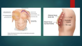
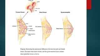
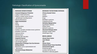
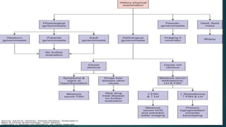
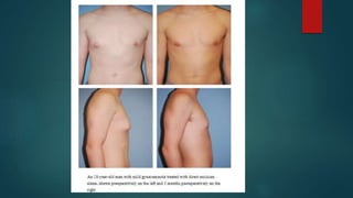
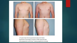
Gynecomastia
DR SAMIK SHARMA
Definition
 Gynecomastia refers to the enlargement of the male breast due to a
proliferation of ductal, stromal, and/or fatty tissue.
 Pseudogynecomastia (lipomastia): Excessive development of the male
breast from subareolar fat deposition without glandular proliferation.
Anatomy of the Gynecomastia Tissue
and Its Clinical Significance
 Unlike the female breast, male glandular tissue contains no lobules.
 Healthy men typically have
 predominantly fatty tissue with few ducts and stroma,
 which is distinctly different from women’s breasts where ducts, stroma, and
glandular tissue predominant
 Gynecomastia is the benign enlargement of this glandular tissue.
 Transient gynecomastia presents as a florid pattern with an increase in
budding ducts and cellular stroma.
Demographics
 It is the most common breast problem in men.
 Most males experience some degree of gynecomastia during their lives,
but definition and reporting are inconsistent.
 Overall incidence is 32%-36% (up to 40% in autopsy series).
 Up to 65% of adolescent boys are affected.
 Up to 75% of cases are bilateral.
Clinical Classification
 Idiopathic (most common)
 Physiologic
 Neonatal: Circulating maternal estrogens via placenta
 Pubertal: Relative excess of plasma estradiol versus testosterone
 Elderly: Decreased circulating testosterone, peripheral aromatization of
testosterone to estrogen
Pathologic Classification of Gynecomastia
Pharmacologic
Histologic Classification
 Degrees of stromal and ductal proliferation
 • Florid: Increased budding ducts and cellular stroma; seen in gynecomastia
that is present for approximately 4 months
 • Intermediate: Overlapping florid and fibrous patterns
 • Fibrous: Extensive stromal fibrosis, minimal ductal proliferation; seen in
gynecomastia thatis present for 1 year
 The duration of gynecomastia is the most important factor in determining
the pathologic picture.
 Florid pattern response well to medical treatment, while fibrous type
needs surgical management.
Risk of Malignant Transformation
 No increased cancer risk for patients without Klinefelter’s syndrome
 Klinefelter’s syndrome: Risk increases 60-fold (1:1000 increases to 1:400)
Preoperative Workup
 History
 Age of onset
 Duration
 Symptoms
 Medications
 Recreational drug
 Physical Examination
 Fat versus glandular predominance, laterality, ptosis, skin excess, masses
 Differentiate true gynecomastia from pseudogynecomastia.
 Rule out breast cancer.
 Testicular examination
 Thyroid, liver, or other abdominal masses
 Lack of male hair distribution
 Feminizing characteristics
Clinical manifestation
 Gynecomastia usually manifests as a palpable, discrete button of tissue
radiating from beneath the nipple and areola.
 Gynecomastia feels “gritty” when the breast is pinched between the thumb
and forefinger.
 Fatty tissue (Pseudogynecomastia), unlike gynecomastia, will not cause
resistance until the nipple is reached. (Difference in clinical examination).
 Additional diagnostic tests
 General labs: Blood testosterone, TSH/free thyroxine, luteinizing hormone (LH),
(LH), human chorionic gonadotropin (hCG)
 Small, firm testes: Karyotyping, because hallmark finding in cases of 47,XXY
 Abnormal testicular examination results or mass: Testicular ultrasound, b-
human chorionic gonadotropin (b-hCG), follicle-stimulating hormone (FSH), LH,
serum testosterone, orestradiol
Classification
 Webster: Based on tissue type
 Type I: Glandular
 Type II: Fatty and glandular mix
 Type III: Simple fatty
 Simon et al: Based on degree of tissue and skin excess
 Type I: Minor breast enlargement without skin excess
 Type II: Moderate breast enlargement
 IIa: Without skin excess
 IIb: With minor skin redundancy
 Type III: Gross breast enlargement with skin excess creating a pendulous
breast
Staging (Rohrich et al)
 Grade I: Minimal hypertrophy (250 g) with no ptosis
 Ia: Primarily glandular
 Ib: Primarily fibrous
 Grade II: Moderate hypertrophy (250-500 g) with no ptosis
 Ia: Primarily glandular
 Ib: Primarily fibrous
 Grade III: Severe hypertrophy (500 g) with grade I ptosis
 Grade IV: Severe hypertrophy with grade II or III ptosis
Management
Idiopathic
 Observation
 Gynecomastia often regresses after 3-18 months of enlargement.
 Gynecomastia that is present for 12 months rarely regresses because of tissue
fibrosis.
 Weight reduction if obese
 Surgery
Physiologic
 Tamoxifen (Nolvadex) is particularly useful for “lump”-type gynecomastia.
 Clomiphene citrate is used with limited success.
 Aromatase inhibitors (letrozole, testolactone) show therapeutic potential
in early trials but efficacy is not confirmed.
 Testosterone has limited ability to induce regression once gynecomastia
established.
 Danazol, a synthetic testosterone derivative, has been used with some
success in pubertal gynecomastia; however, side-effect potential is high.
Pathologic
 Removal of testicular tumors
 Correction of underlying causes or disease
 Remove offending drug in pharmalogical causes
Radiation
 Prophylactic breast irradiation may have some benefit in reducing the
incidence of gynecomastia in patients on long-term antiandrogen therapy
(e.g., prostate cancer).
 Risk of malignancy with this type of exposure is not defined.
 There is no indication for use in cases of idiopathic gynecomastia.
Surgical Options
Techniques
 Periareolar or intraareolar incisions
 Offer direct access for tissue resection
 Transaxillary incisions
 For select cases; limited operative exposure
 All types of dermal and glandular pedicles for nipple relocation
 Free-nipple grafting
 Allows en bloc resection of skin and breast tissue
 Traditional and ultrasound-assisted liposuction (UAL)
 Basic tenets of UAL treatment
 Superwet infiltration
 Stab incisions at inferolateral aspects of intramuscular fat
 Radial pattern across entire chest
 Disruption of intramuscular fat
 Avoid upper lateral pectoral region
 Dressing: Two layers of Topifoam compression vest for 4 weeks continuously,
then 4 more weeks at night
 Arthroscopic shaver
 Allows precise resection of fibrous tissue after liposuction or en bloc resection
Mild to Moderate Gynecomastia (Simon Grades I
and IIa, and Rohrich Grades I and II)
 Milder forms of gynecomastia
 are quite common and 65% of men are thought to have some degree of
gynecomastia,
 the proportion seeking surgical treatment is much lower
 patients with minimal glandular hypertrophy typically have little skin excess
and are treated readily with liposuction, frequently as the definitive treatment
 some patients present with small amounts of fibrous gynecomastia well
localized under the nipple and may be effectively treated with direct excision
using a small periareolar incision
 Power-assisted (PAL) and ultrasound-assisted liposuction (UAL)
technologies have increased the extent of tissue removal capable by
liposuction
 The improved skin retraction often associated with UAL has allowed it to
be used as a definitive treatment modality in many cases.
 Relatively high energy levels are used, with higher levels focused under the
nipple to assist with removing the fibrous glandular tissue.
 Access ports are generally made at the lateral inframammary fold (IMF)
combined with a periareolar or upper anterior axillary incision to allow for
cross-hatching
 Vibration amplification of sound energy at resonance- (VASER) assisted
liposuction
 is a newer form of UAL technology utilizing the application of alternating
ultrasonic energy
 which is considered by many practitioners to be a safer modality when treating
fibrous areas close to the skin surface
 Even in mild forms of gynecomastia, there may be a residual glandular
component that needs to be addressed after liposuction.
 A low threshold for direct excision should be maintained, as residual firm,
subareolar glandular tissue can be a great source of patient dissatisfaction.
 In patients for whom liposuction is not entirely sufficient, glandular tissue
requires direct excision either primarily, or in a staged fashion,
accompanying skin excision.
 “Pull through” technique described by Morselli, which describes blindly
dissecting the breast parenchymal tissue from the skin and pectoral fascia
then grasping and pulling the tissue out through the liposuction incision
for piecemeal excision through liposuction incision
 reinforced or laser-sharpened cannulas, have been explored to allow for
removal of this fibrous component as well.
 Most recently, the use of orthopedic arthroscopic shavers has gained
popularity, patients were treated with liposuction followed by arthroscopic
shaver morcellation to address any residual glandular component
Severe Gynecomastia (Simon Grade IIB
and Rohrich Grade III)
 Patients with severe gynecomastia will usually require some form of skin
resection
 Many techniques utilizing various skin excision patterns and pedicles
similar to those used in female mastopexy and reduction mammoplasty
have been used
 Letterman described the use of an oblique Dufourmentel-Mouly procedure
based on an elliptical incision with a bipedicled dermal areolar flap.
 however, has large oblique extra-areolar scar extending laterally, much like a
traditional mastectomy.
 Wise-pattern scars and glandular pedicles similar to those in traditional
reduction mammoplasty.
 These techniques present many drawbacks for male patients.
 Not only do these procedures often leave excess glandular tissue behind, but
the Wise pattern frequently causes coning of the breast and unacceptable
scarring.
 Circumareolar excision allow for skin excision without extra-areolar
scarring. This technique relies on a central mound with an intercostal
blood supply through the prepectoral fascia.
 Similar techniques, as described by Botta, recognize nipple–areolar
complex (NAC) viability on the subdermal plexus alone and utilize
superiorly based dermo-glandular flaps, allowing for a more uniform
excision of breast tissue
Severe Gynecomastia with Grade II or III Ptosis
(Simon Grade III and Rohrich Grade IV)
 In cases of severe gynecomastia with marked ptosis and extreme skin
excess, other incisions which cause extra-areolar scarring may be required.
 For these cases, the most reliable and simple technique is breast
amputation using an IMF incision with free nipple grafting
 These procedures have become increasingly necessary as the population
of massive-weight-loss patients grows.
 The new NAC is drawn as a horizontal oval ∼3 cm in diameter at the fourth
intercostal space; however, the size varies depending on the patient's
overall body habitus.
 Again, initial liposuction is performed. The incision is made in the IMF and
carried down to the level of the pectoral fascia. The glandular tissue is then
dissected off the fascia to the level of the second intercostal space. The
nipple is removed as a full-thickness graft.
 The superior flap is then pulled inferiorly to estimate and mark the excision
of excess skin and soft tissue. The IMF incision is closed in layers over a
drain. Finally, the nipple is placed onto a de-epithelialized bed and secured
with a bolster dressing.
Complications/Outcomes
 Potential complications from gynecomastia surgery include
 hematoma,
 seroma,
 infection,
 inadequate resection,
 poor scarring,
 contour deformity,
 breast asymmetry,
 sensory changes,
 pain.
 Overall complication rate has been reported between 14.5 to 53%, with
hematoma being the most common.
 The most common late complication is inadequate resection of glandular
tissue or skin.
Summary
 Many patients will obtain adequate correction with less invasive techniques
such as ultrasound-assisted liposuction.
 In patients with significant skin excess or poor skin elasticity, excellent
results can be achieved with a single-stage procedure using a combination
of UAL, direct excision, and periareolar skin excision to flatten the breast
and remove the excess skin and volume.
 Only patients with the most severe excess skin redundancy require
techniques involving an elliptical skin excision or breast amputation with
free nipple grafting.
Thank you

Gynecomastia

  • 1.
  • 2.
    Definition  Gynecomastia refersto the enlargement of the male breast due to a proliferation of ductal, stromal, and/or fatty tissue.  Pseudogynecomastia (lipomastia): Excessive development of the male breast from subareolar fat deposition without glandular proliferation.
  • 3.
    Anatomy of theGynecomastia Tissue and Its Clinical Significance  Unlike the female breast, male glandular tissue contains no lobules.  Healthy men typically have  predominantly fatty tissue with few ducts and stroma,  which is distinctly different from women’s breasts where ducts, stroma, and glandular tissue predominant  Gynecomastia is the benign enlargement of this glandular tissue.  Transient gynecomastia presents as a florid pattern with an increase in budding ducts and cellular stroma.
  • 6.
    Demographics  It isthe most common breast problem in men.  Most males experience some degree of gynecomastia during their lives, but definition and reporting are inconsistent.  Overall incidence is 32%-36% (up to 40% in autopsy series).  Up to 65% of adolescent boys are affected.  Up to 75% of cases are bilateral.
  • 7.
    Clinical Classification  Idiopathic(most common)  Physiologic  Neonatal: Circulating maternal estrogens via placenta  Pubertal: Relative excess of plasma estradiol versus testosterone  Elderly: Decreased circulating testosterone, peripheral aromatization of testosterone to estrogen
  • 8.
  • 9.
  • 10.
    Histologic Classification  Degreesof stromal and ductal proliferation  • Florid: Increased budding ducts and cellular stroma; seen in gynecomastia that is present for approximately 4 months  • Intermediate: Overlapping florid and fibrous patterns  • Fibrous: Extensive stromal fibrosis, minimal ductal proliferation; seen in gynecomastia thatis present for 1 year
  • 11.
     The durationof gynecomastia is the most important factor in determining the pathologic picture.  Florid pattern response well to medical treatment, while fibrous type needs surgical management.
  • 12.
    Risk of MalignantTransformation  No increased cancer risk for patients without Klinefelter’s syndrome  Klinefelter’s syndrome: Risk increases 60-fold (1:1000 increases to 1:400)
  • 13.
    Preoperative Workup  History Age of onset  Duration  Symptoms  Medications  Recreational drug
  • 14.
     Physical Examination Fat versus glandular predominance, laterality, ptosis, skin excess, masses  Differentiate true gynecomastia from pseudogynecomastia.  Rule out breast cancer.  Testicular examination  Thyroid, liver, or other abdominal masses  Lack of male hair distribution  Feminizing characteristics
  • 15.
    Clinical manifestation  Gynecomastiausually manifests as a palpable, discrete button of tissue radiating from beneath the nipple and areola.  Gynecomastia feels “gritty” when the breast is pinched between the thumb and forefinger.  Fatty tissue (Pseudogynecomastia), unlike gynecomastia, will not cause resistance until the nipple is reached. (Difference in clinical examination).
  • 17.
     Additional diagnostictests  General labs: Blood testosterone, TSH/free thyroxine, luteinizing hormone (LH), (LH), human chorionic gonadotropin (hCG)  Small, firm testes: Karyotyping, because hallmark finding in cases of 47,XXY  Abnormal testicular examination results or mass: Testicular ultrasound, b- human chorionic gonadotropin (b-hCG), follicle-stimulating hormone (FSH), LH, serum testosterone, orestradiol
  • 19.
    Classification  Webster: Basedon tissue type  Type I: Glandular  Type II: Fatty and glandular mix  Type III: Simple fatty
  • 20.
     Simon etal: Based on degree of tissue and skin excess  Type I: Minor breast enlargement without skin excess  Type II: Moderate breast enlargement  IIa: Without skin excess  IIb: With minor skin redundancy  Type III: Gross breast enlargement with skin excess creating a pendulous breast
  • 21.
    Staging (Rohrich etal)  Grade I: Minimal hypertrophy (250 g) with no ptosis  Ia: Primarily glandular  Ib: Primarily fibrous  Grade II: Moderate hypertrophy (250-500 g) with no ptosis  Ia: Primarily glandular  Ib: Primarily fibrous  Grade III: Severe hypertrophy (500 g) with grade I ptosis  Grade IV: Severe hypertrophy with grade II or III ptosis
  • 22.
    Management Idiopathic  Observation  Gynecomastiaoften regresses after 3-18 months of enlargement.  Gynecomastia that is present for 12 months rarely regresses because of tissue fibrosis.  Weight reduction if obese  Surgery
  • 23.
    Physiologic  Tamoxifen (Nolvadex)is particularly useful for “lump”-type gynecomastia.  Clomiphene citrate is used with limited success.  Aromatase inhibitors (letrozole, testolactone) show therapeutic potential in early trials but efficacy is not confirmed.  Testosterone has limited ability to induce regression once gynecomastia established.  Danazol, a synthetic testosterone derivative, has been used with some success in pubertal gynecomastia; however, side-effect potential is high.
  • 24.
    Pathologic  Removal oftesticular tumors  Correction of underlying causes or disease  Remove offending drug in pharmalogical causes
  • 25.
    Radiation  Prophylactic breastirradiation may have some benefit in reducing the incidence of gynecomastia in patients on long-term antiandrogen therapy (e.g., prostate cancer).  Risk of malignancy with this type of exposure is not defined.  There is no indication for use in cases of idiopathic gynecomastia.
  • 26.
    Surgical Options Techniques  Periareolaror intraareolar incisions  Offer direct access for tissue resection  Transaxillary incisions  For select cases; limited operative exposure  All types of dermal and glandular pedicles for nipple relocation  Free-nipple grafting  Allows en bloc resection of skin and breast tissue
  • 27.
     Traditional andultrasound-assisted liposuction (UAL)  Basic tenets of UAL treatment  Superwet infiltration  Stab incisions at inferolateral aspects of intramuscular fat  Radial pattern across entire chest  Disruption of intramuscular fat  Avoid upper lateral pectoral region  Dressing: Two layers of Topifoam compression vest for 4 weeks continuously, then 4 more weeks at night
  • 28.
     Arthroscopic shaver Allows precise resection of fibrous tissue after liposuction or en bloc resection
  • 29.
    Mild to ModerateGynecomastia (Simon Grades I and IIa, and Rohrich Grades I and II)  Milder forms of gynecomastia  are quite common and 65% of men are thought to have some degree of gynecomastia,  the proportion seeking surgical treatment is much lower  patients with minimal glandular hypertrophy typically have little skin excess and are treated readily with liposuction, frequently as the definitive treatment  some patients present with small amounts of fibrous gynecomastia well localized under the nipple and may be effectively treated with direct excision using a small periareolar incision
  • 31.
     Power-assisted (PAL)and ultrasound-assisted liposuction (UAL) technologies have increased the extent of tissue removal capable by liposuction  The improved skin retraction often associated with UAL has allowed it to be used as a definitive treatment modality in many cases.  Relatively high energy levels are used, with higher levels focused under the nipple to assist with removing the fibrous glandular tissue.  Access ports are generally made at the lateral inframammary fold (IMF) combined with a periareolar or upper anterior axillary incision to allow for cross-hatching
  • 32.
     Vibration amplificationof sound energy at resonance- (VASER) assisted liposuction  is a newer form of UAL technology utilizing the application of alternating ultrasonic energy  which is considered by many practitioners to be a safer modality when treating fibrous areas close to the skin surface
  • 34.
     Even inmild forms of gynecomastia, there may be a residual glandular component that needs to be addressed after liposuction.  A low threshold for direct excision should be maintained, as residual firm, subareolar glandular tissue can be a great source of patient dissatisfaction.  In patients for whom liposuction is not entirely sufficient, glandular tissue requires direct excision either primarily, or in a staged fashion, accompanying skin excision.  “Pull through” technique described by Morselli, which describes blindly dissecting the breast parenchymal tissue from the skin and pectoral fascia then grasping and pulling the tissue out through the liposuction incision for piecemeal excision through liposuction incision
  • 35.
     reinforced orlaser-sharpened cannulas, have been explored to allow for removal of this fibrous component as well.  Most recently, the use of orthopedic arthroscopic shavers has gained popularity, patients were treated with liposuction followed by arthroscopic shaver morcellation to address any residual glandular component
  • 36.
    Severe Gynecomastia (SimonGrade IIB and Rohrich Grade III)  Patients with severe gynecomastia will usually require some form of skin resection  Many techniques utilizing various skin excision patterns and pedicles similar to those used in female mastopexy and reduction mammoplasty have been used  Letterman described the use of an oblique Dufourmentel-Mouly procedure based on an elliptical incision with a bipedicled dermal areolar flap.  however, has large oblique extra-areolar scar extending laterally, much like a traditional mastectomy.
  • 37.
     Wise-pattern scarsand glandular pedicles similar to those in traditional reduction mammoplasty.  These techniques present many drawbacks for male patients.  Not only do these procedures often leave excess glandular tissue behind, but the Wise pattern frequently causes coning of the breast and unacceptable scarring.
  • 38.
     Circumareolar excisionallow for skin excision without extra-areolar scarring. This technique relies on a central mound with an intercostal blood supply through the prepectoral fascia.  Similar techniques, as described by Botta, recognize nipple–areolar complex (NAC) viability on the subdermal plexus alone and utilize superiorly based dermo-glandular flaps, allowing for a more uniform excision of breast tissue
  • 41.
    Severe Gynecomastia withGrade II or III Ptosis (Simon Grade III and Rohrich Grade IV)
  • 42.
     In casesof severe gynecomastia with marked ptosis and extreme skin excess, other incisions which cause extra-areolar scarring may be required.  For these cases, the most reliable and simple technique is breast amputation using an IMF incision with free nipple grafting  These procedures have become increasingly necessary as the population of massive-weight-loss patients grows.
  • 44.
     The newNAC is drawn as a horizontal oval ∼3 cm in diameter at the fourth intercostal space; however, the size varies depending on the patient's overall body habitus.  Again, initial liposuction is performed. The incision is made in the IMF and carried down to the level of the pectoral fascia. The glandular tissue is then dissected off the fascia to the level of the second intercostal space. The nipple is removed as a full-thickness graft.  The superior flap is then pulled inferiorly to estimate and mark the excision of excess skin and soft tissue. The IMF incision is closed in layers over a drain. Finally, the nipple is placed onto a de-epithelialized bed and secured with a bolster dressing.
  • 45.
    Complications/Outcomes  Potential complicationsfrom gynecomastia surgery include  hematoma,  seroma,  infection,  inadequate resection,  poor scarring,  contour deformity,  breast asymmetry,  sensory changes,  pain.
  • 46.
     Overall complicationrate has been reported between 14.5 to 53%, with hematoma being the most common.  The most common late complication is inadequate resection of glandular tissue or skin.
  • 47.
    Summary  Many patientswill obtain adequate correction with less invasive techniques such as ultrasound-assisted liposuction.  In patients with significant skin excess or poor skin elasticity, excellent results can be achieved with a single-stage procedure using a combination of UAL, direct excision, and periareolar skin excision to flatten the breast and remove the excess skin and volume.  Only patients with the most severe excess skin redundancy require techniques involving an elliptical skin excision or breast amputation with free nipple grafting.
  • 48.