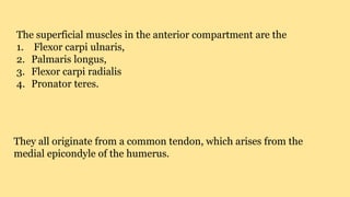
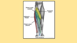
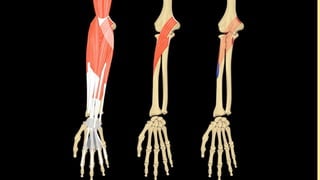
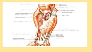
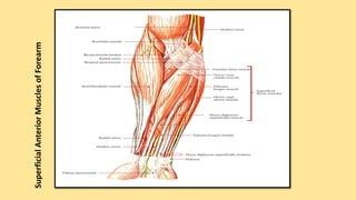
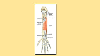
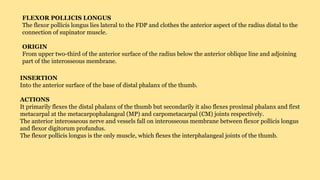
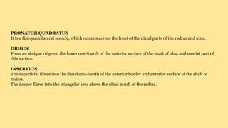
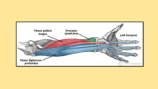
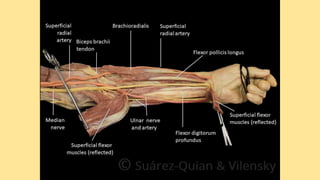
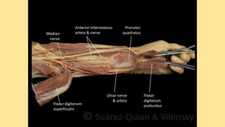
Anterior (Flexor) Compartment
FOREARM MUSCLES
1. superficial,
2. intermediate
3. deep
03 CATEGORIES
SUPERFICIAL COMPARTMENT
The superficial muscles in the anterior compartment are the
1. Flexor carpi ulnaris,
2. Palmaris longus,
3. Flexor carpi radialis
4. Pronator teres.
They all originate from a common tendon, which arises from the
medial epicondyle of the humerus.
PRONATOR TERES
PRONATOR TERES SYNDROME
https://www.youtube.com/watch?v=BjIab-huqgUForearm Muscles
https://www.earthslab.com/anatomy/muscles-anterior-front-
forearm/
Teres โ€“ as appears in ligamentum teres. Teres is a Latin word
that means round and smooth or cylindrical.
Flexor Carpi Radialis
PALMARIS LONGUS
FLEXOR CARPI ULNARIS
SuperficialAnteriorMusclesofForearm
https://www.youtube.com/watch?v=WlDR-5Aiuyspronator teres
https://www.youtube.com/watch?v=sQAUX_5SjRcAnatomy Of The Pronator Teres Muscle
https://www.youtube.com/watch?v=ZqhO1dzqTtYPronator Teres Syndrome
https://www.orthobullets.com/hand/6020/pronator-syndromePronator Syndrome
INTERMEDIATE COMPARTMENT
The flexor digitorum superficialis is the only muscle of the
INTERMEDIATE COMPARTMENT. It can sometimes be classed as a
most cadavers it lies between the deep and superficial muscle layers.
The muscle is a good anatomical landmark in the forearm โ€“ the
median nerve and ulnar artery pass between its two heads, and then travel
ATTACHMENTS: It has two heads โ€“ one originates from the medial
humerus, the other from the radius. The muscle splits into four
tendons at the wrist, which travel through the carpal tunnel, and
attaches to the middle phalanges of the four fingers.
ACTIONS: Flexes the metacarpophalangeal joints and proximal
4 fingers, and flexes at the wrist.
INNERVATION: Median nerve.
FLEXOR DIGITORUM SUPERFICIALIS
Flexor Digitorum Superficialis https://www.youtube.com/watch?v=9lSOE1CbJYw
https://www.earthslab.com/anatomy/flexor-digitorum-
superficialis/
DEEP COMPARTMENT
FLEXOR DIGITORUM PROFUNDUS
The flexor digitorum profundus (FDP) is the most bulky and powerful muscle on
the front of forearm and supplies main gripping power to the hand.
ORIGIN
From upper three-fourth of the anterior and medial surfaces of the shaft of ulna
and nearby medial half of the interosseous membrane.
By an aponeurosis from upper three-fourth of the posterior border of ulna along
with flexor and extensor carpi ulnaris muscles.
From the medial side of olecranon and coronoid process of ulna.
INSERTION
On to the palmar aspect of the bases of distal phalanges of medial four digits. The actual mode of
injection is as follows: the muscle forms four tendons, which enter the palm by passing deep to
the flexor retinaculum. Opposite the proximal phalanx of corresponding digit, the tendon
perforates the tendon of flexor digitorum superficialis and passes forward to be inserted in
palmar surface of the distal phalanx.
NERVE SUPPLY
Medial half by the ulnar nerve.
Lateral half by the anterior interosseous nerve โ€“ a branch of the median nerve.
ACTIONS
FDP flexes the distal interphalangeal (DIP) joints of medial four digits. It also
helps to flex the wrist joint.
CLINICAL TESTING
The flexor digitorum profundus is examined by asking the patient
to flex the DIP joint, while holding the PIP joint in extension.
https://www.youtube.com/watch?v=FAlrNNCmaC0Anatomy Of The Flexor Digitorum Profundus Muscle
FLEXOR POLLICIS LONGUS
The flexor pollicis longus lies lateral to the FDP and clothes the anterior aspect of the radius distal to the
connection of supinator muscle.
ORIGIN
From upper two-third of the anterior surface of the radius below the anterior oblique line and adjoining
part of the interosseous membrane.
INSERTION
Into the anterior surface of the base of distal phalanx of the thumb.
ACTIONS
It primarily flexes the distal phalanx of the thumb but secondarily it also flexes proximal phalanx and first
metacarpal at the metacarpophalangeal (MP) and carpometacarpal (CM) joints respectively.
The anterior interosseous nerve and vessels fall on interosseous membrane between flexor pollicis longus
and flexor digitorum profundus.
The flexor pollicis longus is the only muscle, which flexes the interphalangeal joints of the thumb.
CLINICAL TESTING
The flexor pollicis longus is tested by inquiring the patient to flex the interphalangeal joint of the thumb,
while proximal phalanx of the thumb is kept in extension.
Anatomy Of the Flexor Pollicis Longus Muscle https://www.youtube.com/watch?v=3CJ77o6BvKU
PRONATOR QUADRATUS
It is a flat quadrilateral muscle, which extends across the front of the distal parts of the radius and ulna.
ORIGIN
From an oblique ridge on the lower one-fourth of the anterior surface of the shaft of ulna and medial part of
this surface.
INSERTION
The superficial fibres into the distal one-fourth of the anterior border and anterior surface of the shaft of
radius.
The deeper fibres into the triangular area above the ulnar notch of the radius.
NERVE SUPPLY
By anterior interosseous nerve.
ACTIONS
Pronator quadratus is the primary pronator of the forearm and is supported by pronator teres only in quick and
forceful pronation.
Anatomy Of The Pronator Quadratus https://www.youtube.com/watch?v=WqHYy7bFAA4
Forearm muscles anterior (flexor) compartment

Forearm muscles anterior (flexor) compartment

  • 1.
  • 2.
  • 3.
  • 4.
    The superficial musclesin the anterior compartment are the 1. Flexor carpi ulnaris, 2. Palmaris longus, 3. Flexor carpi radialis 4. Pronator teres. They all originate from a common tendon, which arises from the medial epicondyle of the humerus.
  • 6.
  • 7.
  • 10.
    https://www.youtube.com/watch?v=BjIab-huqgUForearm Muscles https://www.earthslab.com/anatomy/muscles-anterior-front- forearm/ Teres โ€“as appears in ligamentum teres. Teres is a Latin word that means round and smooth or cylindrical.
  • 11.
  • 12.
  • 13.
  • 14.
  • 15.
    https://www.youtube.com/watch?v=WlDR-5Aiuyspronator teres https://www.youtube.com/watch?v=sQAUX_5SjRcAnatomy OfThe Pronator Teres Muscle https://www.youtube.com/watch?v=ZqhO1dzqTtYPronator Teres Syndrome https://www.orthobullets.com/hand/6020/pronator-syndromePronator Syndrome
  • 16.
  • 17.
    The flexor digitorumsuperficialis is the only muscle of the INTERMEDIATE COMPARTMENT. It can sometimes be classed as a most cadavers it lies between the deep and superficial muscle layers. The muscle is a good anatomical landmark in the forearm โ€“ the median nerve and ulnar artery pass between its two heads, and then travel ATTACHMENTS: It has two heads โ€“ one originates from the medial humerus, the other from the radius. The muscle splits into four tendons at the wrist, which travel through the carpal tunnel, and attaches to the middle phalanges of the four fingers. ACTIONS: Flexes the metacarpophalangeal joints and proximal 4 fingers, and flexes at the wrist. INNERVATION: Median nerve.
  • 18.
  • 21.
    Flexor Digitorum Superficialishttps://www.youtube.com/watch?v=9lSOE1CbJYw https://www.earthslab.com/anatomy/flexor-digitorum- superficialis/
  • 23.
  • 24.
    FLEXOR DIGITORUM PROFUNDUS Theflexor digitorum profundus (FDP) is the most bulky and powerful muscle on the front of forearm and supplies main gripping power to the hand. ORIGIN From upper three-fourth of the anterior and medial surfaces of the shaft of ulna and nearby medial half of the interosseous membrane. By an aponeurosis from upper three-fourth of the posterior border of ulna along with flexor and extensor carpi ulnaris muscles. From the medial side of olecranon and coronoid process of ulna. INSERTION On to the palmar aspect of the bases of distal phalanges of medial four digits. The actual mode of injection is as follows: the muscle forms four tendons, which enter the palm by passing deep to the flexor retinaculum. Opposite the proximal phalanx of corresponding digit, the tendon perforates the tendon of flexor digitorum superficialis and passes forward to be inserted in palmar surface of the distal phalanx.
  • 25.
    NERVE SUPPLY Medial halfby the ulnar nerve. Lateral half by the anterior interosseous nerve โ€“ a branch of the median nerve. ACTIONS FDP flexes the distal interphalangeal (DIP) joints of medial four digits. It also helps to flex the wrist joint.
  • 26.
    CLINICAL TESTING The flexordigitorum profundus is examined by asking the patient to flex the DIP joint, while holding the PIP joint in extension.
  • 27.
  • 28.
    FLEXOR POLLICIS LONGUS Theflexor pollicis longus lies lateral to the FDP and clothes the anterior aspect of the radius distal to the connection of supinator muscle. ORIGIN From upper two-third of the anterior surface of the radius below the anterior oblique line and adjoining part of the interosseous membrane. INSERTION Into the anterior surface of the base of distal phalanx of the thumb. ACTIONS It primarily flexes the distal phalanx of the thumb but secondarily it also flexes proximal phalanx and first metacarpal at the metacarpophalangeal (MP) and carpometacarpal (CM) joints respectively. The anterior interosseous nerve and vessels fall on interosseous membrane between flexor pollicis longus and flexor digitorum profundus. The flexor pollicis longus is the only muscle, which flexes the interphalangeal joints of the thumb.
  • 29.
    CLINICAL TESTING The flexorpollicis longus is tested by inquiring the patient to flex the interphalangeal joint of the thumb, while proximal phalanx of the thumb is kept in extension.
  • 31.
    Anatomy Of theFlexor Pollicis Longus Muscle https://www.youtube.com/watch?v=3CJ77o6BvKU
  • 32.
    PRONATOR QUADRATUS It isa flat quadrilateral muscle, which extends across the front of the distal parts of the radius and ulna. ORIGIN From an oblique ridge on the lower one-fourth of the anterior surface of the shaft of ulna and medial part of this surface. INSERTION The superficial fibres into the distal one-fourth of the anterior border and anterior surface of the shaft of radius. The deeper fibres into the triangular area above the ulnar notch of the radius.
  • 33.
    NERVE SUPPLY By anteriorinterosseous nerve. ACTIONS Pronator quadratus is the primary pronator of the forearm and is supported by pronator teres only in quick and forceful pronation.
  • 35.
    Anatomy Of ThePronator Quadratus https://www.youtube.com/watch?v=WqHYy7bFAA4