
Recommended
More Related Content
What's hot
What's hot (20)
Similar to Front of forearm by vidya prashant
Similar to Front of forearm by vidya prashant (20)
Recently uploaded
Recently uploaded (20)
Front of forearm by vidya prashant
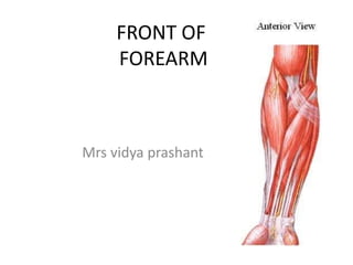
- 1. FRONT OF FOREARM Mrs vidya prashant
- 2. ANTERIOR (FLEXOR) COMPARTMENT Superficial flexors Five(5) in number common origin -- medial epicondyle Pronator Teres Flexor Carpi Radialis Palmaris Longus Flexor Carpi Ulnaris Flexor Digitorum Superficialis Deep Flexors 3 in number confined to radius and Ulna Flexor Pollicis Longus Flexor Digitorum Profundus Pronator Quadratus
- 3. Pronator Teres Origin Humeral Head :medial supracondylar ridge medial epicondyle Ulnar Head :medial border of coronoid process of ulna Insertion : middle of lateral surface of radius Nerve supply : median nerve Action: pronation of forearm weak flexor of elbow joint
- 4. Action : Pronation of Forearm , Weak Flexor of Elbow
- 5. Origin : Medial epicondyle Insertion : Palmer surface of base of 2nd & 3rd metacarpal bones Nerve supply :Median nerve Action: Flexor of wrist Along with ECRL & ECRB – abduction of wrist Flexor Carpi Radialis
- 6. Palmaris longus Origin : medial epicondyle of humerus insertion: continious as palmar aponeurosis in hand Nerve supply : median nerve Action: weak flexor of wrist
- 7. Flexor Carpi Ulnaris Origin Humeral head :Medial epicondyle of humerus Ulnar head: Medial margin of olecranon process &2/3rd of the post border of ulna through deep fascia Insertion : Pisiform bone Nerve supply: ulnar nerve Action: flexor of wrist, along with ECU - adduction of wrist
- 8. Flexor Digitorum Superficialis(Sublimis) Origin Humeral head :medial epicondyle Ulnar head :medial margin of coronoid process of ulna Radial head : anterior oblique line of radius Insertion : gives 4 tendons for medial 4 fingers each tendon splits to enclose FDP ,inserted to the sides of shaft of middle phalanx of medial 4 fingers Nerve supply : median nerve Action: flexion of poximal interphalangeal joint
- 10. Flexor Digitorum Profundus Origin : •Ant.& Medial surface of upper 3/4th of shaft of ulna, • medial surface of coronoid & olecranon processes of ulna, • posterior border of ulna through deep fascia, • interosseous membrane. Insertion: base of terminal phalanx of medial 4 fingers Nerve supply: Medial part – ulnar nerve Lateral part- anterior interosseous nerve branch of median nerve Action: flexes distal interphalangeal joint
- 11. Pronator Quadratus Origin : antero-medial surface of lower1/4th of ulna Insertion: lower 1/4th of radius Nerve supply: anterior interosseous branch of median nerve Action: principal pronator of forearm
- 12. SPACE OF PARONA It is a potential space deep to long flexor tendons of forearm, where the proximal part of synovial sheath of flexor tendon of hand extend Infection may extend to space of parona fron the infected synovial sheaths of flexor tendons Pus is drained by incision along border of forearm.
- 13. Functional Classification OF FLEXOR MUSCLES Flexors of Wrist •Fl. Carpi Radialis •Fl. Carpi Ulnaris Flexors of Middle Phalanges •Fl. Digitorum Superficialis Flexors of Distal Phalanges •Fl. Digitorum Profundus •Fl. Pollicis Longus Pronator of the Forearm •Pronator Teres •Pronator Quadratus
- 14. Flexor Retinaculum Attachment Medially Pisiform Hook of Hamate Laterally Tubercle of Scaphoid Crest of Trapezium
- 15. STRUCTURES PASSING ABOVE THE FLEXOR RETINACULUM Tendon of Palmaris longus Palmer cutaneous branch of Median nerve Palmer cutaneous branch of Ulnar nerve Ulnar vessels Ulnar nerve
- 16. STRUCTURES PASSING BELOW THE FLEXOR RETINACULUM Median nerve Tendon of flexor digitorum superficialis Tendon of flexor digitoum profundus • Tendon of flexor pollicis longus •Radial bursa •Ulnar bursa
- 17. RADIAL ARTERY ORIGIN : at the level of neck of radius COURSE : leaves the apex of cubital fossa Overlapped by brachioradialis Lies in between BR & FCR extend to the styloid process of radius where the pulsation of artery is felt.
- 18. Branches : Radial Recurrent Artery –anastomosis wit radial collateral branch of profunda brachi Palmar & Dorsal Carpal branch – anastomosis wit corr. Carpal branch of ulnar artery to form palmar & dorsal carpal arch Superficial Palmar branch – joins wit ulnar artery to form SUPERFICIAL PALMAR ARCH Muscular branches
- 19. ULNAR ARTERY Origin: larger terminal branch of brachial artery , arises abt 1cm below the bend of elbow Course: lies deep to superficial flexor muscles in lower 2/3rd of forearm , lies in bet. FCU & FDS runs along wit ulnar nerve medially above the flexor retinaculum In palm it lies on lateral side of pisiform bone , here it divides into SUPERFICIAL & DEEP BRANCH FORMS SUPERFICIAL & DEEP PALMAR ARCH
- 20. Branches: 1.Anterior ulnar recurrent artery -anastomosis wit inferior ulnar collateral artery(brachial art.) 2.Posterior ulnar recurrent artery -with s.ulnar collateral & interosseous recurrent arteries
- 21. 3.Common interosseous artery : Anterior interosseous artery- descends in contact wit ant.surface of interosseous membrane accompany by ant.interosseous nerve (median nerve), anastomosis wit posterior interosseou artery & enters in the formation of dorsal carpal arch. It gives descending branch before piercing interosseous memb. to join wit palmar carpal arch. GIVES NUTRIENT BRANCH TO RADIUS & ULNA Posterior interosseous artery: close to post. surface of interosseous membrane acc. by post.interosseous nerve( radial nerve). Joins wit AIA Gives interosseous recurrent – anast. With middle collateral branch of profunda brachi 4.Palmar & dorsal carpal branch 5.Muscular branch
- 23. Median nerve Course: pass inbet.heads of pronator teres undersurface of FDS Abt 5 cm above flexor retinaculum, it emerges from the lateral border of FDS Lies bet. PL &FCR Pass beneath the flexor retinaculum Enters into the palm Supply: superficial group of flexor muscles (except FCU) BRANCHES: 1.Anterior interosseous nerve : deep group of flexor muscles (except medial half of FDP) 2.Articular branch- elbow,sup.&inf.RU ,wrist joint 3.Muscular branches 4.Cutaneous branch –skin of thenar eminance 5.Communicating branches- with ulnar nerve
- 24. ULNAR NERVE COURSE: enters forearm bet.2 heads of FCU in upper half rest on FDP & covered by FCU In lower half it runs lateral to FCU tendon Acc by ulnar artery laterally Enters palm on lateral side of pisiform bone above the flexor retinaculum Divides into SUPERFICIAL & DEEP TERMINAL BRANCH Branches: 1.Muscular branches- FCU & medial half of FDP 2.Articular branch: elbow 3.Dorsal branch 4.Palmar cutaneous branch
- 25. Thank u