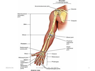
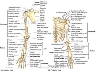
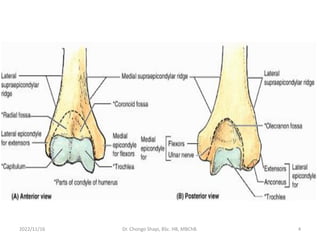
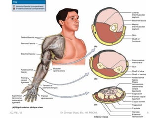
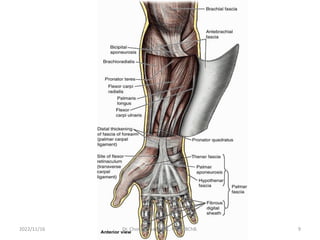
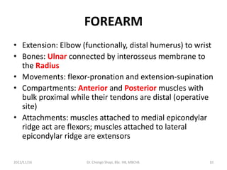
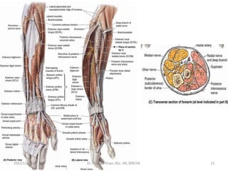
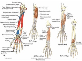
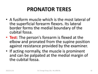
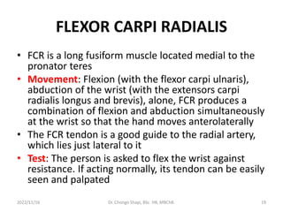
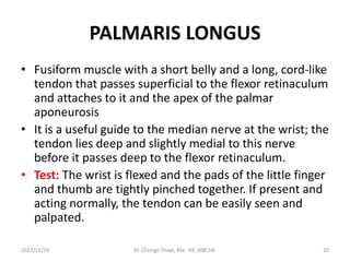
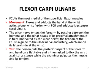
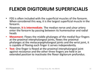
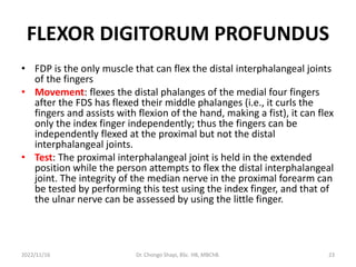
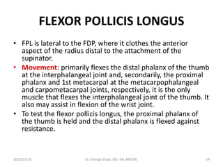
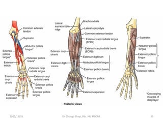
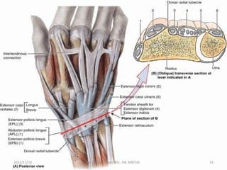
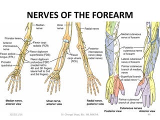
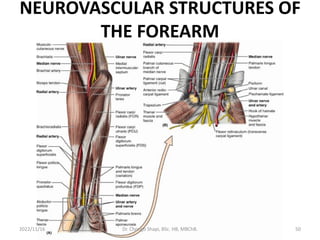
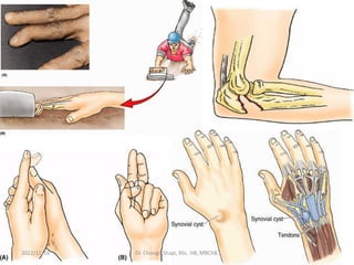
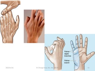
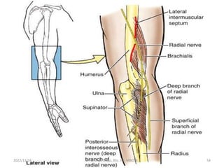
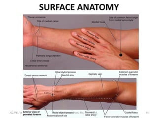
10. FOREARM
• Extension: Elbow (functionally, distal humerus) to wrist
• Bones: Ulnar connected by interosseus membrane to
the Radius
• Movements: flexor-pronation and extension-supination
• Compartments: Anterior and Posterior muscles with
bulk proximal while their tendons are distal (operative
site)
• Attachments: muscles attached to medial epicondylar
ridge act are flexors; muscles attached to lateral
epicondylar ridge are extensors
2022/11/16 Dr. Chongo Shapi, BSc. HB, MBChB. 10
11. FOREARM
• Spiraling gradually over the length of the forearm,
the compartments become truly anterior and
posterior in position in the distal forearm and
wrist
• Because neither boundary is crossed by motor
nerves, they also provide sites for surgical incision
• Innervation: Flexor-pronator muscles (median
nerve and ulna nerve except brachioradialis
supplied by the radial nerve); extensor-supinator
muscles (radial nerve)
2022/11/16 Dr. Chongo Shapi, BSc. HB, MBChB. 11
12. FOREARM
• Fascia: Ends at the joints; Therefore, containing
fluids and infections and cannot readily spread to
other compartments
• The anterior compartment is an exception
because it communicates with the central
compartment of the palm through the carpal
tunnel
• Muscle: The flexor muscles of the anterior
compartment have approximately twice the bulk
and strength of the extensor muscles of the
posterior compartment
2022/11/16 Dr. Chongo Shapi, BSc. HB, MBChB. 12
14. FLEXOR-PRONATOR MUSCLES
• The tendons of most flexor muscles are located
on the anterior surface of the wrist and are held
in place by the palmar carpal ligament and the
flexor retinaculum (transverse carpal ligament),
thickenings of the antebrachial fascia
• Layers: 3- Superficial (pronator teres, flexor carpi
radialis, palmaris longus, and flexor carpi ulnaris),
intermediate (flexor digitorum superficialis) and
deep (flexor digitorum profundus, flexor pollicis
longus, and pronator quadratus)
2022/11/16 Dr. Chongo Shapi, BSc. HB, MBChB. 14
15. FLEXOR-PRONATOR MUSCLES
• The five superficial and intermediate muscles
cross the elbow joint; the three deep muscles do
not. With the exception of the pronator
quadratus
• The more distally placed a muscle's distal
attachment lies, the more distally and deeply
placed is its proximal attachment
• The long flexors of the digits (flexor digitorum
superficialis and flexor digitorum profundus) also
flex the metacarpophalangeal and wrist joint
2022/11/16 Dr. Chongo Shapi, BSc. HB, MBChB. 15
16. FLEXOR-PRONATOR MUSCLES
• The flexor digitorum profundus flexes the fingers
in slow action.
• This action is reinforced by the flexor digitorum
superficialis when speed and flexion against
resistance are required.
• Tendons of the long flexors of the digits pass
through the distal part of the forearm, wrist, and
palm and continue to the medial four fingers.
• The flexor digitorum superficialis flexes the
middle phalanges, and the flexor digitorum
profundus flexes the distal phalanges.
2022/11/16 Dr. Chongo Shapi, BSc. HB, MBChB. 16
18. PRONATOR TERES
• A fusiform muscle which is the most lateral of
the superficial forearm flexors. Its lateral
border forms the medial boundary of the
cubital fossa.
• Test: The person's forearm is flexed at the
elbow and pronated from the supine position
against resistance provided by the examiner.
• If acting normally, the muscle is prominent
and can be palpated at the medial margin of
the cubital fossa.
2022/11/16 Dr. Chongo Shapi, BSc. HB, MBChB. 18
19. FLEXOR CARPI RADIALIS
• FCR is a long fusiform muscle located medial to the
pronator teres
• Movement: Flexion (with the flexor carpi ulnaris),
abduction of the wrist (with the extensors carpi
radialis longus and brevis), alone, FCR produces a
combination of flexion and abduction simultaneously
at the wrist so that the hand moves anterolaterally
• The FCR tendon is a good guide to the radial artery,
which lies just lateral to it
• Test: The person is asked to flex the wrist against
resistance. If acting normally, its tendon can be easily
seen and palpated
2022/11/16 Dr. Chongo Shapi, BSc. HB, MBChB. 19
20. PALMARIS LONGUS
• Fusiform muscle with a short belly and a long, cord-like
tendon that passes superficial to the flexor retinaculum
and attaches to it and the apex of the palmar
aponeurosis
• It is a useful guide to the median nerve at the wrist; the
tendon lies deep and slightly medial to this nerve
before it passes deep to the flexor retinaculum.
• Test: The wrist is flexed and the pads of the little finger
and thumb are tightly pinched together. If present and
acting normally, the tendon can be easily seen and
palpated.
2022/11/16 Dr. Chongo Shapi, BSc. HB, MBChB. 20
21. FLEXOR CARPI ULNARIS
• FCU is the most medial of the superficial flexor muscles
• Movement: Flexes and adducts the hand at the wrist if
acting alone, wrist flexion with FCR and adducts it extensor
carpi ulnaris
• The ulnar nerve enters the forearm by passing between the
humeral and the ulnar heads of its proximal attachment. It
is fully innervated by the ulnar nerve; the tendon of the
FCU is a guide to the ulnar nerve and artery, which are on
its lateral side at the wrist.
• Test: the person puts the posterior aspect of the forearm
and hand on a flat table and is then asked to flex the wrist
against resistance while the examiner palpates the muscle
and its tendon.
2022/11/16 Dr. Chongo Shapi, BSc. HB, MBChB. 21
22. FLEXOR DIGITORUM SUPERFICIALIS
• FDS is often included with the superficial muscles of the forearm.
When considered this way, it is the largest superficial muscle in the
forearm.
• However, it is intermediate. The median nerve and ulnar artery
enter the forearm by passing between its humeroulnar and radial
heads
• Movement: Flexes the middle phalanges of the medial four fingers
at the proximal interphalangeal joints, flexes the proximal
phalanges at the metacarpophalangeal joints and the wrist joint, it
is capable of flexing each finger it serves independently.
• Test: One finger is flexed at the proximal interphalangeal joint
against resistance and the other three fingers are held in an
extended position to inactivate the flexor digitorum profundus.
2022/11/16 Dr. Chongo Shapi, BSc. HB, MBChB. 22
23. FLEXOR DIGITORUM PROFUNDUS
• FDP is the only muscle that can flex the distal interphalangeal joints
of the fingers
• Movement: flexes the distal phalanges of the medial four fingers
after the FDS has flexed their middle phalanges (i.e., it curls the
fingers and assists with flexion of the hand, making a fist), it can flex
only the index finger independently; thus the fingers can be
independently flexed at the proximal but not the distal
interphalangeal joints.
• Test: The proximal interphalangeal joint is held in the extended
position while the person attempts to flex the distal interphalangeal
joint. The integrity of the median nerve in the proximal forearm can
be tested by performing this test using the index finger, and that of
the ulnar nerve can be assessed by using the little finger.
2022/11/16 Dr. Chongo Shapi, BSc. HB, MBChB. 23
24. FLEXOR POLLICIS LONGUS
• FPL is lateral to the FDP, where it clothes the anterior
aspect of the radius distal to the attachment of the
supinator.
• Movement: primarily flexes the distal phalanx of the thumb
at the interphalangeal joint and, secondarily, the proximal
phalanx and 1st metacarpal at the metacarpophalangeal
and carpometacarpal joints, respectively, it is the only
muscle that flexes the interphalangeal joint of the thumb. It
also may assist in flexion of the wrist joint.
• To test the flexor pollicis longus, the proximal phalanx of
the thumb is held and the distal phalanx is flexed against
resistance.
2022/11/16 Dr. Chongo Shapi, BSc. HB, MBChB. 24
25. PRONATOR QUADRATUS
• It is quadrangular and pronates the forearm. It cannot
be palpated or observed, except in dissections,
because it is the deepest muscle in the anterior aspect
of the forearm.
• Movement: It is the prime mover for pronation. The
muscle initiates pronation; It is assisted by the pronator
teres when more speed and power are needed.
• The pronator quadratus also helps the interosseous
membrane hold the radius and ulna together,
particularly when upward thrusts are transmitted
through the wrist (e.g., during a fall on the hand).
2022/11/16 Dr. Chongo Shapi, BSc. HB, MBChB. 25
26. EXTENSOR MUSCLES
• Groups: 3- Muscles that extend and abduct or adduct
the hand at the wrist joint (extensor carpi radialis
longus, extensor carpi radialis brevis, and extensor
carpi ulnaris)
• Muscles that extend the medial four fingers
(extensor digitorum, extensor indicis, and extensor
digiti minimi)
• Muscles that extend or abduct the thumb (abductor
pollicis longus, extensor pollicis brevis, and extensor
pollicis longus).
2022/11/16 Dr. Chongo Shapi, BSc. HB, MBChB. 26
27. EXTENSOR MUSCLES
• The extensor tendons are held in place in the
wrist region by the extensor retinaculum, which
prevents bowstringing of the tendons when the
hand is extended at the wrist joint.
• As the tendons pass over the dorsum of the wrist,
they are provided with synovial tendon sheaths
that reduce friction for the extensor tendons as
they traverse the osseofibrous tunnels formed by
the attachment of the extensor retinaculum to
the distal radius and ulna.
2022/11/16 Dr. Chongo Shapi, BSc. HB, MBChB. 27
28. EXTENSOR MUSCLES
• Anatomical layers: Superficial and deep. Four of
the superficial extensors (extensor carpi radialis
brevis, extensor digitorum, extensor digiti minimi,
and extensor carpi ulnaris) are attached
proximally by a common extensor tendon to the
lateral epicondyle.
• The proximal attachment of the other two
muscles in the superficial group (brachioradialis
and extensor carpi radialis longus) is to the lateral
supraepicondylar ridge of the humerus and
adjacent lateral intermuscular septum.
2022/11/16 Dr. Chongo Shapi, BSc. HB, MBChB. 28
29. EXTENSOR MUSCLES
• The four flat tendons of the extensor
digitorum pass deep to the extensor
retinaculum to the medial four fingers.
• The common tendons of the index and little
fingers are joined on their medial sides near
the knuckles by the respective tendons of the
extensor indicis and extensor digiti minimi
(extensors of the index and little fingers,
respectively).
2022/11/16 Dr. Chongo Shapi, BSc. HB, MBChB. 29
32. BRACHIORADIALIS
• Fusiform muscle, lying superficially on the anterolateral
surface of the forearm. Forms the lateral border of the
cubital fossa.
• It flexes the forearm at the elbow. It is especially active
during quick movements or in the presence of resistance
during flexion of the forearm.
• The brachioradialis and the supinator are the only
muscles of the compartment that do not cross and
therefore are incapable of acting at the wrist.
2022/11/16 Dr. Chongo Shapi, BSc. HB, MBChB. 32
33. BRACHIORADIALIS
• As it descends, the brachioradialis overlies the radial
nerve and artery where they lie together on the
supinator, pronator teres tendon, FDS, and FPL.
• The distal part of the tendon is covered by the abductors
pollicis longus and brevis as they pass to the thumb.
• Test: the elbow joint is flexed against resistance with the
forearm in the midprone position.
• If the brachioradialis is acting normally, the muscle can
be seen and palpated.
2022/11/16 Dr. Chongo Shapi, BSc. HB, MBChB. 33
34. EXTENSOR CARPI RADIALIS LONGUS
• ECRL is a fusiform muscle overlapped by the
brachioradialis, with which it often blends.
• As it passes distally, posterior to the brachioradialis, its
tendon is crossed by the abductor pollicis brevis and
extensor pollicis brevis. The ECRL is indispensable
when clenching the fist
• Test; the wrist is extended and abducted with the
forearm pronated. If acting normally, the muscle can
be palpated inferoposterior to the lateral side of the
elbow. Its tendon can be palpated proximal to the wrist
2022/11/16 Dr. Chongo Shapi, BSc. HB, MBChB. 34
35. EXTENSOR CARPI RADIALIS BREVIS
• ECRB and ECRL pass under the extensor retinaculum
together within the tendinous sheath of the extensor
carpi radiales
• When the two muscles act by themselves, they abduct
the hand as they extend it. Acting with the extensor
carpi ulnaris, they extend the; acting with the FCR they
produce pure abduction.
• Their synergistic action with the extensor carpi ulnaris
is important in steadying the wrist during tight flexion
of the medial four fingers (clenching a fist), a function
in which the longus is more active.
2022/11/16 Dr. Chongo Shapi, BSc. HB, MBChB. 35
36. EXTENSOR DIGITORUM
• It is the principal extensor of the medial four fingers
occupying much of the posterior surface of the forearm.
Normally no finger can remain fully flexed as the other ones
are fully extended.
• Commonly, the fourth tendon is fused initially with the
tendon to the ring finger and reaches the little finger by a
tendinous band.
• Test: The forearm is pronated and the fingers are extended.
The person attempts to keep the fingers extended at the
metacarpophalangeal joints as the examiner exerts pressure
on the proximal phalanges by attempting to flex them.
• If acting normally, the extensor digitorum can be palpated in
the forearm, and its tendons can be seen and palpated on the
dorsum of the hand.
2022/11/16 Dr. Chongo Shapi, BSc. HB, MBChB. 36
37. EXTENSOR DIGITI MINIMI
• EDM, a fusiform slip of muscle, is a partially detached part
of the extensor digitorum.
• The tendon of this extensor of the little finger runs through
a separate compartment of the extensor retinaculum,
posterior to the distal radioulnar joint, within the tendinous
sheath of the extensor digiti minimi.
• The tendon then divides into two slips; the lateral one is
joined to the tendon of the extensor digitorum, with all
three tendons attaching to the dorsal digital expansion of
the little finger.
• After exerting its traction primarily on the 5th finger, it
contributes to extension of the hand.
2022/11/16 Dr. Chongo Shapi, BSc. HB, MBChB. 37
38. EXTENSOR CARPI ULNARIS
• ECU, a long fusiform muscle medially located, has 2
heads: a humeral head and an ulnar head
• Acting with the ECRL and ECRB, it extends the hand;
acting with the FCU, it adducts the hand. Like the ECRL,
it is indispensable when clenching the fist.
• Test: the forearm is pronated and the fingers are
extended. The extended wrist is then adducted against
resistance.
• If acting normally, the muscle can be seen and palpated
in the proximal part of the forearm and its tendon can
be felt proximal to the head of the ulna.
2022/11/16 Dr. Chongo Shapi, BSc. HB, MBChB. 38
39. SUPINATOR
• It lies deep in the cubital fossa and, along with
the brachialis, forms its floor
• The deep branch of the radial nerve passes
between the humeral and ulnar heads as it leaves
the cubital fossa to enter the posterior part of the
arm thereby forming the posterior interosseus
nerve. The supinator rotates the radius
• The deep extensors of the forearm act on the
thumb and the index finger (extensor indicis). The
three muscles acting on the thumb are referred
to as outcropping muscles
2022/11/16 Dr. Chongo Shapi, BSc. HB, MBChB. 39
40. ABDUCTOR POLLICIS LONGUS
• APL is fusiform and lies just distal to the supinator
and is closely related to the EPB
• It acts with the APB during abduction of the
thumb and with the extensor pollicis muscles
during extension of this digit
• Test: the thumb is abducted against resistance at
the metacarpophalangeal joint.
• If acting normally, the tendon of the muscle can
be seen and palpated at the lateral side of the
anatomical snuff box and on the lateral side of
the adjacent extensor pollicis brevis tendon.
2022/11/16 Dr. Chongo Shapi, BSc. HB, MBChB. 40
41. EXTENSOR POLLICIS BREVIS
• In continued action after acting to flex the proximal
phalanx of the thumb, or acting when that joint is fixed
by its antagonists, it helps extend the 1st metacarpal
and extend and abduct the hand.
• When the thumb is fully extended, a hollow called the
anatomical snuff box, can be seen on the radial aspect
of the wrist
• Test: the thumb is extended against resistance at the
metacarpophalangeal joint. If the EPB is acting
normally, the tendon of the muscle can be seen and
palpated at the lateral side of the anatomical snuff box
and on the medial side of the adjacent APL tendon
2022/11/16 Dr. Chongo Shapi, BSc. HB, MBChB. 41
42. EXTENSOR POLLICIS LONGUS
• The tendon passes under the extensor
retinaculum in its own tunnel, within the
tendinous sheath of the extensor pollicis longus,
medial to the dorsal tubercle of the radius.
• It uses the tubercle as a trochlea (pulley) to
change its line of pull as it proceeds to the base
of the distal phalanx of the thumb.
• The gap thus created between the long extensor
tendons of the thumb is the anatomical snuff
box. The EPL also adducts the extended thumb
and rotates it laterally.
2022/11/16 Dr. Chongo Shapi, BSc. HB, MBChB. 42
43. EXTENSOR POLLICIS LONGUS
• To test the extensor pollicis longus, the thumb is
extended against resistance at the interphalangeal
joint.
• If the EPL is acting normally, the tendon of the
muscle can be seen and palpated on the medial side
of the anatomical snuff box.
• The tendons of the APL and EPB bound the
anatomical snuff box anteriorly, and the tendon of
the EPL bounds it posteriorly.
2022/11/16 Dr. Chongo Shapi, BSc. HB, MBChB. 43
44. EXTENSOR POLLICIS LONGUS
• The snuff box is visible when the thumb is fully
extended; this draws the tendons up and produces a
triangular hollow between them. Observe that the:
• Radial artery lies in the floor of the snuff box.
• Radial styloid process can be palpated proximally and
the base of the 1st metacarpal can be palpated
distally in the snuff box.
2022/11/16 Dr. Chongo Shapi, BSc. HB, MBChB. 44
45. EXTENSOR INDICIS
• The extensor indicis has a narrow,
elongated belly that lies medial to and
alongside that of the EPL.
• This muscle confers independence to the
index finger in that the extensor indicis may
act alone or together with the extensor
digitorum to extend the index finger at the
proximal interphalangeal joint, as in
pointing.
• It also helps extend the hand.
2022/11/16 Dr. Chongo Shapi, BSc. HB, MBChB. 45