A protein needs to adopt a final and stable 3-dimensional shape in order to function properly.
• The Tertiary Structure of a protein is the arrangement of the secondary structures into this
final 3-dimensional shape.
Structure of protiens and the applied aspectsMohit Adhikary
The slides explain the structures of proteins, the bond stabilizing the structure of amino acids, the different types of protein structures, the applied aspects and the newer advances in the protein structure.
Tertiary Structure basically of Hydrophobic interactions, (interactions in side chains), hydrogen bonding, salt bridges, Vander Waals interactions.
e.g. Globular proteins & Fibrous Proteins
Proteins have four levels of structural organization: primary, secondary, tertiary, and quaternary. The primary structure refers to the linear sequence of amino acids in the polypeptide chain. Secondary structure involves local folding patterns like alpha helices and beta sheets. Tertiary structure describes the overall 3D shape of a single polypeptide chain. Quaternary structure is the 3D structure formed by the assembly of multiple polypeptide subunits. The structures at each level are stabilized by interactions between the R groups of amino acids in the chain.
This document summarizes the four levels of protein structure: primary, secondary, tertiary, and quaternary. It provides details on each level: primary structure refers to the linear sequence of amino acids in the polypeptide chain. Secondary structure involves hydrogen bonding between amino acids to form regular structures like alpha helices and beta pleated sheets. Tertiary structure describes the overall 3D shape of the protein arising from secondary structures. Quaternary structure involves interactions between two or more polypeptide chains, as seen in hemoglobin which is made of four polypeptide subunits.
The document discusses the determination of the primary structure of proteins. It begins by explaining that proteins are composed of amino acid residues linked by peptide bonds to form a polypeptide chain. The primary structure refers to the specific sequence of amino acids in this chain. Mass spectrometry and tandem mass spectrometry techniques are used to analyze protein fragments obtained through enzymatic or chemical cleavage to determine the amino acid sequence and thereby elucidate the primary structure.
The document summarizes the key forces that stabilize protein structure: covalent interactions such as disulfide bonds between cysteine residues, and non-covalent interactions including hydrophobic interactions between hydrophobic amino acids, van der Waals forces between neighboring amino acid side chains, ionic bonds between oppositely charged residues, and hydrogen bonds between polar residues and water molecules. These various interactions work together to form the tertiary structure of proteins.
Covalent bonds, peptide bonds, and disulfide bridges stabilize protein structures through strong covalent interactions. Non-covalent interactions like van der Waals forces, hydrogen bonds, electrostatic interactions, and hydrophobic effects also contribute to protein stability. These non-covalent interactions are weaker than covalent bonds but work together in large numbers to stabilize a protein's native conformation. Perturbations can disrupt this delicate balance of interactions and cause protein denaturation.
Proteins have a variety of functions in cells including enzymes, structural components, transporters, motors, and signaling molecules. A protein's unique 3D shape, determined by its amino acid sequence, allows it to carry out its specific function. The polypeptide backbone forms secondary structures like alpha helices and beta sheets. Non-covalent interactions further guide protein folding into a stable tertiary structure. Quaternary structure involves interactions between multiple polypeptide chains. Post-translational modifications and ligand binding regulate protein activity.
Some proteins are composed of multiple polypeptide chains that interact with each other through noncovalent bonds like hydrogen bonds and hydrophobic interactions to form a quaternary structure. The quaternary structure stabilizes the overall protein complex and the subunits may function independently or cooperatively, as seen in hemoglobin where the binding of oxygen to one subunit increases oxygen binding in the other subunits.
Protein structure can be described at several levels of organization. The primary structure is the amino acid sequence, while the secondary structure describes local patterns like alpha helices and beta sheets formed by hydrogen bonds. Tertiary structure refers to the overall 3D shape of a single polypeptide chain. Quaternary structure involves the arrangement of multiple protein subunits. Together these organizational levels allow proteins to carry out their diverse functions in the cell.
Biochemistry - Ch4 protein structure , and function Areej Abu Hanieh
This document discusses the structure of proteins at multiple levels. It explains that a protein's amino acid sequence determines its 3D structure, and its structure dictates its function. Noncovalent interactions like hydrogen bonds, hydrophobic interactions, and electrostatic interactions are important forces that stabilize a protein's native structure. The peptide bond is rigid and planar, limiting the possible conformations of the polypeptide backbone. Common secondary structures like alpha helices and beta sheets form due to favorable hydrogen bonding patterns between peptide bonds.
This document discusses the different levels of protein structure:
- Primary structure refers to the specific sequence of amino acids in a polypeptide chain.
- Secondary structure involves localized folding patterns like alpha helices and beta sheets.
- Tertiary structure is the overall three-dimensional shape of a single polypeptide chain. Interactions between amino acid side chains help determine this shape.
- Quaternary structure refers to the arrangement of multiple polypeptide subunits in a multimeric protein, held together by non-covalent bonds. Hemoglobin is given as an example with its a2b2 subunit composition.
1. The document discusses fibrous and globular proteins, focusing on collagen, elastin, myoglobin, and hemoglobin. It provides details on the structure, function, and biosynthesis of these proteins.
2. Collagen is the main fibrous protein in the body. It has a characteristic triple helical structure that gives tissues strength and elasticity. Defects in collagen can lead to conditions like Ehlers-Danlos syndrome and osteogenesis imperfecta.
3. Elastin provides elasticity to tissues like lungs, blood vessels, and skin. It forms cross-links that allow these tissues to stretch and return to their original shape. Mutations can result in Morfan syndrome.
BIOSYNTHESIS OF PHOSPHOLIPIDS
Phospholipids:-
These are compounds containing, in addition to fatty acid and glycerol, phosphoric acid, nitrogenous bases, and another substituent. Polar compounds composed of alcohol attached by phosphodiester bridge to either diacylglycerol or sphingosine.
Amphipathic in nature has a hydrophilic head (phosphate +alcohol
eg., serine, ethanolamine, and choline) and a long, hydrophobic tail
(fatty acids or derivatives ).
- CLASSIFICATION OF PHOSPHOLIPIDS:-
- Glycerophospholipids
- Spingophospholipids or Sphingomyelin
- SYNTHESIS OF PHOSPHOLIPIDS
- FUNCTIONS OF PHOSPHOLIPIDS
- FUNCTIONS OF SPHINGOLIPIDS
Gives in detail primary, secondary, tertiary and Quaternary structure of proteins. Gives classification of secondary structure: alpha helix, beta pleated sheet and different types of tight turns and explains most commonly found tight turn in proteins i.e. beta turn. Briefs about the Ramachandran plot of proteins, dihedral or torsion angles and explains why glycine and proline act as alpha helix breakers. Explains tertiary structure of proteins and different covalent and non covalent bonds in the tertiary structure and relative importance of these bonding interactions. Details about the quaternary structure of proteins and explains why hemoglobin is a quaternary protein and insulin is not.
This document provides an overview of globular proteins, with a focus on hemoglobin and myoglobin. It discusses the following key points:
- Globular proteins have hydrophilic amino acids on the outside and hydrophobic amino acids on the inside, allowing them to be soluble in water. Hemoglobin and myoglobin are important globular proteins.
- Hemoglobin transports oxygen in red blood cells, while myoglobin stores and transports oxygen in muscle cells. Both contain a heme group that reversibly binds oxygen.
- The structures of myoglobin and each subunit of hemoglobin involve alpha helices that form a pocket holding the heme group. Histidine residues help bind the heme iron and oxygen.
- Hemoglobin
The document discusses protein structure and stability. It describes the four levels of protein structure: primary, secondary, tertiary, and quaternary. The primary structure is the amino acid sequence. Secondary structures include alpha helices and beta sheets formed by hydrogen bonding. Tertiary structure involves folding influenced by interactions between R groups. Quaternary structure results from interactions between multiple polypeptide chains, as in hemoglobin. The document also discusses factors that stabilize protein structures such as disulfide bonds and noncovalent interactions, and how denaturation and renaturation can alter protein structure.
This document discusses protein structure and function. It begins by defining proteins and their essential roles in the human body. It then describes the general characteristics of proteins, such as their chemical composition and functions. The document classifies proteins based on their composition, axial ratio, and biological functions. It explains the different levels of protein structure from primary to quaternary, focusing on secondary structure elements like alpha helices and beta sheets. The roles of hydrogen bonding, disulfide bridges, and ionic bonds in tertiary and quaternary structure are also described.
WEAK INTERACTIONS IN AQUEOUS SYSTEMS AND FITNESS OF THE AQUEOUS ENVIRONMENT F...anjusha suki
Weak interactions like hydrogen bonding and van der Waals forces play an important role in maintaining the structures of biological molecules like proteins and DNA. These interactions are weak individually but add up to provide strong conformations. The more complementary the interacting structures, the better they fit together and the more stable the resulting structure. Hydrogen bonding specifically helps dissolve many compounds in water and is important for life's aqueous environment.
Quaternary structure refers to the arrangement of multiple protein subunits into a single protein complex. Hemoglobin is a common example that is made of two alpha and two beta subunits. The subunits interact through hydrophobic interactions, hydrogen bonding, and other bonds. Globular proteins tend to have quaternary structure that clusters the subunits into a spherical shape, while fibrous proteins form long coils or sheets through interactions between subunits. Quaternary structure allows proteins to take on specialized functions beyond what individual subunits could achieve alone.
This document discusses various mechanisms of enzyme regulation in living systems. It begins by explaining that hundreds of enzyme-catalyzed reactions must be precisely controlled for proper cellular functioning. It then describes several key mechanisms by which this regulation can be achieved, including allosteric regulation, isoenzyme expression, zymogen activation, and covalent modification via phosphorylation or glycosylation. Specific examples are provided for each type of regulation, such as feedback inhibition of threonine dehydratase and phosphorylation control of glycogen phosphorylase activity. The document concludes by emphasizing that multiple regulatory strategies acting together ensure survival of the cell and maintenance of homeostasis.
1. Covalent and non-covalent interactions are important for macromolecule structure and function. Covalent bonds strongly bind atomic subunits while non-covalent bonds like hydrogen bonding and hydrophobic interactions more weakly stabilize macromolecule structures.
2. Covalent bonds like peptide bonds link amino acids into protein chains. Non-covalent interactions are crucial for protein folding and binding specificity. Though individually weak, many non-covalent bonds cooperatively bind molecular surfaces.
3. Covalent drugs form irreversible complexes with target proteins, while non-covalent drugs reversibly inhibit enzymes through competitive, noncompetitive, or uncompetitive binding. Examples are covalent penicillin and non-covalent acetylcholinester
Proteins fold into complex 3D structures essential for their function. There are four levels of protein structure - primary, secondary, tertiary, and quaternary. Chaperone proteins help other proteins fold correctly to prevent aggregation. Misfolded proteins can result from changes in temperature, pH, or lack of chaperones and may lead to disease if not degraded. Normally, misfolded proteins are targeted for degradation by the ubiquitin proteasome pathway, but accumulation of misfolded proteins can cause conditions like Alzheimer's disease.
In this ppt competitive inhibition of enzymes is fully explained with its examples. it will be helpful for all the life science students. Non Competitive inhibition , Uncompetitive inhibition & Irreversible inhibition of Enzymes have been well explained in this presentation. it will be helpful for biochemistry, botany, zoology and other life/bio sciences students. I tried to explain Allosteric enzymes, their mechanism of action, Allosteric inhibition, Feedback inhibition in this presentation so that it can be easy to understand the concept for viewers.
PROTEIN STRUCTURE AND FUNCTION PPT(MD MOBARAK HOSSAIN).pptxMDMOBARAKHOSSAIN12
This document provides an overview of protein structure and function. It discusses the four levels of protein structure: primary, secondary, tertiary, and quaternary structure. The primary structure is the amino acid sequence. Secondary structures include alpha helices and beta sheets formed by hydrogen bonding. Tertiary structure describes the overall 3D shape formed by interactions between amino acid side chains. Quaternary structure involves the clustering of multiple peptide chains. Finally, it outlines several key functions of proteins, including structural proteins, transport proteins, and enzymes/receptors.
This document summarizes key concepts about protein structure and collagen. It discusses the forces involved in protein folding like hydrophobic interactions and hydrogen bonding. It describes accessory proteins that assist folding like chaperones. Collagen is introduced as the most abundant protein, composed of tropocollagen triple helices with characteristic Gly-X-Y motifs. Post-translational modifications of collagen including hydroxyproline, hydroxylysine and cross-linking are outlined.
the topic introduction of protein cover their target their primary structure , secondary structure and tertiary structure and their bonding interaction
This document discusses maintaining the native conformation of proteins under cellular conditions. It defines the native conformation as the properly folded state that allows a protein to function. Proteins have multiple levels of structure - primary, secondary, tertiary, and quaternary. Denaturation disrupts these structures by breaking non-covalent bonds, causing proteins to lose their shape and function. Denaturation can be reversible or irreversible and can occur due to heat, chemicals, or other environmental stresses. Maintaining the native conformation is important for protein function in cells.
A protein is an organic compound made up of small molecules called amino acids. There are 20 different amino acids commonly found in the proteins of living organisms. Small proteins may contain just a few hundred amino acids, whereas large proteins may contain thousands of amino acids
Amino acisd structure
Peptide bond formation
Analysis of protein Structure- X-ray Crystallography
Different structural levels of proteins with examples.
Importance of protein structure
Creutzfeldt-Jacob-Disease due to changes in normal protein conformation.
This document provides an overview of protein structure and function. It discusses tertiary structure, which involves interactions between amino acid side chains that cause folds and loops in the polypeptide chain. Supersecondary structures combine different secondary structures. Protein domains consist of structural motifs and can function independently. Quaternary structure involves interactions between polypeptide subunits. The amino acid sequence determines the three-dimensional structure of a protein. Protein folding involves interactions that bury hydrophobic residues in the core and expose hydrophilic residues. Misfolded proteins can accumulate and cause disease.
Protein Structure and levels of protein structurePankajGurra1
This document discusses the different levels of protein structure, including:
1) Primary structure, which is the amino acid sequence that makes up the protein chain.
2) Secondary structure, which involves local folding into structures like alpha helices and beta sheets.
3) Tertiary structure, which describes the overall 3D shape of the protein formed by interactions between different parts of the amino acid chain.
4) Quaternary structure for proteins made of multiple subunits, describing how those subunits are arranged relative to each other.
1) Proteins are made of amino acids joined by peptide bonds and folded into complex shapes that determine their function. There are four levels of protein structure: primary, secondary, tertiary, and quaternary.
2) Nucleic acids are made of nucleotides and transmit hereditary information by determining which proteins are produced. DNA contains genes and is located in the nucleus, while RNA participates in protein synthesis.
3) Biological macromolecules include proteins, nucleic acids, carbohydrates, and lipids, all composed of monomers joined by covalent bonds. These complex molecules have essential functions in cells and living organisms.
The document discusses protein structure and functions. It begins by defining proteins as polymers of amino acids that are essential building blocks of the human body. It then describes the four levels of protein structure - primary, secondary, tertiary, and quaternary. Primary structure refers to the amino acid sequence. Secondary structure involves folding into structures like alpha helices and beta sheets. Tertiary structure describes the 3D conformation determined by interactions between amino acid side chains. Quaternary structure refers to interactions between multiple polypeptide subunits. The document concludes by classifying proteins as either fibrous or globular and providing some examples of different proteins and their functions.
This document discusses the structure of proteins at four levels: primary, secondary, tertiary, and quaternary. The primary structure refers to the amino acid sequence in the polypeptide chain. Secondary structures include alpha helices and beta sheets formed by hydrogen bonding between amino acids. Tertiary structure describes the overall 3D shape of the protein formed by interactions between amino acid side chains. Quaternary structure applies to proteins composed of multiple polypeptide subunits that combine through non-covalent bonds. The structures are determined through techniques like X-ray crystallography and NMR spectroscopy.
Proteins are composed of amino acids and play many essential roles in biology. They exist in primary, secondary, tertiary, and quaternary structures. The primary structure is the amino acid sequence, and secondary structures include alpha helices and beta sheets formed by hydrogen bonding. Tertiary structure involves further folding stabilized by various bonds. Quaternary structure refers to the arrangement of subunits in some proteins. Proteins are classified by their composition, function, shape, and nature. They function as enzymes, hormones, antibodies, and structural components, and are essential for all living processes.
Proteins are composed of amino acids linked together in chains and serve important functions in the body. They exist in complex 3D structures including primary, secondary, tertiary and quaternary forms which determine their function. Proteins can be classified based on their composition, function, shape or nature. They play key roles such as structure, movement, signaling, catalysis and immunity. Their importance includes being enzymes, hormones, structural components and in processes like DNA expression, oxygen transport, homeostasis and immunity.
levels of protein structure , Domains ,motifs & Folds in protein structureAaqib Naseer
Protein structure is hierarchical, with four levels: primary, secondary, tertiary, and quaternary. The primary structure is the amino acid sequence. Secondary structures include alpha helices and beta sheets formed by hydrogen bonding between amino acids in the sequence. Tertiary structure involves folding of the entire chain into a compact 3D structure. Quaternary structure involves the assembly of protein subunits. Other structural features include domains, which are independently folded and functional regions, motifs like loops and barrels formed by secondary structure elements, and folds defined by the arrangement of alpha helices and beta sheets. Understanding protein structure is important for studying protein function and for developing drugs.
Proteins-Classification ,Structure of protein, properties and biological impo...SoniaBajaj10
This document provides an overview of proteins, including their definition, classification, structure, and properties. It discusses how proteins are composed of amino acids and classified based on their chemical nature, structure, shape and solubility. The four levels of protein structure - primary, secondary, tertiary, and quaternary structure - are also summarized. Key properties of proteins like solubility, denaturation and functions in the body are highlighted. The document serves as an introduction to proteins and provides a high-level classification and structural overview.
This document provides an overview of proteins. It defines proteins as macromolecules composed of amino acids that mediate virtually every cellular process. The 20 amino acids that make up proteins can assemble into different structures to form molecules like hormones, enzymes, and muscle fibers. Proteins are synthesized through translation of mRNA in the ribosome. Their primary structure is determined by the linear sequence of amino acids. Secondary structures like alpha helices and beta sheets form through hydrogen bonding between peptide bonds in the polypeptide chain. Tertiary structure arises from folding of the secondary structure into a compact 3D shape stabilized by hydrophobic interactions and disulfide bridges. Some proteins have quaternary structure as multisubunit complexes. The document discusses several
1) The document discusses the structure of proteins from primary to quaternary levels. It explains that proteins fold into secondary and tertiary structures driven by interactions like hydrogen bonding that reduce the protein's energy.
2) Twenty amino acids are the building blocks of proteins. Each amino acid has an amino group, carboxyl group, and unique side chain. The side chains determine each amino acid's properties.
3) Protein folding occurs through a series of steps from primary linear chains to more complex folded structures, driven by interactions that minimize energy. This folding results in the protein's final functional 3D shape.
Proteins are polymers of amino acids linked by peptide bonds that fold into complex three-dimensional structures essential for their functions. There are four levels of protein structure: primary structure is the amino acid sequence; secondary structures include alpha helices and beta sheets formed by hydrogen bonds between amino acids in the backbone. Tertiary structure describes the overall three-dimensional shape including side chains, while quaternary structure refers to the arrangement of multiple protein subunits. The amino acid sequence ultimately determines the three-dimensional structure which is critical for a protein's function.
The binding of cosmological structures by massless topological defectsSérgio Sacani
Assuming spherical symmetry and weak field, it is shown that if one solves the Poisson equation or the Einstein field
equations sourced by a topological defect, i.e. a singularity of a very specific form, the result is a localized gravitational
field capable of driving flat rotation (i.e. Keplerian circular orbits at a constant speed for all radii) of test masses on a thin
spherical shell without any underlying mass. Moreover, a large-scale structure which exploits this solution by assembling
concentrically a number of such topological defects can establish a flat stellar or galactic rotation curve, and can also deflect
light in the same manner as an equipotential (isothermal) sphere. Thus, the need for dark matter or modified gravity theory is
mitigated, at least in part.
The technology uses reclaimed CO₂ as the dyeing medium in a closed loop process. When pressurized, CO₂ becomes supercritical (SC-CO₂). In this state CO₂ has a very high solvent power, allowing the dye to dissolve easily.
The use of Nauplii and metanauplii artemia in aquaculture (brine shrimp).pptxMAGOTI ERNEST
Although Artemia has been known to man for centuries, its use as a food for the culture of larval organisms apparently began only in the 1930s, when several investigators found that it made an excellent food for newly hatched fish larvae (Litvinenko et al., 2023). As aquaculture developed in the 1960s and ‘70s, the use of Artemia also became more widespread, due both to its convenience and to its nutritional value for larval organisms (Arenas-Pardo et al., 2024). The fact that Artemia dormant cysts can be stored for long periods in cans, and then used as an off-the-shelf food requiring only 24 h of incubation makes them the most convenient, least labor-intensive, live food available for aquaculture (Sorgeloos & Roubach, 2021). The nutritional value of Artemia, especially for marine organisms, is not constant, but varies both geographically and temporally. During the last decade, however, both the causes of Artemia nutritional variability and methods to improve poorquality Artemia have been identified (Loufi et al., 2024).
Brine shrimp (Artemia spp.) are used in marine aquaculture worldwide. Annually, more than 2,000 metric tons of dry cysts are used for cultivation of fish, crustacean, and shellfish larva. Brine shrimp are important to aquaculture because newly hatched brine shrimp nauplii (larvae) provide a food source for many fish fry (Mozanzadeh et al., 2021). Culture and harvesting of brine shrimp eggs represents another aspect of the aquaculture industry. Nauplii and metanauplii of Artemia, commonly known as brine shrimp, play a crucial role in aquaculture due to their nutritional value and suitability as live feed for many aquatic species, particularly in larval stages (Sorgeloos & Roubach, 2021).
Authoring a personal GPT for your research and practice: How we created the Q...Leonel Morgado
Thematic analysis in qualitative research is a time-consuming and systematic task, typically done using teams. Team members must ground their activities on common understandings of the major concepts underlying the thematic analysis, and define criteria for its development. However, conceptual misunderstandings, equivocations, and lack of adherence to criteria are challenges to the quality and speed of this process. Given the distributed and uncertain nature of this process, we wondered if the tasks in thematic analysis could be supported by readily available artificial intelligence chatbots. Our early efforts point to potential benefits: not just saving time in the coding process but better adherence to criteria and grounding, by increasing triangulation between humans and artificial intelligence. This tutorial will provide a description and demonstration of the process we followed, as two academic researchers, to develop a custom ChatGPT to assist with qualitative coding in the thematic data analysis process of immersive learning accounts in a survey of the academic literature: QUAL-E Immersive Learning Thematic Analysis Helper. In the hands-on time, participants will try out QUAL-E and develop their ideas for their own qualitative coding ChatGPT. Participants that have the paid ChatGPT Plus subscription can create a draft of their assistants. The organizers will provide course materials and slide deck that participants will be able to utilize to continue development of their custom GPT. The paid subscription to ChatGPT Plus is not required to participate in this workshop, just for trying out personal GPTs during it.
ESPP presentation to EU Waste Water Network, 4th June 2024 “EU policies driving nutrient removal and recycling
and the revised UWWTD (Urban Waste Water Treatment Directive)”
The ability to recreate computational results with minimal effort and actionable metrics provides a solid foundation for scientific research and software development. When people can replicate an analysis at the touch of a button using open-source software, open data, and methods to assess and compare proposals, it significantly eases verification of results, engagement with a diverse range of contributors, and progress. However, we have yet to fully achieve this; there are still many sociotechnical frictions.
Inspired by David Donoho's vision, this talk aims to revisit the three crucial pillars of frictionless reproducibility (data sharing, code sharing, and competitive challenges) with the perspective of deep software variability.
Our observation is that multiple layers — hardware, operating systems, third-party libraries, software versions, input data, compile-time options, and parameters — are subject to variability that exacerbates frictions but is also essential for achieving robust, generalizable results and fostering innovation. I will first review the literature, providing evidence of how the complex variability interactions across these layers affect qualitative and quantitative software properties, thereby complicating the reproduction and replication of scientific studies in various fields.
I will then present some software engineering and AI techniques that can support the strategic exploration of variability spaces. These include the use of abstractions and models (e.g., feature models), sampling strategies (e.g., uniform, random), cost-effective measurements (e.g., incremental build of software configurations), and dimensionality reduction methods (e.g., transfer learning, feature selection, software debloating).
I will finally argue that deep variability is both the problem and solution of frictionless reproducibility, calling the software science community to develop new methods and tools to manage variability and foster reproducibility in software systems.
Exposé invité Journées Nationales du GDR GPL 2024
Immersive Learning That Works: Research Grounding and Paths ForwardLeonel Morgado
We will metaverse into the essence of immersive learning, into its three dimensions and conceptual models. This approach encompasses elements from teaching methodologies to social involvement, through organizational concerns and technologies. Challenging the perception of learning as knowledge transfer, we introduce a 'Uses, Practices & Strategies' model operationalized by the 'Immersive Learning Brain' and ‘Immersion Cube’ frameworks. This approach offers a comprehensive guide through the intricacies of immersive educational experiences and spotlighting research frontiers, along the immersion dimensions of system, narrative, and agency. Our discourse extends to stakeholders beyond the academic sphere, addressing the interests of technologists, instructional designers, and policymakers. We span various contexts, from formal education to organizational transformation to the new horizon of an AI-pervasive society. This keynote aims to unite the iLRN community in a collaborative journey towards a future where immersive learning research and practice coalesce, paving the way for innovative educational research and practice landscapes.
ESR spectroscopy in liquid food and beverages.pptxPRIYANKA PATEL
With increasing population, people need to rely on packaged food stuffs. Packaging of food materials requires the preservation of food. There are various methods for the treatment of food to preserve them and irradiation treatment of food is one of them. It is the most common and the most harmless method for the food preservation as it does not alter the necessary micronutrients of food materials. Although irradiated food doesn’t cause any harm to the human health but still the quality assessment of food is required to provide consumers with necessary information about the food. ESR spectroscopy is the most sophisticated way to investigate the quality of the food and the free radicals induced during the processing of the food. ESR spin trapping technique is useful for the detection of highly unstable radicals in the food. The antioxidant capability of liquid food and beverages in mainly performed by spin trapping technique.
Unlocking the mysteries of reproduction: Exploring fecundity and gonadosomati...AbdullaAlAsif1
The pygmy halfbeak Dermogenys colletei, is known for its viviparous nature, this presents an intriguing case of relatively low fecundity, raising questions about potential compensatory reproductive strategies employed by this species. Our study delves into the examination of fecundity and the Gonadosomatic Index (GSI) in the Pygmy Halfbeak, D. colletei (Meisner, 2001), an intriguing viviparous fish indigenous to Sarawak, Borneo. We hypothesize that the Pygmy halfbeak, D. colletei, may exhibit unique reproductive adaptations to offset its low fecundity, thus enhancing its survival and fitness. To address this, we conducted a comprehensive study utilizing 28 mature female specimens of D. colletei, carefully measuring fecundity and GSI to shed light on the reproductive adaptations of this species. Our findings reveal that D. colletei indeed exhibits low fecundity, with a mean of 16.76 ± 2.01, and a mean GSI of 12.83 ± 1.27, providing crucial insights into the reproductive mechanisms at play in this species. These results underscore the existence of unique reproductive strategies in D. colletei, enabling its adaptation and persistence in Borneo's diverse aquatic ecosystems, and call for further ecological research to elucidate these mechanisms. This study lends to a better understanding of viviparous fish in Borneo and contributes to the broader field of aquatic ecology, enhancing our knowledge of species adaptations to unique ecological challenges.
Travis Hills' Endeavors in Minnesota: Fostering Environmental and Economic Pr...Travis Hills MN
Travis Hills of Minnesota developed a method to convert waste into high-value dry fertilizer, significantly enriching soil quality. By providing farmers with a valuable resource derived from waste, Travis Hills helps enhance farm profitability while promoting environmental stewardship. Travis Hills' sustainable practices lead to cost savings and increased revenue for farmers by improving resource efficiency and reducing waste.
Travis Hills' Endeavors in Minnesota: Fostering Environmental and Economic Pr...
Tertiary Structure of Proteins
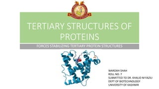
1. TERTIARY STRUCTURES OF
PROTEINS
FORCES STABILIZING TERTIARY PROTEIN STRUCTURES
WARDAH SHAH
ROLL NO. 7
SUBMITTED TO DR. KHALID M FAZILI
DEPT OF BIOTECHNOLOGY
UNIVERSITY OF KASHMIR
3. • A protein needs to adopt a final and stable 3-dimensional shape in order to function properly.
• The Tertiary Structure of a protein is the arrangement of the secondary structures into this
final 3-dimensional shape.
• The sequence of amino acids in a protein (the primary structure) will determine where alpha
helices and beta sheets (the secondary structures) will occur.
• These secondary structure motifs then fold into an overall arrangement that is the final 3-
dimensional fold of the protein (the tertiary structure).Each unique sequence of amino acids
gives rise to a unique protein type, with a unique shape and function.
4. ANFINSENS DOGMA
• Also known as Thermodynamic
Hypothesis
• It states that, at least for a small globular
protein in its standard physiological
environment, the native structure is
determined only by the protein's amino
acid sequence.
• An unfolded protein can go back to its
folded native conformation in favourable
environment due to the properties of its
amino acid sequence.
5. FIBROUS GLOBULAR INTRINSICALLY
DISORDERED
• Fibrous proteins usually consist
of a single type of secondary
structure
• Their tertiary structure is
relatively simple.
• The structures that provide
support, shape, and external
protection to vertebrates are
made of fibrous proteins.
• Globular proteins often
contain several types of
secondary structures.
• Most enzymes are globular
proteins.
• Intrinsically disordered proteins
can lack secondary structure
entirely.
• regulatory proteins can be
globular, disordered, or contain
both globular and disordered
segments.
6. THE STRUCTURE-FUNCTION RELATIONSHIP
• If a protein does not fold correctly it will not function properly. Therefore, researching a protein's
structure is very important when trying to understand what it does and how it works.
• When scientists study a protein they must first determine the sequence of amino acids in the protein
chain (primary structure).
• They use this sequence to predict the presence of any alpha helices or beta sheets (secondary
structure).
• They can then use X-ray crystallography and NMR to determine a protein's full 3-dimensional shape
(tertiary structure).
• Knowing the tertiary structure of a protein is often crucial to understanding how it functions and how
to target it for drug therapy or other medical uses.
• Note: some proteins of similar structure have different functions.
7. FORCES STABILIZING TERTIARY STRUCTURES
•A proteins conformation is
stabilized largely by weak
interactions. These are ~100
folds weaker than covalent
bonds but collectively influence
the 3D structure of proteins
significantly.
•A protein conformation with
the lowest free energy, is the one
with maximum weak
interactions.
8. HYDROPHOBIC FORCE
• Packing of hydrophobic amino acid in the protein core and hydrophilic
amino acids forming the protein surface leads to a favorable increase
in entropy of water by reducing the solvation layer. (solvation layer
formation disrupts waters hydrogen bonding structure which is
energetically unfavorable)
• This protein folding provides maximum hydrogen bonding partners to
water.
9. VAN DER WAALS INTERACTIONS
• The nonpolar side chains in the core are so close together that short-range van der
Waals interactions make a significant contribution to stabilizing interactions.
• It operates over a limited intermolecular distance, I.e., 0.3 nm to 0.6 nm.
• These attractive intermolecular interactions can be of three types:
i. Permanent dipole - dipole interaction (Orientation effect)
ii. Temporary dipole - permanent dipole interaction (Induction effect)
iii. Temporary/Induced dipole – dipole interaction (Dispersion effect)
10. ORIENTATION EFFECT INDUCTION EFFECT DISPERSION EFFECT
• Electrostatic interaction
between to polar molecules
(d+ and d-)
• This is called Keesom force.
• Temporary dipole induced in a
nonpolar molecule by the
permanent dipole of a polar
molecule near it.
• This is called Debye force.
• Temporary dipole formed in a
nonpolar molecule which
leads to temporary dipole in
another nonpolar molecule
near it.
• This is called London force.
11. HYDROGEN BONDING
• The bond in which an electronegative atom shares a
hydrogen atom with another electronegative atom with a
bound hydrogen, is called a hydrogen bond.
• Presence of hydrogen bonding groups without
partners in the hydrophobic core can destabilize the
protein structure. Hence, polar or charged groups in the
protein interior are hydrogen bonded which stabilize the
framework of the protein.
• Hydrogen bonds have an important role in guiding
protein folding process.
• Examples, amide-carbonyl, hydroxyl-carbonyl, hydroxyl-
hydroxyl H-bond.
12. • Energy: 10-40 kJ/mol
• Approx. Length 1.7-3 A
• Strength of the hydrogen
bond varies with angle of the
hydrogen bond interaction.
13. IONIC INTERACTIONS
• Ionic interactions arise from electrostatic attraction
between two groups of opposite charge.
• Ionic bonds are formed as amino acids bearing opposite
electrical charges are juxtaposed in the hydrophobic
core of proteins.
• Although rare, ionic bonds can be important to protein
structure because they are potent electrostatic
attractions that can approach the strength of covalent
bonds.
• The strength of salt bridge increases as it moves to an
environment of lower dielectric constant (in protein
core).
14. DISULFIDE LINKAGE
• Covalent bond formed between thiol
group of two cysteine residues (cystine
formation)
• This plays an important role in protein
folding and stability.
• They are unstable and cytosol as they
require an oxidizing environment. Eg.,
extracellular proteins (insulin).
• It may form the hydrophobic core with
rest of the weak interactions forming
around it.
15. REFERENCES
• Lehninger Principles of Biochemistry
• https://cbm.msoe.edu/teachingResources/proteinStructure
• https://pubs.acs.org/doi/10.1021/acs.jctc.6b00422
• https://www.bionity.com/en/encyclopedia/Hydrophobic_collapse
• https://www.sciencedirect.com/topics/chemistry/van-der-waals-
force
• http://www2.hawaii.edu/~lesaux/621/ewExternalFiles/NJ%20Lecture
%201-1.pdf