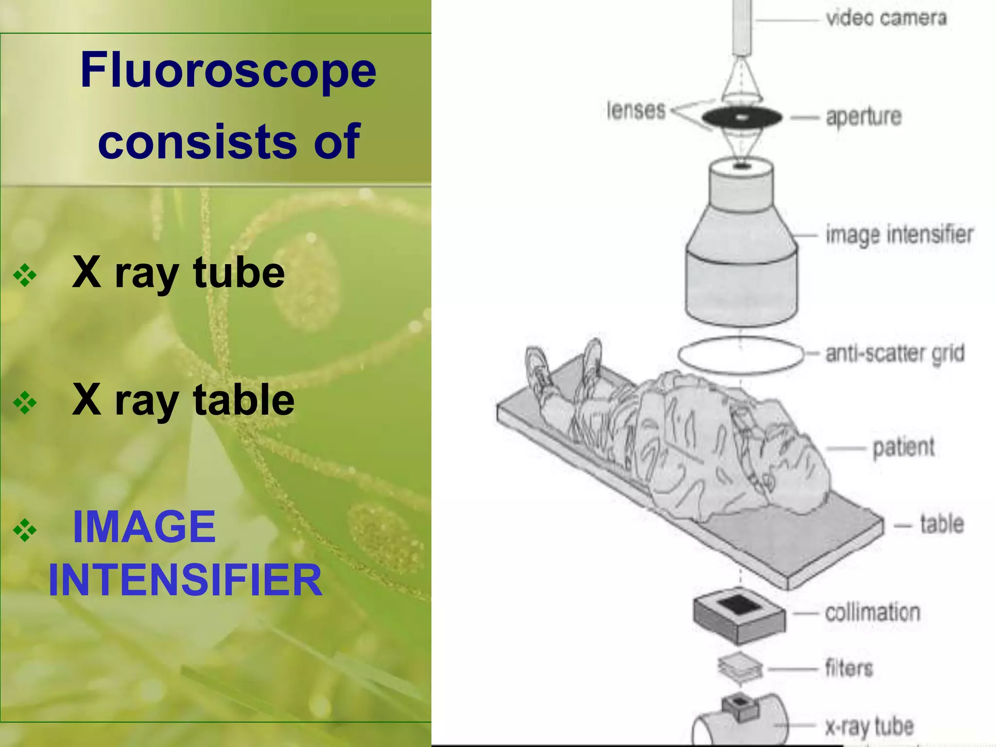
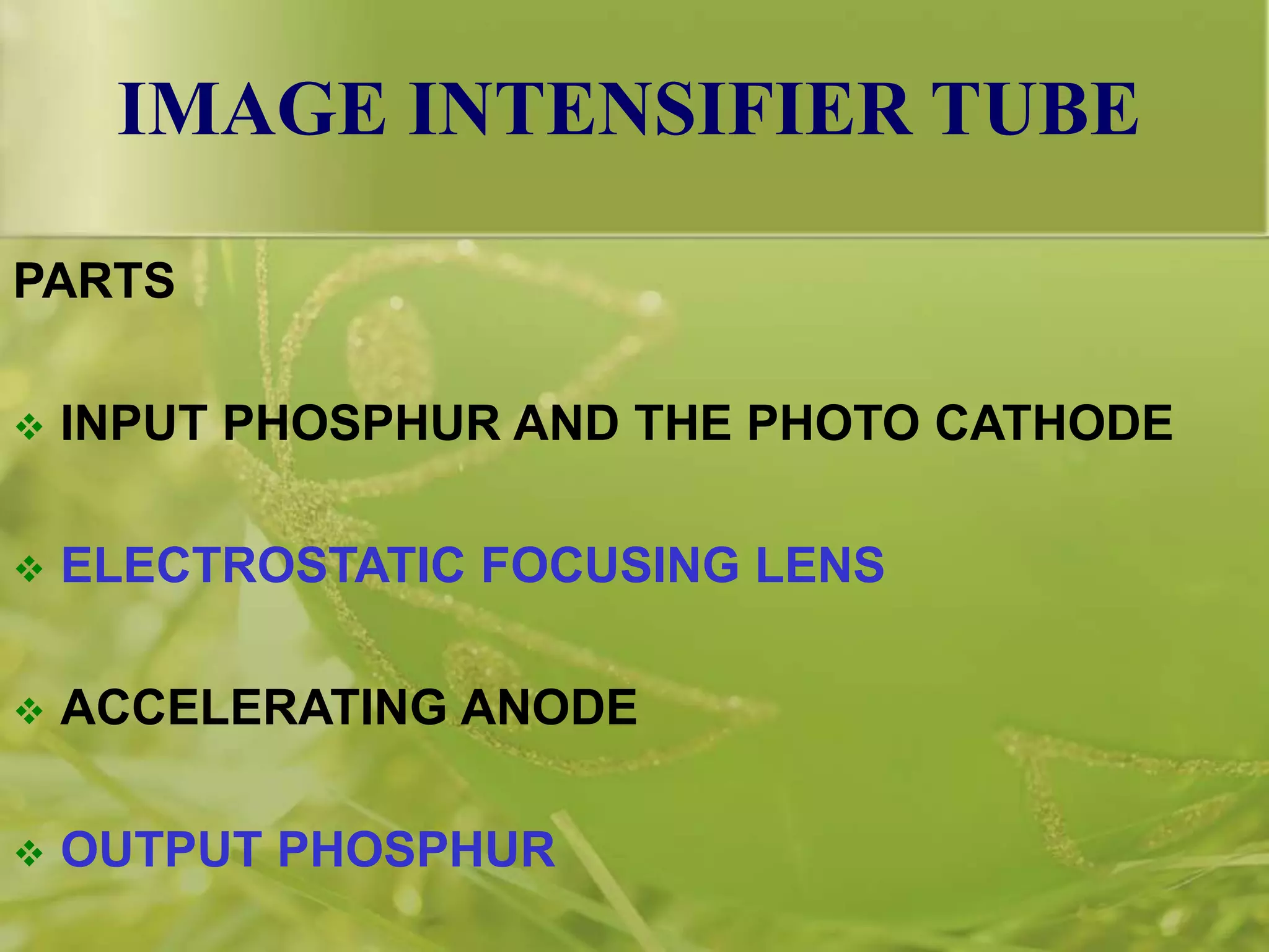
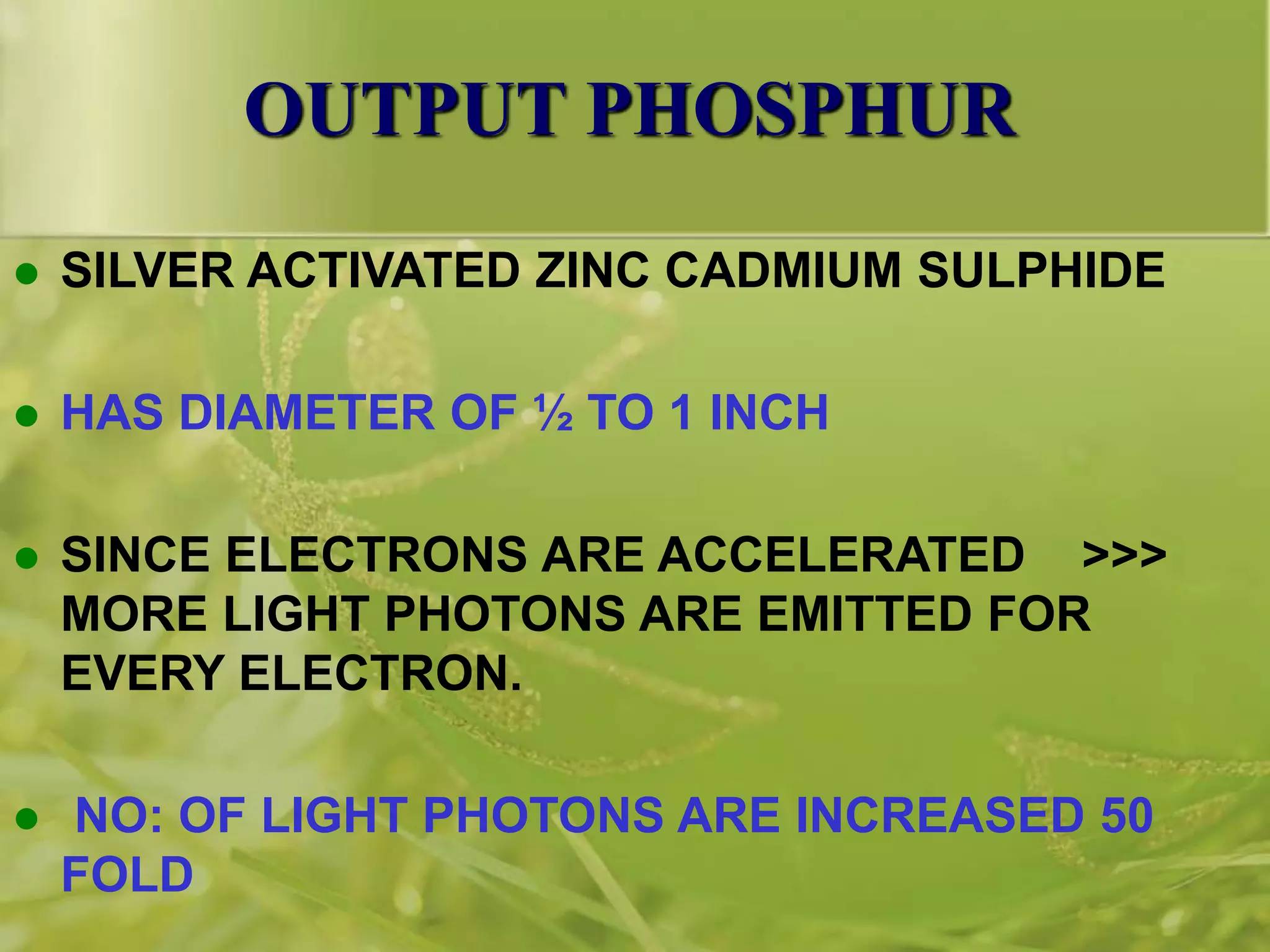
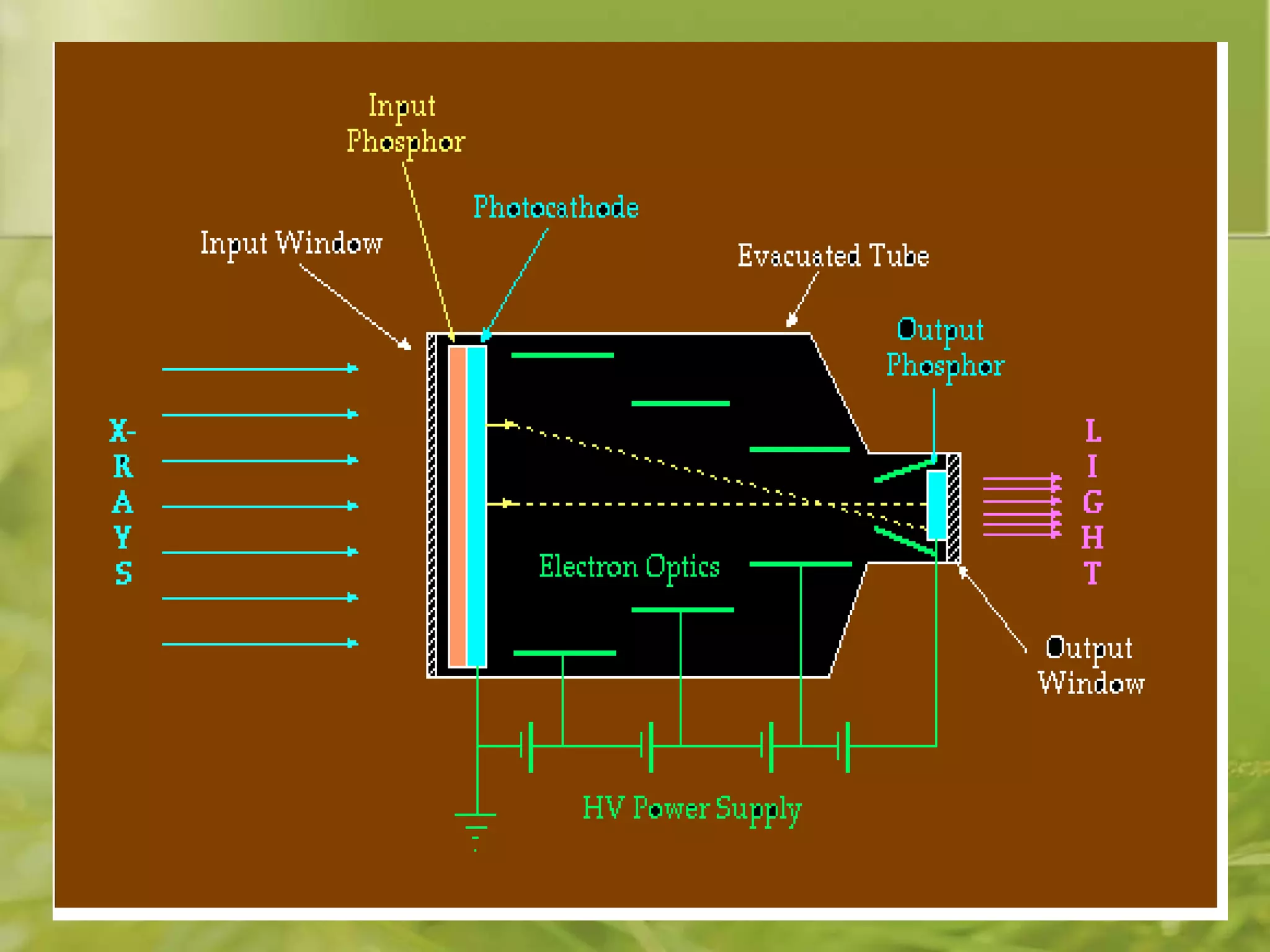
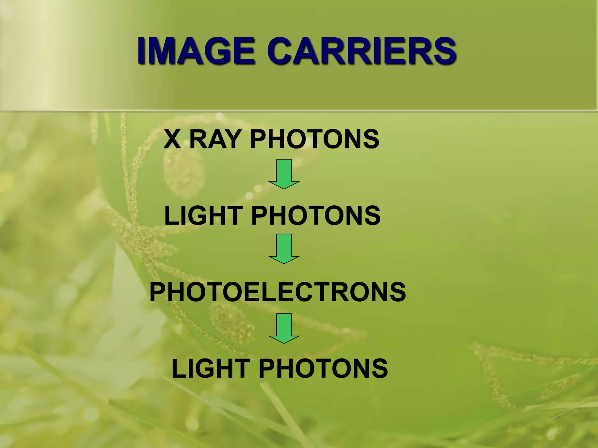
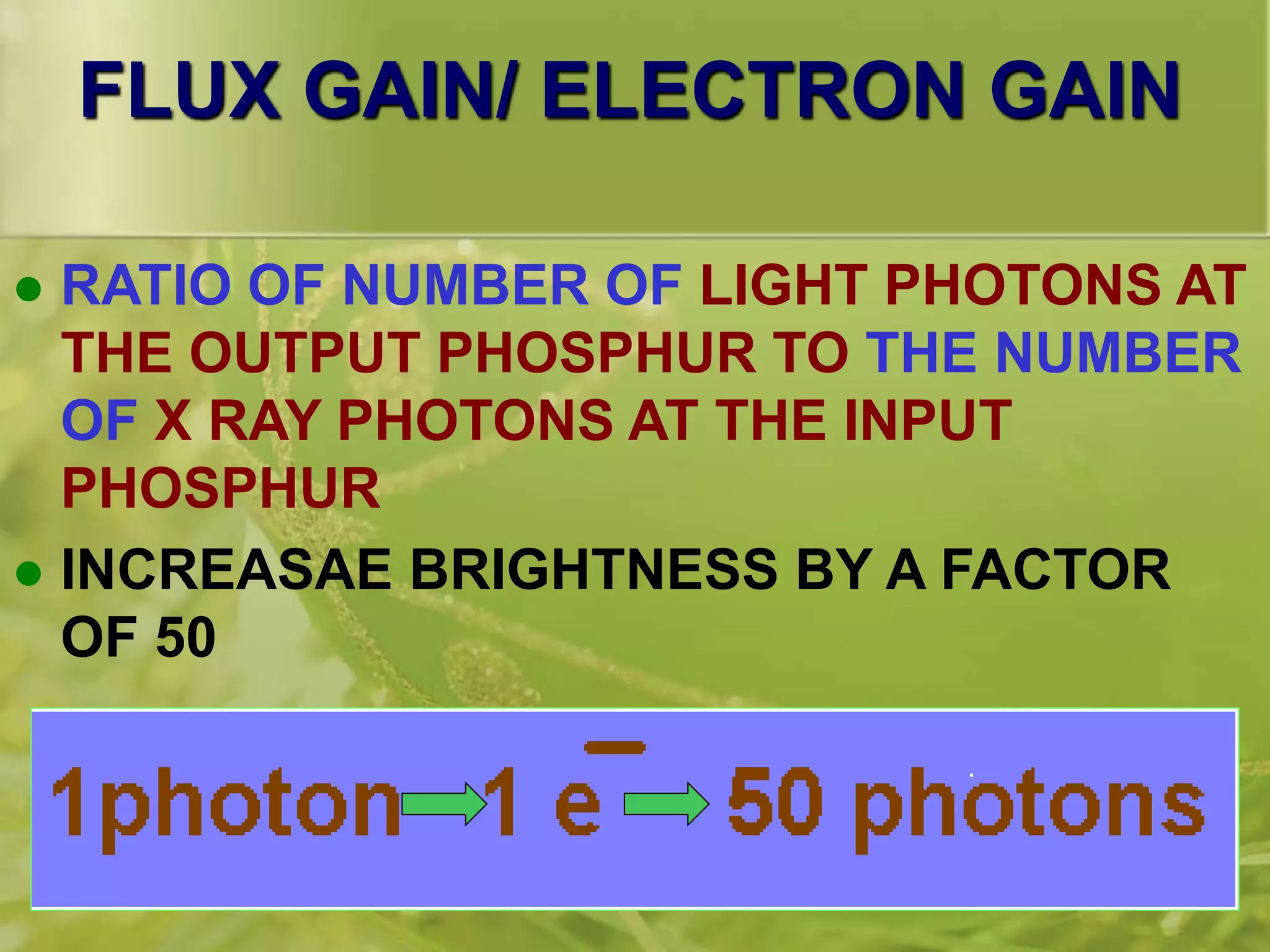
A fluoroscope uses x-rays and a fluorescent screen to enable direct observation of internal organs. It consists of an x-ray tube, table, and image intensifier. The image intensifier converts x-rays into visible light images and amplifies them for viewing. It works by accelerating photoelectrons emitted from a photocathode onto a phosphor screen, producing light photons and gaining brightness. Newer generations of image intensifiers use additional electron multiplication for higher sensitivity. Fluoroscopy provides real-time moving images for procedures while fluorography captures still diagnostic images.


![IMAGE INTENSIFIER DESIGN
VACCUM
GLASS TUBE
[ 2 – 4 mm
THICK ]
ENCLOSED
IN A LEAD
LINED METAL
CONTAINER](https://image.slidesharecdn.com/fluoroscopicimaging-230530110008-07017e6c/75/FLUOROSCOPIC-IMAGING-ppt-8-2048.jpg)


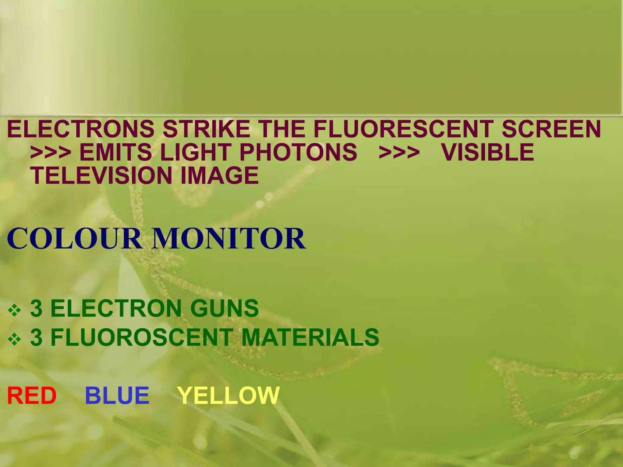
![TV MONITOR
REASSEMBLING OF VIDEO PULSES BACK TO
LIGHT IMAGE IS DONE BY THE TV MONITOR
MAIN PART IS THE PICTURE TUBE
ELECTRONIC VACCUM TUBE
ELECTRON GUN [ CATHODE + CONTROL GRID]
ANODE– 10, 000 V
FLUORESCENT SCREEN
FOCUSING COILS
DEFLECTING COILS](https://image.slidesharecdn.com/fluoroscopicimaging-230530110008-07017e6c/75/FLUOROSCOPIC-IMAGING-ppt-103-2048.jpg)