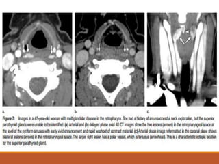
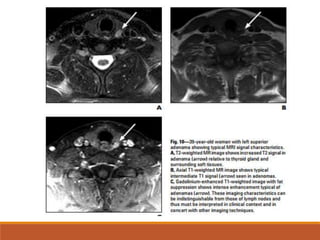
SlideShare a Scribd company logo
1 of 35
ANATOMY
Parathyroid glands are located within the
visceral space of the neck, posterior to the
thyroid gland, in the vicinity of the
tracheoesophageal groove.
They may be located either inside or
outside the thyroid capsule.
SUPERIOR PARATHYROID GLAND
Arises from the 4th branchial pouch, during fetal development,
migrates with the thyroid gland.
Eutopic superior parathyroid glands are located on the posterior
aspect of the superior or middle third of the thyroid lobe in more
than 90% of the general population.
Ectopic superior parathyroid gland may be
above the upper pole of the thyroid lobe (<1%)
posterior to the pharynx or esophagus, in either the neck or the
superior mediastinum (1%–4%)
intrathyroidal (<3%)
INFERIOR PARATHYROID GLAND
Arises from the 3rd branchial pouch, migrates with thymus.
Eutopic inferior parathyroid glands are located at the level
of the inferior pole of the thyroid lobe, in its posterior,
lateral, or anterior aspects (61%).
Ectopic inferior parathyroid glands may be located in the
neck, inferior to the lower pole of the thyroid lobe, in the
anterior mediastinum (4%–5%); intrathyroidal (<1%–2%).
In the anterior mediastinum, inferior parathyroid glands
may be situated within the thymus, at the posterior aspect
of the thymic capsule, or in contact with the great vessels of
the mediastinum
PRIMARY HYPERPARATHYROIDISM
four times more common in women than in men and is
most prevalent in the 5th to 7th decades of life
signs and symptoms of hyperparathyroidism largely
represent hypercalcemia, which may affect multiple systems
Symptomatic patients at presentation can have recurrent
kidney stones, osteopenia, hypertension, peptic ulcer
disease, mental status changes, and fatigue
Most cases of primary hyperparathyroidism are caused
by a solitary parathyroid adenoma (88%).
Other less common causes are multiglandular disease
from double adenoma (4%) and multiple gland
hyperplasia (6%), and rarely, carcinoma ( 1%)
increased incidence of parathyroid hyperplasia in MEN
type I and type IIA.
To select patients for minimally invasive parathyroidectomy
surgery, the surgeon needs precise localization of the
parathyroid adenoma and to exclude multiglandular
disease
IMAGING
ULTRASOUND:
Parathyroid adenomas are
homogeneously hypoechoic to the
overlying thyroid gland.
Larger adenomas can be multilobulated
Color Doppler imaging commonly show a
characteristic extrathyroidal feeding
vessel (typically a branch off the inferior
thyroidal artery), which enters the
parathyroid gland at one of the poles
4D CT TECHNIQUE
The role of four-dimensional (4D) CT is to enable accurate
localization of the parathyroid adenoma and to depict multiglandular
disease
The 4D CT protocol is composed of multiple phases (typically two to
four phases) and reformatted images in three planes.
The first three “dimensions” are multiplanar CT: axial acquisition
with coronal and sagittal reformations.
The fourth “dimension” of 4D CT is change in enhancement overtime
from non– contrast-enhanced, arterial, and delayed (venous) phase
imaging.
The characteristic contrast enhancement pattern of a
parathyroid adenoma is peak enhancement on the arterial
phase, washout of contrast material from the arterial to
delayed phase, and low attenuation on the non–contrast
enhanced images .
Four-dimensional CT has high accuracy in localizing an
adenoma to a quadrant (87%) when used as a firstline
investigation.
DIFFERENTIAL DIAGNOSIS
The two main mimics of parathyroid adenomas- lymph nodes and
thyroid tissue.
Lymph nodes show progressively increasing enhancement after
injection of contrast material, with peak enhancement at 90
seconds corresponding to the delayed phase.
Thyroid tissue can enhance intensely in the arterial phase but will
also have increasing contrast enhancement between the arterial
and the delayed phases when the attenuation is measured .
In addition, thyroid tissue will have intrinsic high attenuation on
the non–contrast-enhanced images because the thyroid gland is
iodine-containing
The feeding artery is usually a branch of the inferior thyroid artery,
and the artery can be tortuous and enlarged in the setting of
adenoma because of its increased blood flow An enlarged adjacent
vessel may also be an enlarged draining vein
The polar vessel sign can be present in up to two-thirds of
parathyroid adenomas on 4D CT images
PARATHYROID SCINTIGRAPHY
Three general techniques of radionuclide scintigraphy are
commonly used for the detection and visualization of
hyperfunctioning parathyroid glands:
1. single-phase dual-isotope subtraction imaging,
2. dual-phase single-isotope imaging, and
3. a combination of the two
Single-isotope dual-phase imaging technique was proposed
on the basis of the difference between the rate of 99mTc
sestamibi washout from the thyroid gland and the rate of
washout from hyperfunctioning parathyroid glands, which
is typically slower.
This technique requires a single injection of 99mTc
sestamibi, followed by imaging at approximately 10–15
minutes and 1.5–3 hours after the injection.
99mTc sestamibi consists of lipophilic cationic molecules.
The detectability of parathyroid adenomas and hyperplastic
parathyroid glands is related to the presence of mitochrondria-
rich oxyphil cells.
99mTc sestamibi normally is distributed to the parotid and
submandibular salivary glands, thyroid gland, heart, and liver;
uptake is not seen in normal parathyroid glands.
Images show delayed washout in a large right inferior
a) parathyroid adenoma that extends from the inferior pole of the right thyroid lobe to
the superior mediastinum.
b) early washout in a large thyroid adenoma, which extends from the lower pole of the
left thyroid lobe to the superior mediastinum
Dual-isotope subtraction imaging techniques is by using
99mTc sestamibi and 123I or 99mTc pertechnetate.
99mTc pertechnetate, which is taken up only by the
thyroid.
subtraction of thyroid images obtained with iodine-123
(123I) or 99mTc pertechnetate from those obtained with
99mTc sestamibi.
Evidence of residual radioactivity on the subtraction
image represents a hyperfunctioning parathyroid gland
MRI
MRI is used in patients with persistent or recurrent
hyperparathyroidism, in whom it has been shown to be
effective in locating remaining abnormal parathyroid tissue.
The T1 and T2 characteristics of abnormal parathyroid
tissue are variable.
The most common tissue characteristics are intermediate-
to low-intensity T1 signal and high-intensity T2 signal.
false-negative studies are most commonly associated with
adenomas that are isointense on T1 and T2 sequences; the
addition of contrast-enhanced images can increase
sensitivity for these cases
SURGICAL TREATMENT
A single parathyroid adenoma is now most commonly
resected through a smaller unilateral incision on one side of
the neck in one quadrant.
The other glands are left unexplored if the venous
intraoperative parathyroid hormone level drops following
resection.
A decrease of more than 50% from the baseline PTH value
at 5–10 minutes after resection is suggestive of a single site
of primary hyperparathyroidism.
The parameters for all three phases are the following:
0.625-mm section thickness;
tube rotation time, 0.4 second; pitch factor, 0.516:1;
field of view, 20 cm; 120 kVp; and automatic tube current
modulation.
Tube current modulation (mA Modulation; GE Healthcare) is used
with a noise index of 8, minimum 100 mA, and maximum 400 mA
for the nonenhanced phase and delayed phase and 600 mA for the
arterial phase.
SYSTEMATIC APPROACH TO 4D CT
Step 1: Review for Lesions in Eutopic Locations in Arterial
Phase.
The superior parathyroid gland has a more consistent location
posterior to the mid-to-upper third of the thyroid gland . The
inferior parathyroid gland lies inferior, lateral, or posterior to
the lower third of the thyroid
The arterial phase should be the focus of the initial review
because this is the phase in which the attenuation of the
parathyroid adenoma will be highest, with reported mean
attenuation ranging from 138 to 180 HU
Step 2: Review for Lesion in Ectopic Locations in Arterial Phase
Ectopic parathyroid adenomas and ectopic parathyroid hyperplasia
account for 20%–25% of cases
The inferior glands -third branchial pouch and the superior glands -
fourth branchial pouch.
These pouches are approximately at the level of the bifurcation of
the carotid artery.
During fetal development, the inferior gland migrates with the
thymus.
So an ectopic inferior gland may be in any location from the carotid
bifurcation to the anterior mediastinum, including within the
thymus or the thyroid gland.
There is less variation in the location of the superior parathyroid,
which migrates with the thyroid gland. Rare sites for an ectopic
superior parathyroid gland are in the retropharyngeal space or
intrathyroid
More commonly, a superior parathyroid adenoma -pseudoectopia
due to the enlarged gland falling caudally and posteriorly in the
tracheoesophageal groove .
Thus a posterior mediastinal parathyroid adenoma is more
characteristic of a superior parathyroid adenoma that has fallen
caudally.
In contrast, an anterior mediastinal parathyroid adenoma will be
from the inferior parathyroid gland and along the thyrothymic
ligament
Step 3: Compare Other Phases for Characteristic Enhancement
Characteristic enhancement for a parathyroid adenoma is vivid
contrast enhancement in the arterial phase, rapid washout of
contrast material in the delayed phase, and lower attenuation
compared with thyroid gland in the nonenhanced phase
The peak enhancement has been described as between 25 and 60
seconds after injection
Step 4: Evaluate for Characteristic Morphology
A parathyroid adenoma is oval or rounded with margins that are
smooth or slightly lobulated
Other shapes of parathyroid adenomas are teardrop, discoid,
tubular, and pyramidal
Lobulated margins and cystic component help differentiate a
candidate lesion from a normal lymph node. Normal lymph nodes
have smooth margins and are solid or have fatty hila
Step 5: Compare CT Findings with Other Modalities and History
If there is a candidate lesion at 4D CT examination, the other
studies should be reviewed to determine if the studies are
concordant
Parathyroid Imaging .pptx

More Related Content

What's hot

Presentation1, radiological imaging of anal carcinoma.
Presentation1, radiological imaging of anal carcinoma.Presentation1, radiological imaging of anal carcinoma.
Presentation1, radiological imaging of anal carcinoma.Abdellah Nazeer
 
GEMC: Systematic Evaluation to Non-Traumatic Head CTs: Resident Training
GEMC: Systematic Evaluation to Non-Traumatic Head CTs: Resident TrainingGEMC: Systematic Evaluation to Non-Traumatic Head CTs: Resident Training
GEMC: Systematic Evaluation to Non-Traumatic Head CTs: Resident TrainingOpen.Michigan
 
Thyroglossal duct cysts
Thyroglossal duct cystsThyroglossal duct cysts
Thyroglossal duct cystsSayan Banerjee
 
Presentation1.pptx, radiological vascular anatomy of the chest and abdomen.
Presentation1.pptx, radiological vascular anatomy of the chest and abdomen.Presentation1.pptx, radiological vascular anatomy of the chest and abdomen.
Presentation1.pptx, radiological vascular anatomy of the chest and abdomen.Abdellah Nazeer
 
Food signs in radiology
Food signs in radiologyFood signs in radiology
Food signs in radiologyMedia Genie
 
Presentation1.pptx, radiological anatomy of the naso, oro and hypopharynx.
Presentation1.pptx, radiological anatomy of the naso, oro and hypopharynx.Presentation1.pptx, radiological anatomy of the naso, oro and hypopharynx.
Presentation1.pptx, radiological anatomy of the naso, oro and hypopharynx.Abdellah Nazeer
 
Presentation1.pptx, radiological imaging of skeletal dysplasia
Presentation1.pptx, radiological imaging of skeletal dysplasiaPresentation1.pptx, radiological imaging of skeletal dysplasia
Presentation1.pptx, radiological imaging of skeletal dysplasiaAbdellah Nazeer
 
Liver Ultrasound
Liver UltrasoundLiver Ultrasound
Liver UltrasoundSafi. Khan
 
Surgical anatomy and approaches to neck
Surgical anatomy and approaches to neckSurgical anatomy and approaches to neck
Surgical anatomy and approaches to necksadaf syed
 
Presentation1.pptx, radiological anatomy of the thigh and leg.
Presentation1.pptx, radiological anatomy of the thigh and leg.Presentation1.pptx, radiological anatomy of the thigh and leg.
Presentation1.pptx, radiological anatomy of the thigh and leg.Abdellah Nazeer
 
Diagnostic Imaging of Adrenal Glands
Diagnostic Imaging of Adrenal GlandsDiagnostic Imaging of Adrenal Glands
Diagnostic Imaging of Adrenal GlandsMohamed M.A. Zaitoun
 
Benign diseases of thyroid
Benign diseases of thyroid Benign diseases of thyroid
Benign diseases of thyroid Praveen RK
 
Oral cavity, pharynx radio-anatomy
Oral cavity, pharynx radio-anatomyOral cavity, pharynx radio-anatomy
Oral cavity, pharynx radio-anatomyDr. Mohit Goel
 
Msk need to know ultrasound lumps &amp; bumps &amp; various joints
Msk need to know ultrasound  lumps &amp; bumps &amp; various jointsMsk need to know ultrasound  lumps &amp; bumps &amp; various joints
Msk need to know ultrasound lumps &amp; bumps &amp; various jointsSuzanneCain2
 
Thyroid final [part 1]
Thyroid final [part 1]Thyroid final [part 1]
Thyroid final [part 1]Anwar Kamal
 

What's hot (20)

Presentation1, radiological imaging of anal carcinoma.
Presentation1, radiological imaging of anal carcinoma.Presentation1, radiological imaging of anal carcinoma.
Presentation1, radiological imaging of anal carcinoma.
 
GEMC: Systematic Evaluation to Non-Traumatic Head CTs: Resident Training
GEMC: Systematic Evaluation to Non-Traumatic Head CTs: Resident TrainingGEMC: Systematic Evaluation to Non-Traumatic Head CTs: Resident Training
GEMC: Systematic Evaluation to Non-Traumatic Head CTs: Resident Training
 
Thyroglossal duct cysts
Thyroglossal duct cystsThyroglossal duct cysts
Thyroglossal duct cysts
 
Presentation1.pptx, radiological vascular anatomy of the chest and abdomen.
Presentation1.pptx, radiological vascular anatomy of the chest and abdomen.Presentation1.pptx, radiological vascular anatomy of the chest and abdomen.
Presentation1.pptx, radiological vascular anatomy of the chest and abdomen.
 
Food signs in radiology
Food signs in radiologyFood signs in radiology
Food signs in radiology
 
Presentation1.pptx, radiological anatomy of the naso, oro and hypopharynx.
Presentation1.pptx, radiological anatomy of the naso, oro and hypopharynx.Presentation1.pptx, radiological anatomy of the naso, oro and hypopharynx.
Presentation1.pptx, radiological anatomy of the naso, oro and hypopharynx.
 
Retroperitoneal space
Retroperitoneal spaceRetroperitoneal space
Retroperitoneal space
 
Presentation1.pptx, radiological imaging of skeletal dysplasia
Presentation1.pptx, radiological imaging of skeletal dysplasiaPresentation1.pptx, radiological imaging of skeletal dysplasia
Presentation1.pptx, radiological imaging of skeletal dysplasia
 
Liver Ultrasound
Liver UltrasoundLiver Ultrasound
Liver Ultrasound
 
Surgical anatomy and approaches to neck
Surgical anatomy and approaches to neckSurgical anatomy and approaches to neck
Surgical anatomy and approaches to neck
 
Spleen[1]
Spleen[1]Spleen[1]
Spleen[1]
 
Presentation1.pptx, radiological anatomy of the thigh and leg.
Presentation1.pptx, radiological anatomy of the thigh and leg.Presentation1.pptx, radiological anatomy of the thigh and leg.
Presentation1.pptx, radiological anatomy of the thigh and leg.
 
Diagnostic Imaging of Adrenal Glands
Diagnostic Imaging of Adrenal GlandsDiagnostic Imaging of Adrenal Glands
Diagnostic Imaging of Adrenal Glands
 
PPT 1
PPT 1PPT 1
PPT 1
 
Radiology of ventricles
Radiology of ventriclesRadiology of ventricles
Radiology of ventricles
 
Benign diseases of thyroid
Benign diseases of thyroid Benign diseases of thyroid
Benign diseases of thyroid
 
Oral cavity, pharynx radio-anatomy
Oral cavity, pharynx radio-anatomyOral cavity, pharynx radio-anatomy
Oral cavity, pharynx radio-anatomy
 
Msk need to know ultrasound lumps &amp; bumps &amp; various joints
Msk need to know ultrasound  lumps &amp; bumps &amp; various jointsMsk need to know ultrasound  lumps &amp; bumps &amp; various joints
Msk need to know ultrasound lumps &amp; bumps &amp; various joints
 
MRI Sequences in Neuroradiology
MRI Sequences in NeuroradiologyMRI Sequences in Neuroradiology
MRI Sequences in Neuroradiology
 
Thyroid final [part 1]
Thyroid final [part 1]Thyroid final [part 1]
Thyroid final [part 1]
 

Similar to Parathyroid Imaging .pptx

Hyperparathyroidism Mancini
Hyperparathyroidism ManciniHyperparathyroidism Mancini
Hyperparathyroidism Mancinishabeel pn
 
NEUROSURGICAL TENETS OF PITUITARY GLAND
NEUROSURGICAL TENETS OF PITUITARY GLANDNEUROSURGICAL TENETS OF PITUITARY GLAND
NEUROSURGICAL TENETS OF PITUITARY GLANDsuresh Bishokarma
 
Radionuclide imaging of parathyroid glands
Radionuclide imaging of parathyroid glandsRadionuclide imaging of parathyroid glands
Radionuclide imaging of parathyroid glandspassant dorgham
 
CANCER DE TIROIDES IMAGENOLOGIA
CANCER DE TIROIDES IMAGENOLOGIACANCER DE TIROIDES IMAGENOLOGIA
CANCER DE TIROIDES IMAGENOLOGIAPedro Proaño T
 
test tum.ppt
test tum.ppttest tum.ppt
test tum.pptT Gupta
 
Approach to adrenal incidentaloma
Approach to adrenal incidentalomaApproach to adrenal incidentaloma
Approach to adrenal incidentalomaDr Dipesh K.K
 
Thyroid Gland - part 1
Thyroid Gland - part 1Thyroid Gland - part 1
Thyroid Gland - part 1Ziyad Salih
 
Imaging of adrenal masses
Imaging of adrenal massesImaging of adrenal masses
Imaging of adrenal massesKusum Pathania
 
Parathyroid gland anatomy, its disorders and manangement
Parathyroid gland anatomy, its disorders and manangementParathyroid gland anatomy, its disorders and manangement
Parathyroid gland anatomy, its disorders and manangementDrAnjaliBansal1
 
Management of ca larynx and hypopharynx
Management of ca larynx and hypopharynxManagement of ca larynx and hypopharynx
Management of ca larynx and hypopharynxVarshu Goel
 

Similar to Parathyroid Imaging .pptx (20)

Parathyroid.pptx
Parathyroid.pptxParathyroid.pptx
Parathyroid.pptx
 
Hyperparathyroidism Mancini
Hyperparathyroidism ManciniHyperparathyroidism Mancini
Hyperparathyroidism Mancini
 
NEUROSURGICAL TENETS OF PITUITARY GLAND
NEUROSURGICAL TENETS OF PITUITARY GLANDNEUROSURGICAL TENETS OF PITUITARY GLAND
NEUROSURGICAL TENETS OF PITUITARY GLAND
 
Radionuclide imaging of parathyroid glands
Radionuclide imaging of parathyroid glandsRadionuclide imaging of parathyroid glands
Radionuclide imaging of parathyroid glands
 
Parathyroids
ParathyroidsParathyroids
Parathyroids
 
CANCER DE TIROIDES IMAGENOLOGIA
CANCER DE TIROIDES IMAGENOLOGIACANCER DE TIROIDES IMAGENOLOGIA
CANCER DE TIROIDES IMAGENOLOGIA
 
testicular tumors
testicular tumorstesticular tumors
testicular tumors
 
test tum.ppt
test tum.ppttest tum.ppt
test tum.ppt
 
Approach to adrenal incidentaloma
Approach to adrenal incidentalomaApproach to adrenal incidentaloma
Approach to adrenal incidentaloma
 
Parathyroid glands
Parathyroid glandsParathyroid glands
Parathyroid glands
 
Thyroid ca
Thyroid caThyroid ca
Thyroid ca
 
Incidental pet ct finding in the neck
Incidental  pet ct finding in the neckIncidental  pet ct finding in the neck
Incidental pet ct finding in the neck
 
Thyroid Gland - part 1
Thyroid Gland - part 1Thyroid Gland - part 1
Thyroid Gland - part 1
 
Imaging of adrenal masses
Imaging of adrenal massesImaging of adrenal masses
Imaging of adrenal masses
 
Staging of bronchogenic carcinoma
Staging of bronchogenic carcinomaStaging of bronchogenic carcinoma
Staging of bronchogenic carcinoma
 
ADRENAL INCDIENTALOMA
ADRENAL INCDIENTALOMAADRENAL INCDIENTALOMA
ADRENAL INCDIENTALOMA
 
revision for first master.pptx
revision for first master.pptxrevision for first master.pptx
revision for first master.pptx
 
Parathyroid gland anatomy, its disorders and manangement
Parathyroid gland anatomy, its disorders and manangementParathyroid gland anatomy, its disorders and manangement
Parathyroid gland anatomy, its disorders and manangement
 
Thyroid ca
Thyroid caThyroid ca
Thyroid ca
 
Management of ca larynx and hypopharynx
Management of ca larynx and hypopharynxManagement of ca larynx and hypopharynx
Management of ca larynx and hypopharynx
 

More from ranjitharadhakrishna3

ANATOMY OF VERTEBRAL COLUMN AND SPINAL CORD.pptx
ANATOMY OF VERTEBRAL COLUMN AND SPINAL CORD.pptxANATOMY OF VERTEBRAL COLUMN AND SPINAL CORD.pptx
ANATOMY OF VERTEBRAL COLUMN AND SPINAL CORD.pptxranjitharadhakrishna3
 
cerebral cisterns for radiology dnb .pptx
cerebral cisterns for radiology dnb .pptxcerebral cisterns for radiology dnb .pptx
cerebral cisterns for radiology dnb .pptxranjitharadhakrishna3
 
brain anatomy radiology ppt for dnbs.ppt
brain anatomy radiology ppt for dnbs.pptbrain anatomy radiology ppt for dnbs.ppt
brain anatomy radiology ppt for dnbs.pptranjitharadhakrishna3
 
pre sacral lesion sept5.pptx RADIOLOGY
pre sacral lesion sept5.pptx RADIOLOGYpre sacral lesion sept5.pptx RADIOLOGY
pre sacral lesion sept5.pptx RADIOLOGYranjitharadhakrishna3
 
Sarcoidosis radiology pulmonary neuro abdominal .ppt
Sarcoidosis radiology pulmonary neuro abdominal .pptSarcoidosis radiology pulmonary neuro abdominal .ppt
Sarcoidosis radiology pulmonary neuro abdominal .pptranjitharadhakrishna3
 
Respiratory system signs final.ppt radiology
Respiratory system signs final.ppt radiologyRespiratory system signs final.ppt radiology
Respiratory system signs final.ppt radiologyranjitharadhakrishna3
 

More from ranjitharadhakrishna3 (20)

ANATOMY OF VERTEBRAL COLUMN AND SPINAL CORD.pptx
ANATOMY OF VERTEBRAL COLUMN AND SPINAL CORD.pptxANATOMY OF VERTEBRAL COLUMN AND SPINAL CORD.pptx
ANATOMY OF VERTEBRAL COLUMN AND SPINAL CORD.pptx
 
cerebral cisterns for radiology dnb .pptx
cerebral cisterns for radiology dnb .pptxcerebral cisterns for radiology dnb .pptx
cerebral cisterns for radiology dnb .pptx
 
brain anatomy radiology ppt for dnbs.ppt
brain anatomy radiology ppt for dnbs.pptbrain anatomy radiology ppt for dnbs.ppt
brain anatomy radiology ppt for dnbs.ppt
 
PANCREATIC ANOMALY radiology.pptx
PANCREATIC ANOMALY radiology.pptxPANCREATIC ANOMALY radiology.pptx
PANCREATIC ANOMALY radiology.pptx
 
FNAC lung (2).ppt
FNAC lung (2).pptFNAC lung (2).ppt
FNAC lung (2).ppt
 
FNAC lung.ppt
FNAC lung.pptFNAC lung.ppt
FNAC lung.ppt
 
pre sacral lesion sept5.pptx RADIOLOGY
pre sacral lesion sept5.pptx RADIOLOGYpre sacral lesion sept5.pptx RADIOLOGY
pre sacral lesion sept5.pptx RADIOLOGY
 
Sarcoidosis radiology pulmonary neuro abdominal .ppt
Sarcoidosis radiology pulmonary neuro abdominal .pptSarcoidosis radiology pulmonary neuro abdominal .ppt
Sarcoidosis radiology pulmonary neuro abdominal .ppt
 
Respiratory system signs final.ppt radiology
Respiratory system signs final.ppt radiologyRespiratory system signs final.ppt radiology
Respiratory system signs final.ppt radiology
 
SOLITARY PULMONARY NODULE.pptx
SOLITARY PULMONARY NODULE.pptxSOLITARY PULMONARY NODULE.pptx
SOLITARY PULMONARY NODULE.pptx
 
barium swallow.pptx
barium swallow.pptxbarium swallow.pptx
barium swallow.pptx
 
mri and ct anatomy of brain-.pptx
mri and ct anatomy of brain-.pptxmri and ct anatomy of brain-.pptx
mri and ct anatomy of brain-.pptx
 
acuteosteomyelitis-.pptx
acuteosteomyelitis-.pptxacuteosteomyelitis-.pptx
acuteosteomyelitis-.pptx
 
retroperitonealmasses-pptx
retroperitonealmasses-pptxretroperitonealmasses-pptx
retroperitonealmasses-pptx
 
ULTRASOUND IN FIRST TRIMESTER.ppt
ULTRASOUND IN FIRST TRIMESTER.pptULTRASOUND IN FIRST TRIMESTER.ppt
ULTRASOUND IN FIRST TRIMESTER.ppt
 
OSTEONECROSIS 2003.ppt
OSTEONECROSIS 2003.pptOSTEONECROSIS 2003.ppt
OSTEONECROSIS 2003.ppt
 
GRIDS.ppt
GRIDS.pptGRIDS.ppt
GRIDS.ppt
 
fluoro1-principles.PPT
fluoro1-principles.PPTfluoro1-principles.PPT
fluoro1-principles.PPT
 
FLUOROSCOPIC IMAGING.ppt
FLUOROSCOPIC IMAGING.pptFLUOROSCOPIC IMAGING.ppt
FLUOROSCOPIC IMAGING.ppt
 
DIFFUSION & PERFUSION r MRI.ppt
DIFFUSION  &   PERFUSION  r MRI.pptDIFFUSION  &   PERFUSION  r MRI.ppt
DIFFUSION & PERFUSION r MRI.ppt
 

Recently uploaded

Disentangling the origin of chemical differences using GHOST
Disentangling the origin of chemical differences using GHOSTDisentangling the origin of chemical differences using GHOST
Disentangling the origin of chemical differences using GHOSTSérgio Sacani
 
Grafana in space: Monitoring Japan's SLIM moon lander in real time
Grafana in space: Monitoring Japan's SLIM moon lander  in real timeGrafana in space: Monitoring Japan's SLIM moon lander  in real time
Grafana in space: Monitoring Japan's SLIM moon lander in real timeSatoshi NAKAHIRA
 
STERILITY TESTING OF PHARMACEUTICALS ppt by DR.C.P.PRINCE
STERILITY TESTING OF PHARMACEUTICALS ppt by DR.C.P.PRINCESTERILITY TESTING OF PHARMACEUTICALS ppt by DR.C.P.PRINCE
STERILITY TESTING OF PHARMACEUTICALS ppt by DR.C.P.PRINCEPRINCE C P
 
Call Girls in Munirka Delhi 💯Call Us 🔝8264348440🔝
Call Girls in Munirka Delhi 💯Call Us 🔝8264348440🔝Call Girls in Munirka Delhi 💯Call Us 🔝8264348440🔝
Call Girls in Munirka Delhi 💯Call Us 🔝8264348440🔝soniya singh
 
zoogeography of pakistan.pptx fauna of Pakistan
zoogeography of pakistan.pptx fauna of Pakistanzoogeography of pakistan.pptx fauna of Pakistan
zoogeography of pakistan.pptx fauna of Pakistanzohaibmir069
 
Unlocking the Potential: Deep dive into ocean of Ceramic Magnets.pptx
Unlocking  the Potential: Deep dive into ocean of Ceramic Magnets.pptxUnlocking  the Potential: Deep dive into ocean of Ceramic Magnets.pptx
Unlocking the Potential: Deep dive into ocean of Ceramic Magnets.pptxanandsmhk
 
Bentham & Hooker's Classification. along with the merits and demerits of the ...
Bentham & Hooker's Classification. along with the merits and demerits of the ...Bentham & Hooker's Classification. along with the merits and demerits of the ...
Bentham & Hooker's Classification. along with the merits and demerits of the ...Nistarini College, Purulia (W.B) India
 
Cultivation of KODO MILLET . made by Ghanshyam pptx
Cultivation of KODO MILLET . made by Ghanshyam pptxCultivation of KODO MILLET . made by Ghanshyam pptx
Cultivation of KODO MILLET . made by Ghanshyam pptxpradhanghanshyam7136
 
Call Girls in Munirka Delhi 💯Call Us 🔝9953322196🔝 💯Escort.
Call Girls in Munirka Delhi 💯Call Us 🔝9953322196🔝 💯Escort.Call Girls in Munirka Delhi 💯Call Us 🔝9953322196🔝 💯Escort.
Call Girls in Munirka Delhi 💯Call Us 🔝9953322196🔝 💯Escort.aasikanpl
 
Spermiogenesis or Spermateleosis or metamorphosis of spermatid
Spermiogenesis or Spermateleosis or metamorphosis of spermatidSpermiogenesis or Spermateleosis or metamorphosis of spermatid
Spermiogenesis or Spermateleosis or metamorphosis of spermatidSarthak Sekhar Mondal
 
Natural Polymer Based Nanomaterials
Natural Polymer Based NanomaterialsNatural Polymer Based Nanomaterials
Natural Polymer Based NanomaterialsAArockiyaNisha
 
GFP in rDNA Technology (Biotechnology).pptx
GFP in rDNA Technology (Biotechnology).pptxGFP in rDNA Technology (Biotechnology).pptx
GFP in rDNA Technology (Biotechnology).pptxAleenaTreesaSaji
 
Physiochemical properties of nanomaterials and its nanotoxicity.pptx
Physiochemical properties of nanomaterials and its nanotoxicity.pptxPhysiochemical properties of nanomaterials and its nanotoxicity.pptx
Physiochemical properties of nanomaterials and its nanotoxicity.pptxAArockiyaNisha
 
Lucknow 💋 Russian Call Girls Lucknow Finest Escorts Service 8923113531 Availa...
Lucknow 💋 Russian Call Girls Lucknow Finest Escorts Service 8923113531 Availa...Lucknow 💋 Russian Call Girls Lucknow Finest Escorts Service 8923113531 Availa...
Lucknow 💋 Russian Call Girls Lucknow Finest Escorts Service 8923113531 Availa...anilsa9823
 
Nightside clouds and disequilibrium chemistry on the hot Jupiter WASP-43b
Nightside clouds and disequilibrium chemistry on the hot Jupiter WASP-43bNightside clouds and disequilibrium chemistry on the hot Jupiter WASP-43b
Nightside clouds and disequilibrium chemistry on the hot Jupiter WASP-43bSérgio Sacani
 
PossibleEoarcheanRecordsoftheGeomagneticFieldPreservedintheIsuaSupracrustalBe...
PossibleEoarcheanRecordsoftheGeomagneticFieldPreservedintheIsuaSupracrustalBe...PossibleEoarcheanRecordsoftheGeomagneticFieldPreservedintheIsuaSupracrustalBe...
PossibleEoarcheanRecordsoftheGeomagneticFieldPreservedintheIsuaSupracrustalBe...Sérgio Sacani
 
Discovery of an Accretion Streamer and a Slow Wide-angle Outflow around FUOri...
Discovery of an Accretion Streamer and a Slow Wide-angle Outflow around FUOri...Discovery of an Accretion Streamer and a Slow Wide-angle Outflow around FUOri...
Discovery of an Accretion Streamer and a Slow Wide-angle Outflow around FUOri...Sérgio Sacani
 
Isotopic evidence of long-lived volcanism on Io
Isotopic evidence of long-lived volcanism on IoIsotopic evidence of long-lived volcanism on Io
Isotopic evidence of long-lived volcanism on IoSérgio Sacani
 

Recently uploaded (20)

Disentangling the origin of chemical differences using GHOST
Disentangling the origin of chemical differences using GHOSTDisentangling the origin of chemical differences using GHOST
Disentangling the origin of chemical differences using GHOST
 
Grafana in space: Monitoring Japan's SLIM moon lander in real time
Grafana in space: Monitoring Japan's SLIM moon lander  in real timeGrafana in space: Monitoring Japan's SLIM moon lander  in real time
Grafana in space: Monitoring Japan's SLIM moon lander in real time
 
The Philosophy of Science
The Philosophy of ScienceThe Philosophy of Science
The Philosophy of Science
 
STERILITY TESTING OF PHARMACEUTICALS ppt by DR.C.P.PRINCE
STERILITY TESTING OF PHARMACEUTICALS ppt by DR.C.P.PRINCESTERILITY TESTING OF PHARMACEUTICALS ppt by DR.C.P.PRINCE
STERILITY TESTING OF PHARMACEUTICALS ppt by DR.C.P.PRINCE
 
Call Girls in Munirka Delhi 💯Call Us 🔝8264348440🔝
Call Girls in Munirka Delhi 💯Call Us 🔝8264348440🔝Call Girls in Munirka Delhi 💯Call Us 🔝8264348440🔝
Call Girls in Munirka Delhi 💯Call Us 🔝8264348440🔝
 
zoogeography of pakistan.pptx fauna of Pakistan
zoogeography of pakistan.pptx fauna of Pakistanzoogeography of pakistan.pptx fauna of Pakistan
zoogeography of pakistan.pptx fauna of Pakistan
 
Unlocking the Potential: Deep dive into ocean of Ceramic Magnets.pptx
Unlocking  the Potential: Deep dive into ocean of Ceramic Magnets.pptxUnlocking  the Potential: Deep dive into ocean of Ceramic Magnets.pptx
Unlocking the Potential: Deep dive into ocean of Ceramic Magnets.pptx
 
Bentham & Hooker's Classification. along with the merits and demerits of the ...
Bentham & Hooker's Classification. along with the merits and demerits of the ...Bentham & Hooker's Classification. along with the merits and demerits of the ...
Bentham & Hooker's Classification. along with the merits and demerits of the ...
 
Cultivation of KODO MILLET . made by Ghanshyam pptx
Cultivation of KODO MILLET . made by Ghanshyam pptxCultivation of KODO MILLET . made by Ghanshyam pptx
Cultivation of KODO MILLET . made by Ghanshyam pptx
 
Call Girls in Munirka Delhi 💯Call Us 🔝9953322196🔝 💯Escort.
Call Girls in Munirka Delhi 💯Call Us 🔝9953322196🔝 💯Escort.Call Girls in Munirka Delhi 💯Call Us 🔝9953322196🔝 💯Escort.
Call Girls in Munirka Delhi 💯Call Us 🔝9953322196🔝 💯Escort.
 
Spermiogenesis or Spermateleosis or metamorphosis of spermatid
Spermiogenesis or Spermateleosis or metamorphosis of spermatidSpermiogenesis or Spermateleosis or metamorphosis of spermatid
Spermiogenesis or Spermateleosis or metamorphosis of spermatid
 
Natural Polymer Based Nanomaterials
Natural Polymer Based NanomaterialsNatural Polymer Based Nanomaterials
Natural Polymer Based Nanomaterials
 
GFP in rDNA Technology (Biotechnology).pptx
GFP in rDNA Technology (Biotechnology).pptxGFP in rDNA Technology (Biotechnology).pptx
GFP in rDNA Technology (Biotechnology).pptx
 
Physiochemical properties of nanomaterials and its nanotoxicity.pptx
Physiochemical properties of nanomaterials and its nanotoxicity.pptxPhysiochemical properties of nanomaterials and its nanotoxicity.pptx
Physiochemical properties of nanomaterials and its nanotoxicity.pptx
 
Lucknow 💋 Russian Call Girls Lucknow Finest Escorts Service 8923113531 Availa...
Lucknow 💋 Russian Call Girls Lucknow Finest Escorts Service 8923113531 Availa...Lucknow 💋 Russian Call Girls Lucknow Finest Escorts Service 8923113531 Availa...
Lucknow 💋 Russian Call Girls Lucknow Finest Escorts Service 8923113531 Availa...
 
Nightside clouds and disequilibrium chemistry on the hot Jupiter WASP-43b
Nightside clouds and disequilibrium chemistry on the hot Jupiter WASP-43bNightside clouds and disequilibrium chemistry on the hot Jupiter WASP-43b
Nightside clouds and disequilibrium chemistry on the hot Jupiter WASP-43b
 
Engler and Prantl system of classification in plant taxonomy
Engler and Prantl system of classification in plant taxonomyEngler and Prantl system of classification in plant taxonomy
Engler and Prantl system of classification in plant taxonomy
 
PossibleEoarcheanRecordsoftheGeomagneticFieldPreservedintheIsuaSupracrustalBe...
PossibleEoarcheanRecordsoftheGeomagneticFieldPreservedintheIsuaSupracrustalBe...PossibleEoarcheanRecordsoftheGeomagneticFieldPreservedintheIsuaSupracrustalBe...
PossibleEoarcheanRecordsoftheGeomagneticFieldPreservedintheIsuaSupracrustalBe...
 
Discovery of an Accretion Streamer and a Slow Wide-angle Outflow around FUOri...
Discovery of an Accretion Streamer and a Slow Wide-angle Outflow around FUOri...Discovery of an Accretion Streamer and a Slow Wide-angle Outflow around FUOri...
Discovery of an Accretion Streamer and a Slow Wide-angle Outflow around FUOri...
 
Isotopic evidence of long-lived volcanism on Io
Isotopic evidence of long-lived volcanism on IoIsotopic evidence of long-lived volcanism on Io
Isotopic evidence of long-lived volcanism on Io
 

Parathyroid Imaging .pptx

  • 1.
  • 2. ANATOMY Parathyroid glands are located within the visceral space of the neck, posterior to the thyroid gland, in the vicinity of the tracheoesophageal groove. They may be located either inside or outside the thyroid capsule.
  • 3. SUPERIOR PARATHYROID GLAND Arises from the 4th branchial pouch, during fetal development, migrates with the thyroid gland. Eutopic superior parathyroid glands are located on the posterior aspect of the superior or middle third of the thyroid lobe in more than 90% of the general population. Ectopic superior parathyroid gland may be above the upper pole of the thyroid lobe (<1%) posterior to the pharynx or esophagus, in either the neck or the superior mediastinum (1%–4%) intrathyroidal (<3%)
  • 4. INFERIOR PARATHYROID GLAND Arises from the 3rd branchial pouch, migrates with thymus. Eutopic inferior parathyroid glands are located at the level of the inferior pole of the thyroid lobe, in its posterior, lateral, or anterior aspects (61%). Ectopic inferior parathyroid glands may be located in the neck, inferior to the lower pole of the thyroid lobe, in the anterior mediastinum (4%–5%); intrathyroidal (<1%–2%). In the anterior mediastinum, inferior parathyroid glands may be situated within the thymus, at the posterior aspect of the thymic capsule, or in contact with the great vessels of the mediastinum
  • 5. PRIMARY HYPERPARATHYROIDISM four times more common in women than in men and is most prevalent in the 5th to 7th decades of life signs and symptoms of hyperparathyroidism largely represent hypercalcemia, which may affect multiple systems Symptomatic patients at presentation can have recurrent kidney stones, osteopenia, hypertension, peptic ulcer disease, mental status changes, and fatigue
  • 6. Most cases of primary hyperparathyroidism are caused by a solitary parathyroid adenoma (88%). Other less common causes are multiglandular disease from double adenoma (4%) and multiple gland hyperplasia (6%), and rarely, carcinoma ( 1%) increased incidence of parathyroid hyperplasia in MEN type I and type IIA.
  • 7. To select patients for minimally invasive parathyroidectomy surgery, the surgeon needs precise localization of the parathyroid adenoma and to exclude multiglandular disease
  • 9. ULTRASOUND: Parathyroid adenomas are homogeneously hypoechoic to the overlying thyroid gland. Larger adenomas can be multilobulated Color Doppler imaging commonly show a characteristic extrathyroidal feeding vessel (typically a branch off the inferior thyroidal artery), which enters the parathyroid gland at one of the poles
  • 10. 4D CT TECHNIQUE The role of four-dimensional (4D) CT is to enable accurate localization of the parathyroid adenoma and to depict multiglandular disease The 4D CT protocol is composed of multiple phases (typically two to four phases) and reformatted images in three planes. The first three “dimensions” are multiplanar CT: axial acquisition with coronal and sagittal reformations. The fourth “dimension” of 4D CT is change in enhancement overtime from non– contrast-enhanced, arterial, and delayed (venous) phase imaging.
  • 11. The characteristic contrast enhancement pattern of a parathyroid adenoma is peak enhancement on the arterial phase, washout of contrast material from the arterial to delayed phase, and low attenuation on the non–contrast enhanced images . Four-dimensional CT has high accuracy in localizing an adenoma to a quadrant (87%) when used as a firstline investigation.
  • 12.
  • 13. DIFFERENTIAL DIAGNOSIS The two main mimics of parathyroid adenomas- lymph nodes and thyroid tissue. Lymph nodes show progressively increasing enhancement after injection of contrast material, with peak enhancement at 90 seconds corresponding to the delayed phase. Thyroid tissue can enhance intensely in the arterial phase but will also have increasing contrast enhancement between the arterial and the delayed phases when the attenuation is measured . In addition, thyroid tissue will have intrinsic high attenuation on the non–contrast-enhanced images because the thyroid gland is iodine-containing
  • 14. The feeding artery is usually a branch of the inferior thyroid artery, and the artery can be tortuous and enlarged in the setting of adenoma because of its increased blood flow An enlarged adjacent vessel may also be an enlarged draining vein The polar vessel sign can be present in up to two-thirds of parathyroid adenomas on 4D CT images
  • 15.
  • 16. PARATHYROID SCINTIGRAPHY Three general techniques of radionuclide scintigraphy are commonly used for the detection and visualization of hyperfunctioning parathyroid glands: 1. single-phase dual-isotope subtraction imaging, 2. dual-phase single-isotope imaging, and 3. a combination of the two
  • 17. Single-isotope dual-phase imaging technique was proposed on the basis of the difference between the rate of 99mTc sestamibi washout from the thyroid gland and the rate of washout from hyperfunctioning parathyroid glands, which is typically slower. This technique requires a single injection of 99mTc sestamibi, followed by imaging at approximately 10–15 minutes and 1.5–3 hours after the injection.
  • 18. 99mTc sestamibi consists of lipophilic cationic molecules. The detectability of parathyroid adenomas and hyperplastic parathyroid glands is related to the presence of mitochrondria- rich oxyphil cells. 99mTc sestamibi normally is distributed to the parotid and submandibular salivary glands, thyroid gland, heart, and liver; uptake is not seen in normal parathyroid glands.
  • 19.
  • 20. Images show delayed washout in a large right inferior a) parathyroid adenoma that extends from the inferior pole of the right thyroid lobe to the superior mediastinum. b) early washout in a large thyroid adenoma, which extends from the lower pole of the left thyroid lobe to the superior mediastinum
  • 21. Dual-isotope subtraction imaging techniques is by using 99mTc sestamibi and 123I or 99mTc pertechnetate. 99mTc pertechnetate, which is taken up only by the thyroid. subtraction of thyroid images obtained with iodine-123 (123I) or 99mTc pertechnetate from those obtained with 99mTc sestamibi. Evidence of residual radioactivity on the subtraction image represents a hyperfunctioning parathyroid gland
  • 22. MRI MRI is used in patients with persistent or recurrent hyperparathyroidism, in whom it has been shown to be effective in locating remaining abnormal parathyroid tissue. The T1 and T2 characteristics of abnormal parathyroid tissue are variable. The most common tissue characteristics are intermediate- to low-intensity T1 signal and high-intensity T2 signal. false-negative studies are most commonly associated with adenomas that are isointense on T1 and T2 sequences; the addition of contrast-enhanced images can increase sensitivity for these cases
  • 23.
  • 24. SURGICAL TREATMENT A single parathyroid adenoma is now most commonly resected through a smaller unilateral incision on one side of the neck in one quadrant. The other glands are left unexplored if the venous intraoperative parathyroid hormone level drops following resection. A decrease of more than 50% from the baseline PTH value at 5–10 minutes after resection is suggestive of a single site of primary hyperparathyroidism.
  • 25.
  • 26. The parameters for all three phases are the following: 0.625-mm section thickness; tube rotation time, 0.4 second; pitch factor, 0.516:1; field of view, 20 cm; 120 kVp; and automatic tube current modulation. Tube current modulation (mA Modulation; GE Healthcare) is used with a noise index of 8, minimum 100 mA, and maximum 400 mA for the nonenhanced phase and delayed phase and 600 mA for the arterial phase.
  • 27. SYSTEMATIC APPROACH TO 4D CT Step 1: Review for Lesions in Eutopic Locations in Arterial Phase. The superior parathyroid gland has a more consistent location posterior to the mid-to-upper third of the thyroid gland . The inferior parathyroid gland lies inferior, lateral, or posterior to the lower third of the thyroid The arterial phase should be the focus of the initial review because this is the phase in which the attenuation of the parathyroid adenoma will be highest, with reported mean attenuation ranging from 138 to 180 HU
  • 28. Step 2: Review for Lesion in Ectopic Locations in Arterial Phase Ectopic parathyroid adenomas and ectopic parathyroid hyperplasia account for 20%–25% of cases The inferior glands -third branchial pouch and the superior glands - fourth branchial pouch. These pouches are approximately at the level of the bifurcation of the carotid artery.
  • 29. During fetal development, the inferior gland migrates with the thymus. So an ectopic inferior gland may be in any location from the carotid bifurcation to the anterior mediastinum, including within the thymus or the thyroid gland.
  • 30. There is less variation in the location of the superior parathyroid, which migrates with the thyroid gland. Rare sites for an ectopic superior parathyroid gland are in the retropharyngeal space or intrathyroid More commonly, a superior parathyroid adenoma -pseudoectopia due to the enlarged gland falling caudally and posteriorly in the tracheoesophageal groove . Thus a posterior mediastinal parathyroid adenoma is more characteristic of a superior parathyroid adenoma that has fallen caudally. In contrast, an anterior mediastinal parathyroid adenoma will be from the inferior parathyroid gland and along the thyrothymic ligament
  • 31. Step 3: Compare Other Phases for Characteristic Enhancement Characteristic enhancement for a parathyroid adenoma is vivid contrast enhancement in the arterial phase, rapid washout of contrast material in the delayed phase, and lower attenuation compared with thyroid gland in the nonenhanced phase The peak enhancement has been described as between 25 and 60 seconds after injection
  • 32.
  • 33. Step 4: Evaluate for Characteristic Morphology A parathyroid adenoma is oval or rounded with margins that are smooth or slightly lobulated Other shapes of parathyroid adenomas are teardrop, discoid, tubular, and pyramidal Lobulated margins and cystic component help differentiate a candidate lesion from a normal lymph node. Normal lymph nodes have smooth margins and are solid or have fatty hila
  • 34. Step 5: Compare CT Findings with Other Modalities and History If there is a candidate lesion at 4D CT examination, the other studies should be reviewed to determine if the studies are concordant