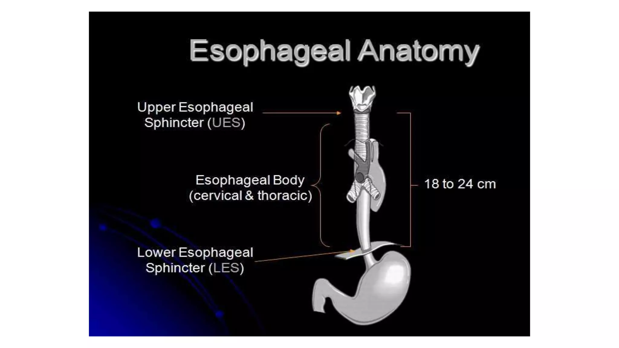
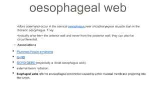
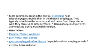
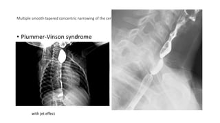
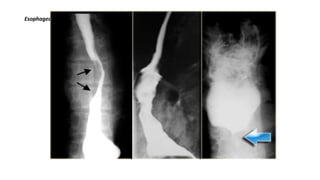
SlideShare a Scribd company logo
•More commonly occur in the cervical oesophagus near cricopharyngeus muscle than in the
thoracic oesophagus. They
•typically arise from the anterior wall and never from the posterior wall; they can also be
circumferential.
• Associations
• Plummer-Vinson syndrome
• GvHD
• GORD/GERD (especially a distal oesophagus web)
• external beam radiation.
• Esophageal webs refer to an esophageal constriction caused by a thin mucosal membrane projecting into
the lumen.
oesophageal web
• More commonly occur in the cervical esophagus near
cricopharyngeus muscle than in the thoracic esophagus. They
typically arise from the anterior wall and never from the posterior
wall; they can also be circumferential 4. Occasionally, multiple webs
are visualized during maximal distension.
• Associations
• Plummer-Vinson syndrome
• graft-versus-host disease
• gastro-esophageal reflux disease (especially a distal esophagus web) 7
• external beam radiation
Multiple smooth tapered concentric narrowing of the cervical esophagus in keeping with esophageal webs.
• Plummer-Vinson syndrome
with jet effect
• To detect the level of obstruction in case of radiolucent foreign body in esophagus,marsh mellow coated
with barium is swallowed
• Passage of marsh mellow will be
hindered
• at the level of obstruction
• Barium swallow shows irregular areas
of narrowing and dilatation ----“Shish
kebab”
Foreign Body Impaction
Diffuse oesophageal spasm
• “corkscrew” “rosary bead" esophagus The esophageal muscle is hypertrophied, but histologically normal
CA ESOPHAGUS
• Preferably high viscosity with normal density barium is used.
• Classical finding in carcinoma-rat tail appearance
ACHALASIA CARDIA
• Barium swallow showing dilatation of the esophageal body
• With short segment stricture.
• A “bird- peak " like tapering of the esophagus at the GE junction.
• Achalasia (primary achalasia) is a failure of organized esophageal peristalsis causing impaired relaxation of
the lower esophageal sphincter, and resulting in food stasis and often marked dilatation of the esophagus.
• Obstruction of the distal esophagus from other non-functional etiologies, notably malignancy, may have a
similar presentation and has been termed "secondary achalasia" or "pseudoachalasia".
• The lower esophageal sphincter fails to relax, either partially or completely, with elevated pressures
demonstrated manometrically 4. This appears to be due to loss/destruction of neurons in
the Auerbach/myenteric plexus. Early in the course of achalasia, the lower esophageal sphincter tone may
be normal or changes may be subtle.
• Peristalsis in the distal smooth muscle segment of the esophagus is eventually lost due to a combination of
damage to the Auerbach plexus and vagus nerve (possibly partly due to damage at the dorsal motor nucleus
of the vagus nerve).
HIATUS HERNIA
• High abdominal pressure is required to demonstrate.
• Pt has to strain.
• Lie down,straighten legs & then raise them up.
• Manual compression of abdomen.
• Pt stands upright,ask him to bend downward with leg straight.
• Stomach should be distended to demonstrate HH.
• Barium meal in Trendlenberg position. Displacement of the cardio-esophageal junction above the
esophageal hiatus . Part of the stomach is present in the chest
• Reflux of barium into the esophagus
Sliding hernia
Para-esophageal hernia
ESOPHAGEAL VARICES
• Varices are best demonstrated in mucosal relief study after using Buscopan/ valsalva maneuver.
• Mild dilatation of the esophagus with multiple persistent filling defects in the lower third of the
esophagus and/or longitudinal furrows.
ESOPHAGITIS
Air-contrast esophagram shows thick esophageal mucosal
folds (arrows) and an ulcer (arrowhead) due to GERD.
Single contrast esophagram shows stricture (arrow) and
sliding hiatus hernia
Gastro esophageal reflux
• SIPHON TEST
• Fill the stomach with 50% barium(150-200ml)
• Follow this 1-2 mouthful of water to remove traces of barium in esophagus
• Pt in supine with left side raised 15% up
• Keep one mouthful of water in pt mouth
• Ask pt to swallow water-a jet of barium will shoot into water column as it enter GO junction
• Alternatively with full stomach,ask pt to roll side to side • Reflux will be seen
• The water siphon test may be performed as part of a barium swallow to assess for gastro-esophageal
reflux. It is performed in the supine RPO position with the patient drinking water continuously. The test is
said to be positive if there is visible barium reflux in the esophagus, and is more sensitive for gastro-
esophageal reflux than observation spontaneous reflux
Oesophageal reflux
• reflux oesophagitis with a deep ulcer (straight arrow). There is also asymmetric
narrowing of the distal esophagus with the distal esophagus with a relatively abrupt
cutoff (curved arrow) at the proximal border of the narrowed segment
INFECTIOUS ESOPHAGITIS
•Increasingly common because of the use of steroid and cytotoxic drugs, disseminated
malignancy,
•and increasing incidence of acquired immunodeficiency syndrome
• CANDIDA ESOPHAGITIS
CANDIDA ESOPHAGITIS
• Radiographic findings include
1. Abnormql esophageal motility ( dilated, atonic esophagus ) is often an early stage
2. Irregular, nodular, plaque-like mucosal pattern ( arrow), irregular folds(arrowhead) with
marginal serrations ( shaggy appearance )
3. Multiple ulcerations of various sizes
4. Frequently involve the entire thoracic esophagus
CANDIDA ESOPHAGITIS.
Cytomegalovirus esophagitis. Cohn's esophagitis.
T.B esophagitis. Eosinophilic esophagitis
Candida esophagitis
• Shaggy esophagus associated with Candida infection , image "A" depicts the longitudinally oriented
plaque-like lesions visible in Candida esophagitis , image "B" depicts the granular appearance of
the esophageal mucosa secondary to edema and inflammation
Cytomegalovirus esophagitis
• Cytomegalovirus esophagitis in a patient with AIDS
• Double contrast esophagram shows a large flat ulcer in profile (large arrows) in the midesophagus
with a cluster of small satellite ulcers (small arrows)
•
Eosinophilic esophagitis
• small calibre oesophagus.
• • transient or fixed circular rings are
seen.
CORROSIVE ESOPHAGITIS.
• Most severe corrosive injuries are caused by alkalis Barium study is unnecessary during
acute phase.
• Radiographic findings
• Diffuse superficial or deep ulceration involving long portion of the distal esophaguS
1. Abnormal motility
2. Fibrotic healing results in a long esophageal stricture ( arrow) that extends down to the
cardioesophageal junction.
• Note : barium was aspirated into left main bronchus(green arrow)
Post-corrosive stricture.
Barrett’s oesophagus
• The reticular mucosa is characteristic of Barrett's columnar metaplasia, especially with the associated web
like (arrow) stricture.
Zenker’s Diverticulum
KILLIAN JAMIESON DIVERTICULUM
• Killian-Jamieson diverticulum is a pulsion diverticulum, that protrudes through a lateral anatomic
weak site of the cervical esophagus below the cricopharyngeus muscle. AP view shows
diverticulum (arrow) originating laterally.
• Lateral view confirms diverticulum does not originate posteriorly as a Zenkers diverticulum would.
Killian-Jamieson diverticulum: AP and lateral view
THORACIC DIVERTICULUM
•Arises in the distal of the esophagus, just above diaphragm
••Pulsion diverticulum (arrow) that probably related to incoordination of esophageal
peristalsis and relaxation of the lower esophageal sphincter
Feline Oesophagus
• The appearance is almost always associated with active gastro-
esophageal reflux 2,3 and is thought to be due to contraction of the
muscularis mucosae with resultant shortening of the esophagus and
'bunching up' of the mucosa in the lumen 2.
• Feline esophagus also known as esophageal shiver, refers to the
transient transverse bands seen in the mid and lower esophagus on a
double-contrast barium swallow.
• Transient horizontal ridges throughout the esophagus (they disappear
with a subsequent swallow).
• These features are typical of the so-called feline esophagus.
Schatzki rings
• A Schatzki ring, also called a Schatzki-Gary ring, is a symptomatic, narrow esophageal B-
ring occurring in the distal esophagus and usually associated with a hiatus hernia.
• The pathogenesis of the Schatzki ring is unclear with conflicting hypotheses that include
redundant pleats of mucosa, congenital abnormalities and modified peptic strictures.
Interestingly, there is a reduced incidence of Barrett esophagus in patients with a Schatzki ring.
• Depending on its luminal diameter, an esophageal B-ring may be symptomatic or asymptomatic 4:
• <13 mm: almost always symptomatic
• 13-20 mm: sometimes symptomatic
• >20 mm: rarely symptomatic
• When it is symptomatic, it is termed a "Schatzki ring" ref.
Boerhaave syndrome:
Boerhaave's syndrome is rupture of the esophageal wall. It is
most often caused by excessive vomiting in eating disorders
such as bulimia although it may rarely occur in extremely
forceful coughing or other situations, such as obstruction by
food.
Boerhaave's syndrome is a transmural or full-thickness
perforation of the esophagus, distinct from MalloryWeiss
syndrome, a non-transmural esophageal tear also associated
with vomiting.
These syndromes are distinct from iatrogenic perforation,
which accounts for 85-90% of cases of esophageal rupture,
typically as a complication of an endoscopic procedure,
feeding tube, or unrelated surgery.
Mallory-Weiss tear
A Mallory-Weiss tear results from prolonged and forceful
vomiting, coughing or convulsions. Typically the mucous
membrane at the junction of the esophagus and the stomach
develops lacerations which bleed, evident by bright red blood
in vomitus, or bloody stools.
It may occur as a result of excessive alcohol ingestion.
This is an acute condition which usually resolves within 10
days without special treatment.
Esophageal hematoma:
These unusual lesions have been associated with increased
esophageal intraluminal pressure, most often vomiting,
instrumentation, and anticoagulation or bleeding disorders.
Some are spontaneous.
Blunt trauma is a rare cause.
Hematomas are self-limited and almost never progress to
perforation.
Most esophageal hematomas resolve in 1-2 weeks with
conservative treatment.
Dissecting intramural hematoma Intramural extravasation (arrow) resulting
Dysphagia lusoria
• The oesophagus may be compressed by a congenitally aberrant right subclavian artery.
• If this is symptomatic a diagnosis of dysphagia lusoria is made
• Here it is seen as oblique tubular extrinsic compression in upper oesophagus.
Leiomyomas
Leiomyomas are the most common benign esophageal
neoplasm and are often large yet nonobstructive.
Gastrointestinal stromal tumors (GIST) are least common in
the esophagus.
A calcified esophageal mass is almost always a leiomyoma.
On the left a patient with a calcified esophageal lesion
(arrows) protrudes into azygoesophageal recess on
radiograph.
ranular cell myoblastomas, an uncommon benign tumor.
Fibrovascular polyp
A foregut
duplication cyst Esophageal duplication. is a congenital cyst.
MALIGNANT
LEFT: Small polypoid carcinoma. RIGHT: Large polypoid lesion.
Adenocarcinoma of the distal esophagus.
• A 4 cm segmental narrowing with an irregular margin and shouldering
appearance at the distal half of the esophagus suggests tumoral
infiltration.
• Evidence of prior sternotomy is noted.
Gastric cardia cancer invading the distal esophagus
(barium swallow)
• Severe stricture with shouldering appearance is present at the distal
portion of the esophagus and gastric cardia that causes pre-stricture
dilatation and contrast media stasis compatible with esophageal
pseudoachalasia.
Submucosal or intramural mass.
Malignant mass at the distal esophagus.
Leiomyosarcoma of the esophagus
Esophageal obstruction due to metastatic mediastinal lymph nodes.
Cricopharyngeal bar
• Cricopharyngeal bar refers to the radiographic appearance of a
prominent cricopharyngeus muscle contour on barium swallow.
• Causes include 1,2:
• idiopathic (i.e. normal variant)
• cricopharyngeal muscle spasm/achalasia (i.e. failed relaxation)
• cricopharyngeus muscle hypertrophy and/or fibrosis
There is smooth and prominent
impression of the cricopharyngeus
muscle at the level of C5-C6 results
in a stenosis.
COMPLICATION
• Leakage of barium from unsuspected perforation
• ASPIRATION
• Difficulty in swallowing, long history of GERD and recent unintentional loss of weight.
• During the rapid-drinking phase, the patient accidentally aspirated a small amount of barium (without any
elicited cough reflex). Contrast outlines the trachea and right main bronchus and smaller amount reached
bronchioles.
• Barium aspiration is an indication to terminate a study, and we sent the patient to be under close
observation in the hospital for 24 hours.

More Related Content

Similar to barium swallow.pptx

VOLVULUS.pptx
VOLVULUS.pptxVOLVULUS.pptx
VOLVULUS.pptx
DR.P.S SUDHAKAR
 
Presentation1.pptx, radiological imaging of esophageal lesions.
Presentation1.pptx, radiological imaging of esophageal lesions.Presentation1.pptx, radiological imaging of esophageal lesions.
Presentation1.pptx, radiological imaging of esophageal lesions.
Abdellah Nazeer
 
Presentation main surgery 123456nhnhnhnahko
Presentation main surgery 123456nhnhnhnahkoPresentation main surgery 123456nhnhnhnahko
Presentation main surgery 123456nhnhnhnahko
HardikSiwach1
 
INTESTINAL OBSTRUCTION
INTESTINAL OBSTRUCTIONINTESTINAL OBSTRUCTION
INTESTINAL OBSTRUCTION
shahadatsurg
 
barium studies in gi pathologies
barium studies in gi pathologiesbarium studies in gi pathologies
barium studies in gi pathologies
Ahmad Jawad
 
GASTRIC OUTLET OBSTRUCTION.pptx
GASTRIC OUTLET OBSTRUCTION.pptxGASTRIC OUTLET OBSTRUCTION.pptx
GASTRIC OUTLET OBSTRUCTION.pptx
Thlamuana Knox
 
Barium swallow diseases
Barium swallow diseasesBarium swallow diseases
Barium swallow diseases
Shrunga Tejasvi
 
Git anomalies
Git anomaliesGit anomalies
Git anomalies
Dr Varun Bansal
 
Presentation1, radiological imaging of barium studies.
Presentation1, radiological imaging of barium studies.Presentation1, radiological imaging of barium studies.
Presentation1, radiological imaging of barium studies.
Abdellah Nazeer
 
IMAGING IN NEONATAL GIT
IMAGING IN NEONATAL GITIMAGING IN NEONATAL GIT
IMAGING IN NEONATAL GIT
Thiyagarajan Shanmugam
 
Malrotation
MalrotationMalrotation
Malrotation
HusseinSigat
 
Barium meal
Barium mealBarium meal
Barium meal
dypradio
 
GERD / GASTRO ESOPHAGEAL REFLUX
GERD / GASTRO ESOPHAGEAL REFLUX GERD / GASTRO ESOPHAGEAL REFLUX
GERD / GASTRO ESOPHAGEAL REFLUX
Arun Karmakar
 
Esophageal diseases .pdf by university of kufa college of medicine
Esophageal diseases .pdf by university of kufa college of medicineEsophageal diseases .pdf by university of kufa college of medicine
Esophageal diseases .pdf by university of kufa college of medicine
zahraa934924
 
Hiatal hernia
Hiatal herniaHiatal hernia
Hiatal hernia
MutahirRehman
 
Barium meal PPT Slide PK
Barium meal PPT Slide  PKBarium meal PPT Slide  PK
Barium meal PPT Slide PK
Dr pradeep Kumar
 
Group 4 dysphagia 2016 version 3.1 validated
Group 4   dysphagia 2016 version 3.1 validatedGroup 4   dysphagia 2016 version 3.1 validated
Group 4 dysphagia 2016 version 3.1 validated
Dennis Lee
 
I.o Intestinal
I.o IntestinalI.o Intestinal
I.o Intestinal
MochiManja
 
Intestinal obstruction, Ileus, and volvulus
Intestinal obstruction, Ileus, and volvulusIntestinal obstruction, Ileus, and volvulus
Intestinal obstruction, Ileus, and volvulus
Khaled AlKhodari
 
Dr. kawa bilateral
Dr. kawa bilateralDr. kawa bilateral
Dr. kawa bilateral
Rzgar Tayeb
 

Similar to barium swallow.pptx (20)

VOLVULUS.pptx
VOLVULUS.pptxVOLVULUS.pptx
VOLVULUS.pptx
 
Presentation1.pptx, radiological imaging of esophageal lesions.
Presentation1.pptx, radiological imaging of esophageal lesions.Presentation1.pptx, radiological imaging of esophageal lesions.
Presentation1.pptx, radiological imaging of esophageal lesions.
 
Presentation main surgery 123456nhnhnhnahko
Presentation main surgery 123456nhnhnhnahkoPresentation main surgery 123456nhnhnhnahko
Presentation main surgery 123456nhnhnhnahko
 
INTESTINAL OBSTRUCTION
INTESTINAL OBSTRUCTIONINTESTINAL OBSTRUCTION
INTESTINAL OBSTRUCTION
 
barium studies in gi pathologies
barium studies in gi pathologiesbarium studies in gi pathologies
barium studies in gi pathologies
 
GASTRIC OUTLET OBSTRUCTION.pptx
GASTRIC OUTLET OBSTRUCTION.pptxGASTRIC OUTLET OBSTRUCTION.pptx
GASTRIC OUTLET OBSTRUCTION.pptx
 
Barium swallow diseases
Barium swallow diseasesBarium swallow diseases
Barium swallow diseases
 
Git anomalies
Git anomaliesGit anomalies
Git anomalies
 
Presentation1, radiological imaging of barium studies.
Presentation1, radiological imaging of barium studies.Presentation1, radiological imaging of barium studies.
Presentation1, radiological imaging of barium studies.
 
IMAGING IN NEONATAL GIT
IMAGING IN NEONATAL GITIMAGING IN NEONATAL GIT
IMAGING IN NEONATAL GIT
 
Malrotation
MalrotationMalrotation
Malrotation
 
Barium meal
Barium mealBarium meal
Barium meal
 
GERD / GASTRO ESOPHAGEAL REFLUX
GERD / GASTRO ESOPHAGEAL REFLUX GERD / GASTRO ESOPHAGEAL REFLUX
GERD / GASTRO ESOPHAGEAL REFLUX
 
Esophageal diseases .pdf by university of kufa college of medicine
Esophageal diseases .pdf by university of kufa college of medicineEsophageal diseases .pdf by university of kufa college of medicine
Esophageal diseases .pdf by university of kufa college of medicine
 
Hiatal hernia
Hiatal herniaHiatal hernia
Hiatal hernia
 
Barium meal PPT Slide PK
Barium meal PPT Slide  PKBarium meal PPT Slide  PK
Barium meal PPT Slide PK
 
Group 4 dysphagia 2016 version 3.1 validated
Group 4   dysphagia 2016 version 3.1 validatedGroup 4   dysphagia 2016 version 3.1 validated
Group 4 dysphagia 2016 version 3.1 validated
 
I.o Intestinal
I.o IntestinalI.o Intestinal
I.o Intestinal
 
Intestinal obstruction, Ileus, and volvulus
Intestinal obstruction, Ileus, and volvulusIntestinal obstruction, Ileus, and volvulus
Intestinal obstruction, Ileus, and volvulus
 
Dr. kawa bilateral
Dr. kawa bilateralDr. kawa bilateral
Dr. kawa bilateral
 

More from ranjitharadhakrishna3

ANATOMY OF VERTEBRAL COLUMN AND SPINAL CORD.pptx
ANATOMY OF VERTEBRAL COLUMN AND SPINAL CORD.pptxANATOMY OF VERTEBRAL COLUMN AND SPINAL CORD.pptx
ANATOMY OF VERTEBRAL COLUMN AND SPINAL CORD.pptx
ranjitharadhakrishna3
 
cerebral cisterns for radiology dnb .pptx
cerebral cisterns for radiology dnb .pptxcerebral cisterns for radiology dnb .pptx
cerebral cisterns for radiology dnb .pptx
ranjitharadhakrishna3
 
brain anatomy radiology ppt for dnbs.ppt
brain anatomy radiology ppt for dnbs.pptbrain anatomy radiology ppt for dnbs.ppt
brain anatomy radiology ppt for dnbs.ppt
ranjitharadhakrishna3
 
PANCREATIC ANOMALY radiology.pptx
PANCREATIC ANOMALY radiology.pptxPANCREATIC ANOMALY radiology.pptx
PANCREATIC ANOMALY radiology.pptx
ranjitharadhakrishna3
 
FNAC lung (2).ppt
FNAC lung (2).pptFNAC lung (2).ppt
FNAC lung (2).ppt
ranjitharadhakrishna3
 
FNAC lung.ppt
FNAC lung.pptFNAC lung.ppt
FNAC lung.ppt
ranjitharadhakrishna3
 
pre sacral lesion sept5.pptx RADIOLOGY
pre sacral lesion sept5.pptx RADIOLOGYpre sacral lesion sept5.pptx RADIOLOGY
pre sacral lesion sept5.pptx RADIOLOGY
ranjitharadhakrishna3
 
Sarcoidosis radiology pulmonary neuro abdominal .ppt
Sarcoidosis radiology pulmonary neuro abdominal .pptSarcoidosis radiology pulmonary neuro abdominal .ppt
Sarcoidosis radiology pulmonary neuro abdominal .ppt
ranjitharadhakrishna3
 
Respiratory system signs final.ppt radiology
Respiratory system signs final.ppt radiologyRespiratory system signs final.ppt radiology
Respiratory system signs final.ppt radiology
ranjitharadhakrishna3
 
SOLITARY PULMONARY NODULE.pptx
SOLITARY PULMONARY NODULE.pptxSOLITARY PULMONARY NODULE.pptx
SOLITARY PULMONARY NODULE.pptx
ranjitharadhakrishna3
 
Parathyroid Imaging .pptx
Parathyroid Imaging .pptxParathyroid Imaging .pptx
Parathyroid Imaging .pptx
ranjitharadhakrishna3
 
mri and ct anatomy of brain-.pptx
mri and ct anatomy of brain-.pptxmri and ct anatomy of brain-.pptx
mri and ct anatomy of brain-.pptx
ranjitharadhakrishna3
 
acuteosteomyelitis-.pptx
acuteosteomyelitis-.pptxacuteosteomyelitis-.pptx
acuteosteomyelitis-.pptx
ranjitharadhakrishna3
 
retroperitonealmasses-pptx
retroperitonealmasses-pptxretroperitonealmasses-pptx
retroperitonealmasses-pptx
ranjitharadhakrishna3
 
ULTRASOUND IN FIRST TRIMESTER.ppt
ULTRASOUND IN FIRST TRIMESTER.pptULTRASOUND IN FIRST TRIMESTER.ppt
ULTRASOUND IN FIRST TRIMESTER.ppt
ranjitharadhakrishna3
 
OSTEONECROSIS 2003.ppt
OSTEONECROSIS 2003.pptOSTEONECROSIS 2003.ppt
OSTEONECROSIS 2003.ppt
ranjitharadhakrishna3
 
GRIDS.ppt
GRIDS.pptGRIDS.ppt
fluoro1-principles.PPT
fluoro1-principles.PPTfluoro1-principles.PPT
fluoro1-principles.PPT
ranjitharadhakrishna3
 
FLUOROSCOPIC IMAGING.ppt
FLUOROSCOPIC IMAGING.pptFLUOROSCOPIC IMAGING.ppt
FLUOROSCOPIC IMAGING.ppt
ranjitharadhakrishna3
 
DIFFUSION & PERFUSION r MRI.ppt
DIFFUSION  &   PERFUSION  r MRI.pptDIFFUSION  &   PERFUSION  r MRI.ppt
DIFFUSION & PERFUSION r MRI.ppt
ranjitharadhakrishna3
 

More from ranjitharadhakrishna3 (20)

ANATOMY OF VERTEBRAL COLUMN AND SPINAL CORD.pptx
ANATOMY OF VERTEBRAL COLUMN AND SPINAL CORD.pptxANATOMY OF VERTEBRAL COLUMN AND SPINAL CORD.pptx
ANATOMY OF VERTEBRAL COLUMN AND SPINAL CORD.pptx
 
cerebral cisterns for radiology dnb .pptx
cerebral cisterns for radiology dnb .pptxcerebral cisterns for radiology dnb .pptx
cerebral cisterns for radiology dnb .pptx
 
brain anatomy radiology ppt for dnbs.ppt
brain anatomy radiology ppt for dnbs.pptbrain anatomy radiology ppt for dnbs.ppt
brain anatomy radiology ppt for dnbs.ppt
 
PANCREATIC ANOMALY radiology.pptx
PANCREATIC ANOMALY radiology.pptxPANCREATIC ANOMALY radiology.pptx
PANCREATIC ANOMALY radiology.pptx
 
FNAC lung (2).ppt
FNAC lung (2).pptFNAC lung (2).ppt
FNAC lung (2).ppt
 
FNAC lung.ppt
FNAC lung.pptFNAC lung.ppt
FNAC lung.ppt
 
pre sacral lesion sept5.pptx RADIOLOGY
pre sacral lesion sept5.pptx RADIOLOGYpre sacral lesion sept5.pptx RADIOLOGY
pre sacral lesion sept5.pptx RADIOLOGY
 
Sarcoidosis radiology pulmonary neuro abdominal .ppt
Sarcoidosis radiology pulmonary neuro abdominal .pptSarcoidosis radiology pulmonary neuro abdominal .ppt
Sarcoidosis radiology pulmonary neuro abdominal .ppt
 
Respiratory system signs final.ppt radiology
Respiratory system signs final.ppt radiologyRespiratory system signs final.ppt radiology
Respiratory system signs final.ppt radiology
 
SOLITARY PULMONARY NODULE.pptx
SOLITARY PULMONARY NODULE.pptxSOLITARY PULMONARY NODULE.pptx
SOLITARY PULMONARY NODULE.pptx
 
Parathyroid Imaging .pptx
Parathyroid Imaging .pptxParathyroid Imaging .pptx
Parathyroid Imaging .pptx
 
mri and ct anatomy of brain-.pptx
mri and ct anatomy of brain-.pptxmri and ct anatomy of brain-.pptx
mri and ct anatomy of brain-.pptx
 
acuteosteomyelitis-.pptx
acuteosteomyelitis-.pptxacuteosteomyelitis-.pptx
acuteosteomyelitis-.pptx
 
retroperitonealmasses-pptx
retroperitonealmasses-pptxretroperitonealmasses-pptx
retroperitonealmasses-pptx
 
ULTRASOUND IN FIRST TRIMESTER.ppt
ULTRASOUND IN FIRST TRIMESTER.pptULTRASOUND IN FIRST TRIMESTER.ppt
ULTRASOUND IN FIRST TRIMESTER.ppt
 
OSTEONECROSIS 2003.ppt
OSTEONECROSIS 2003.pptOSTEONECROSIS 2003.ppt
OSTEONECROSIS 2003.ppt
 
GRIDS.ppt
GRIDS.pptGRIDS.ppt
GRIDS.ppt
 
fluoro1-principles.PPT
fluoro1-principles.PPTfluoro1-principles.PPT
fluoro1-principles.PPT
 
FLUOROSCOPIC IMAGING.ppt
FLUOROSCOPIC IMAGING.pptFLUOROSCOPIC IMAGING.ppt
FLUOROSCOPIC IMAGING.ppt
 
DIFFUSION & PERFUSION r MRI.ppt
DIFFUSION  &   PERFUSION  r MRI.pptDIFFUSION  &   PERFUSION  r MRI.ppt
DIFFUSION & PERFUSION r MRI.ppt
 

Recently uploaded

Travis Hills' Endeavors in Minnesota: Fostering Environmental and Economic Pr...
Travis Hills' Endeavors in Minnesota: Fostering Environmental and Economic Pr...Travis Hills' Endeavors in Minnesota: Fostering Environmental and Economic Pr...
Travis Hills' Endeavors in Minnesota: Fostering Environmental and Economic Pr...
Travis Hills MN
 
THEMATIC APPERCEPTION TEST(TAT) cognitive abilities, creativity, and critic...
THEMATIC  APPERCEPTION  TEST(TAT) cognitive abilities, creativity, and critic...THEMATIC  APPERCEPTION  TEST(TAT) cognitive abilities, creativity, and critic...
THEMATIC APPERCEPTION TEST(TAT) cognitive abilities, creativity, and critic...
Abdul Wali Khan University Mardan,kP,Pakistan
 
Eukaryotic Transcription Presentation.pptx
Eukaryotic Transcription Presentation.pptxEukaryotic Transcription Presentation.pptx
Eukaryotic Transcription Presentation.pptx
RitabrataSarkar3
 
Deep Software Variability and Frictionless Reproducibility
Deep Software Variability and Frictionless ReproducibilityDeep Software Variability and Frictionless Reproducibility
Deep Software Variability and Frictionless Reproducibility
University of Rennes, INSA Rennes, Inria/IRISA, CNRS
 
NuGOweek 2024 Ghent programme overview flyer
NuGOweek 2024 Ghent programme overview flyerNuGOweek 2024 Ghent programme overview flyer
NuGOweek 2024 Ghent programme overview flyer
pablovgd
 
DERIVATION OF MODIFIED BERNOULLI EQUATION WITH VISCOUS EFFECTS AND TERMINAL V...
DERIVATION OF MODIFIED BERNOULLI EQUATION WITH VISCOUS EFFECTS AND TERMINAL V...DERIVATION OF MODIFIED BERNOULLI EQUATION WITH VISCOUS EFFECTS AND TERMINAL V...
DERIVATION OF MODIFIED BERNOULLI EQUATION WITH VISCOUS EFFECTS AND TERMINAL V...
Wasswaderrick3
 
The debris of the ‘last major merger’ is dynamically young
The debris of the ‘last major merger’ is dynamically youngThe debris of the ‘last major merger’ is dynamically young
The debris of the ‘last major merger’ is dynamically young
Sérgio Sacani
 
ESR spectroscopy in liquid food and beverages.pptx
ESR spectroscopy in liquid food and beverages.pptxESR spectroscopy in liquid food and beverages.pptx
ESR spectroscopy in liquid food and beverages.pptx
PRIYANKA PATEL
 
Nucleic Acid-its structural and functional complexity.
Nucleic Acid-its structural and functional complexity.Nucleic Acid-its structural and functional complexity.
Nucleic Acid-its structural and functional complexity.
Nistarini College, Purulia (W.B) India
 
Nucleophilic Addition of carbonyl compounds.pptx
Nucleophilic Addition of carbonyl  compounds.pptxNucleophilic Addition of carbonyl  compounds.pptx
Nucleophilic Addition of carbonyl compounds.pptx
SSR02
 
3D Hybrid PIC simulation of the plasma expansion (ISSS-14)
3D Hybrid PIC simulation of the plasma expansion (ISSS-14)3D Hybrid PIC simulation of the plasma expansion (ISSS-14)
3D Hybrid PIC simulation of the plasma expansion (ISSS-14)
David Osipyan
 
Topic: SICKLE CELL DISEASE IN CHILDREN-3.pdf
Topic: SICKLE CELL DISEASE IN CHILDREN-3.pdfTopic: SICKLE CELL DISEASE IN CHILDREN-3.pdf
Topic: SICKLE CELL DISEASE IN CHILDREN-3.pdf
TinyAnderson
 
Oedema_types_causes_pathophysiology.pptx
Oedema_types_causes_pathophysiology.pptxOedema_types_causes_pathophysiology.pptx
Oedema_types_causes_pathophysiology.pptx
muralinath2
 
Unveiling the Energy Potential of Marshmallow Deposits.pdf
Unveiling the Energy Potential of Marshmallow Deposits.pdfUnveiling the Energy Potential of Marshmallow Deposits.pdf
Unveiling the Energy Potential of Marshmallow Deposits.pdf
Erdal Coalmaker
 
Shallowest Oil Discovery of Turkiye.pptx
Shallowest Oil Discovery of Turkiye.pptxShallowest Oil Discovery of Turkiye.pptx
Shallowest Oil Discovery of Turkiye.pptx
Gokturk Mehmet Dilci
 
What is greenhouse gasses and how many gasses are there to affect the Earth.
What is greenhouse gasses and how many gasses are there to affect the Earth.What is greenhouse gasses and how many gasses are there to affect the Earth.
What is greenhouse gasses and how many gasses are there to affect the Earth.
moosaasad1975
 
Chapter 12 - climate change and the energy crisis
Chapter 12 - climate change and the energy crisisChapter 12 - climate change and the energy crisis
Chapter 12 - climate change and the energy crisis
tonzsalvador2222
 
Comparing Evolved Extractive Text Summary Scores of Bidirectional Encoder Rep...
Comparing Evolved Extractive Text Summary Scores of Bidirectional Encoder Rep...Comparing Evolved Extractive Text Summary Scores of Bidirectional Encoder Rep...
Comparing Evolved Extractive Text Summary Scores of Bidirectional Encoder Rep...
University of Maribor
 
DMARDs Pharmacolgy Pharm D 5th Semester.pdf
DMARDs Pharmacolgy Pharm D 5th Semester.pdfDMARDs Pharmacolgy Pharm D 5th Semester.pdf
DMARDs Pharmacolgy Pharm D 5th Semester.pdf
fafyfskhan251kmf
 
Orion Air Quality Monitoring Systems - CWS
Orion Air Quality Monitoring Systems - CWSOrion Air Quality Monitoring Systems - CWS
Orion Air Quality Monitoring Systems - CWS
Columbia Weather Systems
 

Recently uploaded (20)

Travis Hills' Endeavors in Minnesota: Fostering Environmental and Economic Pr...
Travis Hills' Endeavors in Minnesota: Fostering Environmental and Economic Pr...Travis Hills' Endeavors in Minnesota: Fostering Environmental and Economic Pr...
Travis Hills' Endeavors in Minnesota: Fostering Environmental and Economic Pr...
 
THEMATIC APPERCEPTION TEST(TAT) cognitive abilities, creativity, and critic...
THEMATIC  APPERCEPTION  TEST(TAT) cognitive abilities, creativity, and critic...THEMATIC  APPERCEPTION  TEST(TAT) cognitive abilities, creativity, and critic...
THEMATIC APPERCEPTION TEST(TAT) cognitive abilities, creativity, and critic...
 
Eukaryotic Transcription Presentation.pptx
Eukaryotic Transcription Presentation.pptxEukaryotic Transcription Presentation.pptx
Eukaryotic Transcription Presentation.pptx
 
Deep Software Variability and Frictionless Reproducibility
Deep Software Variability and Frictionless ReproducibilityDeep Software Variability and Frictionless Reproducibility
Deep Software Variability and Frictionless Reproducibility
 
NuGOweek 2024 Ghent programme overview flyer
NuGOweek 2024 Ghent programme overview flyerNuGOweek 2024 Ghent programme overview flyer
NuGOweek 2024 Ghent programme overview flyer
 
DERIVATION OF MODIFIED BERNOULLI EQUATION WITH VISCOUS EFFECTS AND TERMINAL V...
DERIVATION OF MODIFIED BERNOULLI EQUATION WITH VISCOUS EFFECTS AND TERMINAL V...DERIVATION OF MODIFIED BERNOULLI EQUATION WITH VISCOUS EFFECTS AND TERMINAL V...
DERIVATION OF MODIFIED BERNOULLI EQUATION WITH VISCOUS EFFECTS AND TERMINAL V...
 
The debris of the ‘last major merger’ is dynamically young
The debris of the ‘last major merger’ is dynamically youngThe debris of the ‘last major merger’ is dynamically young
The debris of the ‘last major merger’ is dynamically young
 
ESR spectroscopy in liquid food and beverages.pptx
ESR spectroscopy in liquid food and beverages.pptxESR spectroscopy in liquid food and beverages.pptx
ESR spectroscopy in liquid food and beverages.pptx
 
Nucleic Acid-its structural and functional complexity.
Nucleic Acid-its structural and functional complexity.Nucleic Acid-its structural and functional complexity.
Nucleic Acid-its structural and functional complexity.
 
Nucleophilic Addition of carbonyl compounds.pptx
Nucleophilic Addition of carbonyl  compounds.pptxNucleophilic Addition of carbonyl  compounds.pptx
Nucleophilic Addition of carbonyl compounds.pptx
 
3D Hybrid PIC simulation of the plasma expansion (ISSS-14)
3D Hybrid PIC simulation of the plasma expansion (ISSS-14)3D Hybrid PIC simulation of the plasma expansion (ISSS-14)
3D Hybrid PIC simulation of the plasma expansion (ISSS-14)
 
Topic: SICKLE CELL DISEASE IN CHILDREN-3.pdf
Topic: SICKLE CELL DISEASE IN CHILDREN-3.pdfTopic: SICKLE CELL DISEASE IN CHILDREN-3.pdf
Topic: SICKLE CELL DISEASE IN CHILDREN-3.pdf
 
Oedema_types_causes_pathophysiology.pptx
Oedema_types_causes_pathophysiology.pptxOedema_types_causes_pathophysiology.pptx
Oedema_types_causes_pathophysiology.pptx
 
Unveiling the Energy Potential of Marshmallow Deposits.pdf
Unveiling the Energy Potential of Marshmallow Deposits.pdfUnveiling the Energy Potential of Marshmallow Deposits.pdf
Unveiling the Energy Potential of Marshmallow Deposits.pdf
 
Shallowest Oil Discovery of Turkiye.pptx
Shallowest Oil Discovery of Turkiye.pptxShallowest Oil Discovery of Turkiye.pptx
Shallowest Oil Discovery of Turkiye.pptx
 
What is greenhouse gasses and how many gasses are there to affect the Earth.
What is greenhouse gasses and how many gasses are there to affect the Earth.What is greenhouse gasses and how many gasses are there to affect the Earth.
What is greenhouse gasses and how many gasses are there to affect the Earth.
 
Chapter 12 - climate change and the energy crisis
Chapter 12 - climate change and the energy crisisChapter 12 - climate change and the energy crisis
Chapter 12 - climate change and the energy crisis
 
Comparing Evolved Extractive Text Summary Scores of Bidirectional Encoder Rep...
Comparing Evolved Extractive Text Summary Scores of Bidirectional Encoder Rep...Comparing Evolved Extractive Text Summary Scores of Bidirectional Encoder Rep...
Comparing Evolved Extractive Text Summary Scores of Bidirectional Encoder Rep...
 
DMARDs Pharmacolgy Pharm D 5th Semester.pdf
DMARDs Pharmacolgy Pharm D 5th Semester.pdfDMARDs Pharmacolgy Pharm D 5th Semester.pdf
DMARDs Pharmacolgy Pharm D 5th Semester.pdf
 
Orion Air Quality Monitoring Systems - CWS
Orion Air Quality Monitoring Systems - CWSOrion Air Quality Monitoring Systems - CWS
Orion Air Quality Monitoring Systems - CWS
 

barium swallow.pptx

  • 1.
  • 2. •More commonly occur in the cervical oesophagus near cricopharyngeus muscle than in the thoracic oesophagus. They •typically arise from the anterior wall and never from the posterior wall; they can also be circumferential. • Associations • Plummer-Vinson syndrome • GvHD • GORD/GERD (especially a distal oesophagus web) • external beam radiation. • Esophageal webs refer to an esophageal constriction caused by a thin mucosal membrane projecting into the lumen. oesophageal web
  • 3. • More commonly occur in the cervical esophagus near cricopharyngeus muscle than in the thoracic esophagus. They typically arise from the anterior wall and never from the posterior wall; they can also be circumferential 4. Occasionally, multiple webs are visualized during maximal distension. • Associations • Plummer-Vinson syndrome • graft-versus-host disease • gastro-esophageal reflux disease (especially a distal esophagus web) 7 • external beam radiation
  • 4. Multiple smooth tapered concentric narrowing of the cervical esophagus in keeping with esophageal webs. • Plummer-Vinson syndrome with jet effect
  • 5. • To detect the level of obstruction in case of radiolucent foreign body in esophagus,marsh mellow coated with barium is swallowed • Passage of marsh mellow will be hindered • at the level of obstruction • Barium swallow shows irregular areas of narrowing and dilatation ----“Shish kebab” Foreign Body Impaction
  • 6. Diffuse oesophageal spasm • “corkscrew” “rosary bead" esophagus The esophageal muscle is hypertrophied, but histologically normal
  • 7.
  • 8. CA ESOPHAGUS • Preferably high viscosity with normal density barium is used. • Classical finding in carcinoma-rat tail appearance
  • 9. ACHALASIA CARDIA • Barium swallow showing dilatation of the esophageal body • With short segment stricture. • A “bird- peak " like tapering of the esophagus at the GE junction. • Achalasia (primary achalasia) is a failure of organized esophageal peristalsis causing impaired relaxation of the lower esophageal sphincter, and resulting in food stasis and often marked dilatation of the esophagus. • Obstruction of the distal esophagus from other non-functional etiologies, notably malignancy, may have a similar presentation and has been termed "secondary achalasia" or "pseudoachalasia". • The lower esophageal sphincter fails to relax, either partially or completely, with elevated pressures demonstrated manometrically 4. This appears to be due to loss/destruction of neurons in the Auerbach/myenteric plexus. Early in the course of achalasia, the lower esophageal sphincter tone may be normal or changes may be subtle. • Peristalsis in the distal smooth muscle segment of the esophagus is eventually lost due to a combination of damage to the Auerbach plexus and vagus nerve (possibly partly due to damage at the dorsal motor nucleus of the vagus nerve).
  • 10.
  • 11.
  • 12. HIATUS HERNIA • High abdominal pressure is required to demonstrate. • Pt has to strain. • Lie down,straighten legs & then raise them up. • Manual compression of abdomen. • Pt stands upright,ask him to bend downward with leg straight. • Stomach should be distended to demonstrate HH. • Barium meal in Trendlenberg position. Displacement of the cardio-esophageal junction above the esophageal hiatus . Part of the stomach is present in the chest • Reflux of barium into the esophagus
  • 15.
  • 16. ESOPHAGEAL VARICES • Varices are best demonstrated in mucosal relief study after using Buscopan/ valsalva maneuver. • Mild dilatation of the esophagus with multiple persistent filling defects in the lower third of the esophagus and/or longitudinal furrows.
  • 17.
  • 18.
  • 20. Air-contrast esophagram shows thick esophageal mucosal folds (arrows) and an ulcer (arrowhead) due to GERD. Single contrast esophagram shows stricture (arrow) and sliding hiatus hernia
  • 21. Gastro esophageal reflux • SIPHON TEST • Fill the stomach with 50% barium(150-200ml) • Follow this 1-2 mouthful of water to remove traces of barium in esophagus • Pt in supine with left side raised 15% up • Keep one mouthful of water in pt mouth • Ask pt to swallow water-a jet of barium will shoot into water column as it enter GO junction • Alternatively with full stomach,ask pt to roll side to side • Reflux will be seen • The water siphon test may be performed as part of a barium swallow to assess for gastro-esophageal reflux. It is performed in the supine RPO position with the patient drinking water continuously. The test is said to be positive if there is visible barium reflux in the esophagus, and is more sensitive for gastro- esophageal reflux than observation spontaneous reflux
  • 22. Oesophageal reflux • reflux oesophagitis with a deep ulcer (straight arrow). There is also asymmetric narrowing of the distal esophagus with the distal esophagus with a relatively abrupt cutoff (curved arrow) at the proximal border of the narrowed segment
  • 23. INFECTIOUS ESOPHAGITIS •Increasingly common because of the use of steroid and cytotoxic drugs, disseminated malignancy, •and increasing incidence of acquired immunodeficiency syndrome • CANDIDA ESOPHAGITIS
  • 24. CANDIDA ESOPHAGITIS • Radiographic findings include 1. Abnormql esophageal motility ( dilated, atonic esophagus ) is often an early stage 2. Irregular, nodular, plaque-like mucosal pattern ( arrow), irregular folds(arrowhead) with marginal serrations ( shaggy appearance ) 3. Multiple ulcerations of various sizes 4. Frequently involve the entire thoracic esophagus
  • 28. Candida esophagitis • Shaggy esophagus associated with Candida infection , image "A" depicts the longitudinally oriented plaque-like lesions visible in Candida esophagitis , image "B" depicts the granular appearance of the esophageal mucosa secondary to edema and inflammation
  • 29. Cytomegalovirus esophagitis • Cytomegalovirus esophagitis in a patient with AIDS • Double contrast esophagram shows a large flat ulcer in profile (large arrows) in the midesophagus with a cluster of small satellite ulcers (small arrows) •
  • 30. Eosinophilic esophagitis • small calibre oesophagus. • • transient or fixed circular rings are seen.
  • 31. CORROSIVE ESOPHAGITIS. • Most severe corrosive injuries are caused by alkalis Barium study is unnecessary during acute phase. • Radiographic findings • Diffuse superficial or deep ulceration involving long portion of the distal esophaguS 1. Abnormal motility 2. Fibrotic healing results in a long esophageal stricture ( arrow) that extends down to the cardioesophageal junction. • Note : barium was aspirated into left main bronchus(green arrow)
  • 33.
  • 34.
  • 35. Barrett’s oesophagus • The reticular mucosa is characteristic of Barrett's columnar metaplasia, especially with the associated web like (arrow) stricture.
  • 36.
  • 37.
  • 39. KILLIAN JAMIESON DIVERTICULUM • Killian-Jamieson diverticulum is a pulsion diverticulum, that protrudes through a lateral anatomic weak site of the cervical esophagus below the cricopharyngeus muscle. AP view shows diverticulum (arrow) originating laterally. • Lateral view confirms diverticulum does not originate posteriorly as a Zenkers diverticulum would.
  • 41. THORACIC DIVERTICULUM •Arises in the distal of the esophagus, just above diaphragm ••Pulsion diverticulum (arrow) that probably related to incoordination of esophageal peristalsis and relaxation of the lower esophageal sphincter
  • 42. Feline Oesophagus • The appearance is almost always associated with active gastro- esophageal reflux 2,3 and is thought to be due to contraction of the muscularis mucosae with resultant shortening of the esophagus and 'bunching up' of the mucosa in the lumen 2. • Feline esophagus also known as esophageal shiver, refers to the transient transverse bands seen in the mid and lower esophagus on a double-contrast barium swallow. • Transient horizontal ridges throughout the esophagus (they disappear with a subsequent swallow). • These features are typical of the so-called feline esophagus.
  • 43. Schatzki rings • A Schatzki ring, also called a Schatzki-Gary ring, is a symptomatic, narrow esophageal B- ring occurring in the distal esophagus and usually associated with a hiatus hernia. • The pathogenesis of the Schatzki ring is unclear with conflicting hypotheses that include redundant pleats of mucosa, congenital abnormalities and modified peptic strictures. Interestingly, there is a reduced incidence of Barrett esophagus in patients with a Schatzki ring. • Depending on its luminal diameter, an esophageal B-ring may be symptomatic or asymptomatic 4: • <13 mm: almost always symptomatic • 13-20 mm: sometimes symptomatic • >20 mm: rarely symptomatic • When it is symptomatic, it is termed a "Schatzki ring" ref.
  • 44.
  • 45.
  • 46. Boerhaave syndrome: Boerhaave's syndrome is rupture of the esophageal wall. It is most often caused by excessive vomiting in eating disorders such as bulimia although it may rarely occur in extremely forceful coughing or other situations, such as obstruction by food. Boerhaave's syndrome is a transmural or full-thickness perforation of the esophagus, distinct from MalloryWeiss syndrome, a non-transmural esophageal tear also associated with vomiting. These syndromes are distinct from iatrogenic perforation, which accounts for 85-90% of cases of esophageal rupture, typically as a complication of an endoscopic procedure, feeding tube, or unrelated surgery.
  • 47.
  • 48. Mallory-Weiss tear A Mallory-Weiss tear results from prolonged and forceful vomiting, coughing or convulsions. Typically the mucous membrane at the junction of the esophagus and the stomach develops lacerations which bleed, evident by bright red blood in vomitus, or bloody stools. It may occur as a result of excessive alcohol ingestion. This is an acute condition which usually resolves within 10 days without special treatment.
  • 49.
  • 50. Esophageal hematoma: These unusual lesions have been associated with increased esophageal intraluminal pressure, most often vomiting, instrumentation, and anticoagulation or bleeding disorders. Some are spontaneous. Blunt trauma is a rare cause. Hematomas are self-limited and almost never progress to perforation. Most esophageal hematomas resolve in 1-2 weeks with conservative treatment.
  • 51.
  • 52. Dissecting intramural hematoma Intramural extravasation (arrow) resulting
  • 53. Dysphagia lusoria • The oesophagus may be compressed by a congenitally aberrant right subclavian artery. • If this is symptomatic a diagnosis of dysphagia lusoria is made • Here it is seen as oblique tubular extrinsic compression in upper oesophagus.
  • 54.
  • 55. Leiomyomas Leiomyomas are the most common benign esophageal neoplasm and are often large yet nonobstructive. Gastrointestinal stromal tumors (GIST) are least common in the esophagus.
  • 56. A calcified esophageal mass is almost always a leiomyoma. On the left a patient with a calcified esophageal lesion (arrows) protrudes into azygoesophageal recess on radiograph.
  • 57. ranular cell myoblastomas, an uncommon benign tumor. Fibrovascular polyp
  • 58. A foregut duplication cyst Esophageal duplication. is a congenital cyst.
  • 60. LEFT: Small polypoid carcinoma. RIGHT: Large polypoid lesion.
  • 61. Adenocarcinoma of the distal esophagus.
  • 62. • A 4 cm segmental narrowing with an irregular margin and shouldering appearance at the distal half of the esophagus suggests tumoral infiltration. • Evidence of prior sternotomy is noted.
  • 63. Gastric cardia cancer invading the distal esophagus (barium swallow) • Severe stricture with shouldering appearance is present at the distal portion of the esophagus and gastric cardia that causes pre-stricture dilatation and contrast media stasis compatible with esophageal pseudoachalasia.
  • 65. Malignant mass at the distal esophagus.
  • 66.
  • 68. Esophageal obstruction due to metastatic mediastinal lymph nodes.
  • 69. Cricopharyngeal bar • Cricopharyngeal bar refers to the radiographic appearance of a prominent cricopharyngeus muscle contour on barium swallow. • Causes include 1,2: • idiopathic (i.e. normal variant) • cricopharyngeal muscle spasm/achalasia (i.e. failed relaxation) • cricopharyngeus muscle hypertrophy and/or fibrosis There is smooth and prominent impression of the cricopharyngeus muscle at the level of C5-C6 results in a stenosis.
  • 70. COMPLICATION • Leakage of barium from unsuspected perforation
  • 71. • ASPIRATION • Difficulty in swallowing, long history of GERD and recent unintentional loss of weight. • During the rapid-drinking phase, the patient accidentally aspirated a small amount of barium (without any elicited cough reflex). Contrast outlines the trachea and right main bronchus and smaller amount reached bronchioles. • Barium aspiration is an indication to terminate a study, and we sent the patient to be under close observation in the hospital for 24 hours.