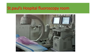
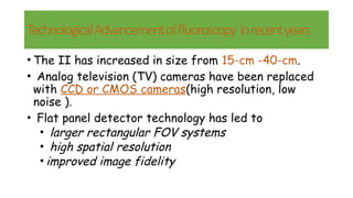
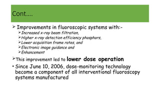
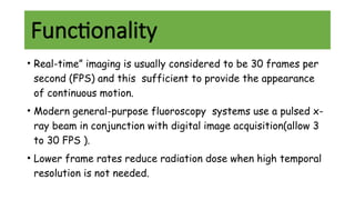
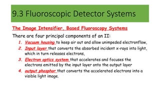
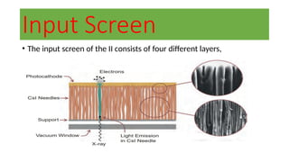
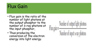
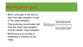
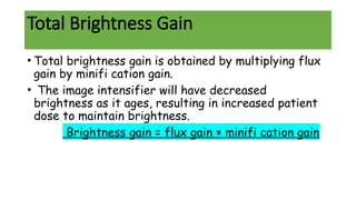
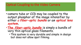
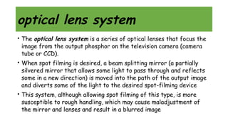
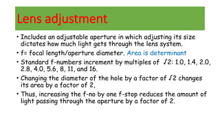
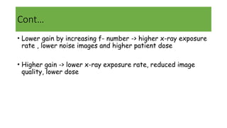
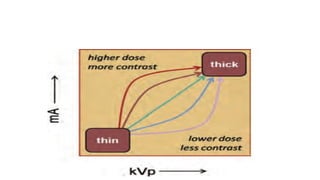
Fluoroscopy
Eyasu Altamo(PCR1)
Moderator :Dr Seife.T Medical Physicist
Content of session
Introduction
Fluoroscopy imaging chain components
Fluoroscopy detector system
Automatic exposure rate control
Fluoroscopy modes of operation
Imaging quality in fluoroscopy
Fluoroscopy suites
Radiation dose
Introduction
Fluoroscopy is an imaging modality that allows real-time x-
ray viewing of a patient with high temporal resolution.
 Its works based on an x-ray image intensifier coupled to a
still/video camera.
But curently ,Flat panel detectors have been replacing the
image intensifiers(II).
Introduction …
Fluoroscopy are commonly used
To position the imaging system for the recording of
images (e.g., angiography)
To provide imaging guidance for interventional
procedures (e.g., angioplasty).
St.paul’s Hospital fluoroscopy room
TechnologicalAdvancementoffluoroscopy inrecentyears
• The II has increased in size from 15-cm -40-cm.
• Analog television (TV) cameras have been replaced
with CCD or CMOS cameras(high resolution, low
noise ).
• Flat panel detector technology has led to
• larger rectangular FOV systems
• high spatial resolution
• improved image fidelity
Cont….
 Improvements in fluoroscopic systems with:-
Increased x-ray beam filtration,
Higher x-ray detection efficiency phosphors,
Lower acquisition frame rates, and
Electronic image guidance and
Enhancement
This improvement led to lower dose operation
• Since June 10, 2006, dose-monitoring technology
become a component of all interventional fluoroscopy
systems manufactured
Functionality
• Real-time” imaging is usually considered to be 30 frames per
second (FPS) and this sufficient to provide the appearance
of continuous motion.
• Modern general-purpose fluoroscopy systems use a pulsed x-
ray beam in conjunction with digital image acquisition(allow 3
to 30 FPS ).
• Lower frame rates reduce radiation dose when high temporal
resolution is not needed.
Cont..
• Digital fluoroscopy systems allow the recording of a real-
time sequence of digital images.
• Unrecorded fluoroscopy sequences are used for advancing a
catheter (positioning) during angiographic procedures
• In gastrointestinal fluoroscopy, much of the diagnosis is
formulated during the unrecorded fluoroscopy sequence.
• Fluoroscopy produce real-time x-ray images with high frame rates
and a low-dose of radiation per image.
• In general, five configurations of fluoroscopy systems are available:
I. Gastrointestinal/genitourinary (GI/GU) systems (with the x-ray tube under the
patient table);
II. Remote fluoroscopy (over-table x-ray tube);
III.Interventional vascular (vascular surgery, radiology);
IV. Cardiology and interventional electrophysiology, and
V. Mobile C-arm configurations (surgery, pain medicine).
All configurations have the same basic components.
9.2 Fluoroscopic Imaging Chain Components
Fluoroscopic Imaging Chain Components
X-ray tube
Filters :Copper filtration combined with lower kV values allows
angiography systems to provide lower dose operation while still
delivering high image contrast for angiographic applications.
Collimators (circular diaphragms for II and rectangular for
FPF)
operator adjustable attenuating wedges
image intensifiers(II)
flat panel detectors operate
several thousand times more sensitive than a
standard radiographic detector and, in principle,
can produce images using several thousand
times less radiation
9.3 Fluoroscopic Detector Systems
The Image Intensifier, Based Fluoroscopy Systems
There are four principal components of an II:
1. Vacuum housing to keep air out and allow unimpeded electronflow,
2. Input layer that converts the absorbed incident x-rays into light,
which in turn releases electrons,
3. Electron optics system that accelerates and focuses the
electrons emitted by the input layer onto the output layer
4. output phosphor that converts the accelerated electrons into a
visible light image.
A Diagram of the modern II
Input Screen
• The input screen of the II consists of four different layers,
1,vacuum window
• The first layer is the vacuum window, a thin (typically 1 mm)
aluminum window that is part of the vacuum containment
vessel.
• Its keeps the air out of the II,
• Curvature is designed to withstand the force of the air
pressing against it.
• The vacuum window of a large FOV (35-cm) II supports over a
ton of force from atmospheric air pressure.
• A vacuum is necessary in all devices in which electrons are
accelerated across open space.
2,Support layer
•Its supports the input phosphor and
photocathode layers.
•Commonly 0.5 mm of aluminum
• The first component in the electronic lens
system.
• Its curvature is designed for accurate
electron focusing.
3,Input phosphor
• Function is to absorb the x-rays and convert their energy
into visible light.
• All modern IIs use cesium iodide (CsI) for the input
phosphor.
• The CsI crystals have a trace amount of sodium, causing it
to emit bluelight.
• For a 60-keV x-ray photon absorbed in the phosphor,
approximately 3,000 light photons (at about 420 nm
wavelength) are emitted.
4,Photocathode
• The fourth layer of the input screen is the photocathode,
• Thin layer of antimony and alkali metals (such as Sb2 S3 ) that
emits electrons when struck by visible light.
• Approximately 400 electrons are released from the photocathode
for each 60-keV x-ray photon absorbed in the phosphor.
• With 10% to 20% conversion efficiency
III, Electron Optics
• Focusing the electrons is accomplished by the several
electrodes in the electron optics chain
• Five-component electronic lens system:
• G1, G2, and G3 electrodes
• Input phosphor substrate (the cathode) and
• The anode just proximal to the output phosphor,
• The kinetic energy of each electron is dramatically
increased by acceleration due to the voltage difference
between the cathode and anode, resulting in electronic gain
IV, The Output Phosphor
Anode
• The anode is a very thin (0.2 mm) coating of aluminum on the
vacuum side of the output phosphor.
• which is electrically conductive to carry away the electrons
once they deposit their kinetic energy in the phosphor.
output phosphor
• Made of zinc cadmium sulfide doped with silver (ZnCdS: Ag),
• ZnCdS: Ag has a green (530 nm) emission spectrum.
• The ZnCdS phosphor particles are very small (1 to 2 mm),
and the output phosphor is quite thin (4 to 8 mm), to
preserve high spatial resolution.
• Each electron causes the emission of approximately
1,000 light photons from the output phosphor.
output window
• The last stage in the II that the image signal passes through is
the output window
• The output window is part of the vacuum enclosure and must be
transparent to the emission of light from the output phosphor.
• Some fraction of the light emitted by the output phosphor is
reflected inside the glass window.
• This contribute veiling glare, which can reduce image contrast.
• This glare is reduced by using a thick (about 14 mm) clear glass
window
Brightness Gain
• Brightness gain is a measurement of the increase in
image brightness or intensification achieved by the
conversions in the image intensification tube.
• Two factors are used to determine the total
brightness gain: flux gain and minifi cation gain.
• The total gain in brightness comes from a
combination of acceleration of the
photoelectrons, called flux gain, and compression
of the image size, called minification gain.
Flux Gain
• Flux gain is the ratio of the
number of light photons at
the output phosphor to the
number of x-ray photons at
the input phosphor,
• Thus producing the
conversion of the electron
energy into light energy.
Minification gain
• Minifi cation gain is the ratio of
light from input phosphor to light
at the output phosphor,
• Thus producing concentrated light
from the larger input phosphor
onto the smaller output phosphor.
• Minification is an increase in
brightness or intensity of the
image;
Total Brightness Gain
• Total brightness gain is obtained by multiplying flux
gain by minifi cation gain.
• The image intensifier will have decreased
brightness as it ages, resulting in increased patient
dose to maintain brightness.
Brightness gain = flux gain × minifi cation gain
Optical Coupling to the Video Camera
•camera tube or CCD may be coupled to the
output phosphor of the image intensifier by
either a fiber-optic bundle or an optical lens
system.
•The fiber-optic bundle is simply a bundle of
very thin optical glass filaments.
• This system is very durable and simple in design
but does not allow spot filming.
optical lens system
• The optical lens system is a series of optical lenses that focus the
image from the output phosphor on the television camera (camera
tube or CCD).
• When spot filming is desired, a beam splitting mirror (a partially
silvered mirror that allows some light to pass through and reflects
some in a new direction) is moved into the path of the output image
and diverts some of the light to the desired spot-filming device
• This system, although allowing spot filming of this type, is more
susceptible to rough handling, which may cause maladjustment of
the mirror and lenses and result in a blurred image
Lens adjustment
• Includes an adjustable aperture in which adjusting its size
dictates how much light gets through the lens system.
• f= focal length/aperture diameter. Area is determinant
• Standard f-numbers increment by multiples of √2: 1.0, 1.4, 2.0,
2.8, 4.0, 5.6, 8, 11, and 16.
• Changing the diameter of the hole by a factor of √2 changes
its area by a factor of 2,
• Thus, increasing the f-no by one f-stop reduces the amount of
light passing through the aperture by a factor of 2.
Cont…
• Lower gain by increasing f- number -> higher x-ray exposure
rate , lower noise images and higher patient dose
• Higher gain -> lower x-ray exposure rate, reduced image
quality, lower dose
Video Cameras
•A light-sensitive camera such as :
1.Analog vidicon
2. Solid-state CCD (Charge coupled device)
3. CMOS system (complementary metal oxide
semiconductor)
•They optically coupled to the output screen of
the II and is used to relay the output image to
a video monitor for viewing by the operator.
Analog vidicon
• Its function on the principle
of photocondutivity,where
resistance of photoconductive
surface decrease with
increase in light intensity
• Vidicon is short tube with
length of 12 to 20cm and
diameter between 1.5to 4cm.
• Its no longer used in modern
fluoroscopy
2,CCD (Charge coupled device)&CMOS system
(complementary metal oxide semiconductor
• They are solid-state electronic arrays that convert a
projection image into a digital image and then into a video
signal.
• Such a device uses photodiode semiconductor materials in an
array to convert incident light intensity incident into
corresponding electronic charge (electrons), and locally
store the charge for electronic readout.
Cont..
• Both CCD and CMOS cameras have very small pixel
dimensions, on the order of 10–20 micron (0.01 to 0.02 mm),
incorporated into a 2.5 by 2.5 cm area that replaces an
analog TV camera.
• The typical matrix comprises 1,000 by 1,000 or 2,000 by
2,000 detector elements, but there is a wide variety and
selection of sizes that a manufacturer might choose to
include in a system.
Cont..
• In either output (CCD or CMOS) a digital projection image
is produced, and then converted to a video signal that is
used to drive one or more monitors in the fluoroscopy suite.
• Solid-state cameras are preferred because of their
stability, reproducibility, and linearity.
charge-coupled device (CCD)
• The CCD consists of a series of semiconductor capacitors, with
each capacitor representing a pixel.
• Each pixel is composed of photosensitive material that
dislodges electrons when stimulated by light photons.
• The CCD reads out the stored charge in a “bucket brigade” fashion,
causing locally stored electrons to be transferred from one row to the
next row, along defined columns, to eventually be deposited onto a charge
amplifier at the end of each column.
CMOS system (complementary metal oxide
semiconductor
• The CMOS device has
independent detector elements,
each comprised of a storage
capacitor and transistor switch.
• Charge to voltage conversion as
well as amplification is carried
out in the pixel
• itself : speed is higher.
• The signal from a CMOS device
is accessed, as is the signal from
a TFT flat panel detector.
Characteristics Unique to Image Intensifier Systems
•1,Brightness Gain
•2 ,Pincushion Distortion
•3,S Distortion
1,Brightness Gain
• The brightness gain is the product of the electronic and
minification gains of the II.
• The electronic gain of an II is roughly about 50,
• The minification gain is variable depending on the electronic
magnification
• As the effective diameter (FOV) of the input phosphor
decreases ,the brightness gain decreases.
2 ,Pincushion Distortion
• It is the result of projecting the image with a
curved input phosphor to the flat output phosphor .
• Pincushion distortion worps the image by stretching
the physical dimensions in the periphery of the
image.
• Therefore, for improved accuracy with distance
measurements,
• it is best to position the desired anatomy in the central
area of the FOV
3,S Distortion
• S distortion is a spatial warping of the image in an S shape
through the image.
• This type of distortion is usually subtle.
• It is the result of stray magnetic fields and the earth’s
magnetic field affecting the electron trajectory from the
cathode to the anode inside the II.
Cont..
On fluoroscopic systems
capable of rotation.
the S distortion will
generally shift in the image
during rotation due to the
change in the II’s
orientation with respect to
the earth’s magnetic field.
Flat Panel Detector Based Digital Fluoroscopy
Systems
• Flat panel detectors are comprised of thin film transistor (TFT) arrays of individual
detector elements (dexels) that are packaged in a square or rectangular area.
• Each detector element has :-
Capacitor, which accumulates and stores the signal as an electrical charge,
Transistor which serves as a switch
• There are two modes of TFT:-
• Indirect
• Direct x-ray conversion modes
cont’d
Indirect detection TFT systems,
The absorbed x-rays are initially converted to light in the adjacent
layered phosphor
Each dexel has a transistor and a capacitor, in addition to a
photodiode, which converts the x-ray induced light from the
phosphor into a corresponding charge.
Direct detection fluoroscopy detectors
 Semiconductor (selenium) produces x-ray induced charge directly,
which is collected under a voltage to ensure that the signal is
captured within the same dexel as the x-ray absorption event.
Cont..
• For fluoroscopic applications, the flat panel image receptor
replaces the II, lenses, and camera system, and directly
records the real-time fluoroscopic image sequence.
• The size of a detector element (dexel) in fluoroscopy is usually
larger than in radiography.
• some flat panel systems have the ability to adjust the dexel
size by binning four dexels into one larger dexel.
9.4 Automatic exposure rate Control
• In modern fluoroscopic systems the exposure rates are controlled
automatically.
• This enabled by Automatic exposure rate control (AERC) circuit
(formerly referred to as Automatic Brightness Control – ABC)
• This system help to provide a consistent optical density/
signal-to-noise ratio between images, regardless of patient-centric
factors such as size and density.
• It does this by regulating the x-ray exposure rate incident on the
input phosphor of the II or flat panel detector.
• When the system is panned from a region of low attenuation to one of
greater attenuation of the patient, fewer x-rays strike the detector.
An AERC sensor measures the x-ray intensity and the AERC sends a
signal to the x-ray generator to increase the x-ray exposure rate .
Cont…
cont...
• Its maintain a consistent overall appearance of the image by
automatically adjusting the kVp and/or mAs.
• How the mA and kV change as a function of patient thickness has an
important influence on the compromise between patient dose and
image quality
• When the fluoroscopist pans to a thicker region of the patient, the
AERC circuit requests more x-rays from the generator.
AERC
• When the generator responds by increasing the kV, the
subject contrast decreases, but the dose to the patient is
kept low because more x-rays penetrate the patient at
higher kV.
• In situations where contrast is crucial (e.g., angiography),
the generator can increase the mA instead of the kV; this
preserves subject contrast at the expense of higher patient
dose
Reference

Fluoroscopy finalcccvvvvvvvvvvvvvvvv.pptx

  • 1.
  • 2.
    Content of session Introduction Fluoroscopyimaging chain components Fluoroscopy detector system Automatic exposure rate control Fluoroscopy modes of operation Imaging quality in fluoroscopy Fluoroscopy suites Radiation dose
  • 3.
    Introduction Fluoroscopy is animaging modality that allows real-time x- ray viewing of a patient with high temporal resolution.  Its works based on an x-ray image intensifier coupled to a still/video camera. But curently ,Flat panel detectors have been replacing the image intensifiers(II).
  • 4.
    Introduction … Fluoroscopy arecommonly used To position the imaging system for the recording of images (e.g., angiography) To provide imaging guidance for interventional procedures (e.g., angioplasty).
  • 5.
  • 6.
  • 7.
  • 8.
  • 9.
    TechnologicalAdvancementoffluoroscopy inrecentyears • TheII has increased in size from 15-cm -40-cm. • Analog television (TV) cameras have been replaced with CCD or CMOS cameras(high resolution, low noise ). • Flat panel detector technology has led to • larger rectangular FOV systems • high spatial resolution • improved image fidelity
  • 10.
    Cont….  Improvements influoroscopic systems with:- Increased x-ray beam filtration, Higher x-ray detection efficiency phosphors, Lower acquisition frame rates, and Electronic image guidance and Enhancement This improvement led to lower dose operation • Since June 10, 2006, dose-monitoring technology become a component of all interventional fluoroscopy systems manufactured
  • 11.
    Functionality • Real-time” imagingis usually considered to be 30 frames per second (FPS) and this sufficient to provide the appearance of continuous motion. • Modern general-purpose fluoroscopy systems use a pulsed x- ray beam in conjunction with digital image acquisition(allow 3 to 30 FPS ). • Lower frame rates reduce radiation dose when high temporal resolution is not needed.
  • 12.
    Cont.. • Digital fluoroscopysystems allow the recording of a real- time sequence of digital images. • Unrecorded fluoroscopy sequences are used for advancing a catheter (positioning) during angiographic procedures • In gastrointestinal fluoroscopy, much of the diagnosis is formulated during the unrecorded fluoroscopy sequence.
  • 13.
    • Fluoroscopy producereal-time x-ray images with high frame rates and a low-dose of radiation per image. • In general, five configurations of fluoroscopy systems are available: I. Gastrointestinal/genitourinary (GI/GU) systems (with the x-ray tube under the patient table); II. Remote fluoroscopy (over-table x-ray tube); III.Interventional vascular (vascular surgery, radiology); IV. Cardiology and interventional electrophysiology, and V. Mobile C-arm configurations (surgery, pain medicine). All configurations have the same basic components. 9.2 Fluoroscopic Imaging Chain Components
  • 14.
  • 15.
    Fluoroscopic Imaging ChainComponents X-ray tube Filters :Copper filtration combined with lower kV values allows angiography systems to provide lower dose operation while still delivering high image contrast for angiographic applications. Collimators (circular diaphragms for II and rectangular for FPF) operator adjustable attenuating wedges image intensifiers(II) flat panel detectors operate several thousand times more sensitive than a standard radiographic detector and, in principle, can produce images using several thousand times less radiation
  • 16.
    9.3 Fluoroscopic DetectorSystems The Image Intensifier, Based Fluoroscopy Systems There are four principal components of an II: 1. Vacuum housing to keep air out and allow unimpeded electronflow, 2. Input layer that converts the absorbed incident x-rays into light, which in turn releases electrons, 3. Electron optics system that accelerates and focuses the electrons emitted by the input layer onto the output layer 4. output phosphor that converts the accelerated electrons into a visible light image.
  • 17.
    A Diagram ofthe modern II
  • 18.
    Input Screen • Theinput screen of the II consists of four different layers,
  • 19.
    1,vacuum window • Thefirst layer is the vacuum window, a thin (typically 1 mm) aluminum window that is part of the vacuum containment vessel. • Its keeps the air out of the II, • Curvature is designed to withstand the force of the air pressing against it. • The vacuum window of a large FOV (35-cm) II supports over a ton of force from atmospheric air pressure. • A vacuum is necessary in all devices in which electrons are accelerated across open space.
  • 20.
    2,Support layer •Its supportsthe input phosphor and photocathode layers. •Commonly 0.5 mm of aluminum • The first component in the electronic lens system. • Its curvature is designed for accurate electron focusing.
  • 21.
    3,Input phosphor • Functionis to absorb the x-rays and convert their energy into visible light. • All modern IIs use cesium iodide (CsI) for the input phosphor. • The CsI crystals have a trace amount of sodium, causing it to emit bluelight. • For a 60-keV x-ray photon absorbed in the phosphor, approximately 3,000 light photons (at about 420 nm wavelength) are emitted.
  • 22.
    4,Photocathode • The fourthlayer of the input screen is the photocathode, • Thin layer of antimony and alkali metals (such as Sb2 S3 ) that emits electrons when struck by visible light. • Approximately 400 electrons are released from the photocathode for each 60-keV x-ray photon absorbed in the phosphor. • With 10% to 20% conversion efficiency
  • 23.
    III, Electron Optics •Focusing the electrons is accomplished by the several electrodes in the electron optics chain • Five-component electronic lens system: • G1, G2, and G3 electrodes • Input phosphor substrate (the cathode) and • The anode just proximal to the output phosphor, • The kinetic energy of each electron is dramatically increased by acceleration due to the voltage difference between the cathode and anode, resulting in electronic gain
  • 24.
  • 25.
    Anode • The anodeis a very thin (0.2 mm) coating of aluminum on the vacuum side of the output phosphor. • which is electrically conductive to carry away the electrons once they deposit their kinetic energy in the phosphor.
  • 26.
    output phosphor • Madeof zinc cadmium sulfide doped with silver (ZnCdS: Ag), • ZnCdS: Ag has a green (530 nm) emission spectrum. • The ZnCdS phosphor particles are very small (1 to 2 mm), and the output phosphor is quite thin (4 to 8 mm), to preserve high spatial resolution. • Each electron causes the emission of approximately 1,000 light photons from the output phosphor.
  • 27.
    output window • Thelast stage in the II that the image signal passes through is the output window • The output window is part of the vacuum enclosure and must be transparent to the emission of light from the output phosphor. • Some fraction of the light emitted by the output phosphor is reflected inside the glass window. • This contribute veiling glare, which can reduce image contrast. • This glare is reduced by using a thick (about 14 mm) clear glass window
  • 28.
    Brightness Gain • Brightnessgain is a measurement of the increase in image brightness or intensification achieved by the conversions in the image intensification tube. • Two factors are used to determine the total brightness gain: flux gain and minifi cation gain. • The total gain in brightness comes from a combination of acceleration of the photoelectrons, called flux gain, and compression of the image size, called minification gain.
  • 29.
    Flux Gain • Fluxgain is the ratio of the number of light photons at the output phosphor to the number of x-ray photons at the input phosphor, • Thus producing the conversion of the electron energy into light energy.
  • 30.
    Minification gain • Minification gain is the ratio of light from input phosphor to light at the output phosphor, • Thus producing concentrated light from the larger input phosphor onto the smaller output phosphor. • Minification is an increase in brightness or intensity of the image;
  • 31.
    Total Brightness Gain •Total brightness gain is obtained by multiplying flux gain by minifi cation gain. • The image intensifier will have decreased brightness as it ages, resulting in increased patient dose to maintain brightness. Brightness gain = flux gain × minifi cation gain
  • 32.
    Optical Coupling tothe Video Camera •camera tube or CCD may be coupled to the output phosphor of the image intensifier by either a fiber-optic bundle or an optical lens system. •The fiber-optic bundle is simply a bundle of very thin optical glass filaments. • This system is very durable and simple in design but does not allow spot filming.
  • 34.
    optical lens system •The optical lens system is a series of optical lenses that focus the image from the output phosphor on the television camera (camera tube or CCD). • When spot filming is desired, a beam splitting mirror (a partially silvered mirror that allows some light to pass through and reflects some in a new direction) is moved into the path of the output image and diverts some of the light to the desired spot-filming device • This system, although allowing spot filming of this type, is more susceptible to rough handling, which may cause maladjustment of the mirror and lenses and result in a blurred image
  • 35.
    Lens adjustment • Includesan adjustable aperture in which adjusting its size dictates how much light gets through the lens system. • f= focal length/aperture diameter. Area is determinant • Standard f-numbers increment by multiples of √2: 1.0, 1.4, 2.0, 2.8, 4.0, 5.6, 8, 11, and 16. • Changing the diameter of the hole by a factor of √2 changes its area by a factor of 2, • Thus, increasing the f-no by one f-stop reduces the amount of light passing through the aperture by a factor of 2.
  • 36.
    Cont… • Lower gainby increasing f- number -> higher x-ray exposure rate , lower noise images and higher patient dose • Higher gain -> lower x-ray exposure rate, reduced image quality, lower dose
  • 37.
    Video Cameras •A light-sensitivecamera such as : 1.Analog vidicon 2. Solid-state CCD (Charge coupled device) 3. CMOS system (complementary metal oxide semiconductor) •They optically coupled to the output screen of the II and is used to relay the output image to a video monitor for viewing by the operator.
  • 38.
    Analog vidicon • Itsfunction on the principle of photocondutivity,where resistance of photoconductive surface decrease with increase in light intensity • Vidicon is short tube with length of 12 to 20cm and diameter between 1.5to 4cm. • Its no longer used in modern fluoroscopy
  • 39.
    2,CCD (Charge coupleddevice)&CMOS system (complementary metal oxide semiconductor • They are solid-state electronic arrays that convert a projection image into a digital image and then into a video signal. • Such a device uses photodiode semiconductor materials in an array to convert incident light intensity incident into corresponding electronic charge (electrons), and locally store the charge for electronic readout.
  • 40.
    Cont.. • Both CCDand CMOS cameras have very small pixel dimensions, on the order of 10–20 micron (0.01 to 0.02 mm), incorporated into a 2.5 by 2.5 cm area that replaces an analog TV camera. • The typical matrix comprises 1,000 by 1,000 or 2,000 by 2,000 detector elements, but there is a wide variety and selection of sizes that a manufacturer might choose to include in a system.
  • 41.
    Cont.. • In eitheroutput (CCD or CMOS) a digital projection image is produced, and then converted to a video signal that is used to drive one or more monitors in the fluoroscopy suite. • Solid-state cameras are preferred because of their stability, reproducibility, and linearity.
  • 42.
    charge-coupled device (CCD) •The CCD consists of a series of semiconductor capacitors, with each capacitor representing a pixel. • Each pixel is composed of photosensitive material that dislodges electrons when stimulated by light photons. • The CCD reads out the stored charge in a “bucket brigade” fashion, causing locally stored electrons to be transferred from one row to the next row, along defined columns, to eventually be deposited onto a charge amplifier at the end of each column.
  • 45.
    CMOS system (complementarymetal oxide semiconductor • The CMOS device has independent detector elements, each comprised of a storage capacitor and transistor switch. • Charge to voltage conversion as well as amplification is carried out in the pixel • itself : speed is higher. • The signal from a CMOS device is accessed, as is the signal from a TFT flat panel detector.
  • 46.
    Characteristics Unique toImage Intensifier Systems •1,Brightness Gain •2 ,Pincushion Distortion •3,S Distortion
  • 47.
    1,Brightness Gain • Thebrightness gain is the product of the electronic and minification gains of the II. • The electronic gain of an II is roughly about 50, • The minification gain is variable depending on the electronic magnification • As the effective diameter (FOV) of the input phosphor decreases ,the brightness gain decreases.
  • 48.
    2 ,Pincushion Distortion •It is the result of projecting the image with a curved input phosphor to the flat output phosphor . • Pincushion distortion worps the image by stretching the physical dimensions in the periphery of the image. • Therefore, for improved accuracy with distance measurements, • it is best to position the desired anatomy in the central area of the FOV
  • 50.
    3,S Distortion • Sdistortion is a spatial warping of the image in an S shape through the image. • This type of distortion is usually subtle. • It is the result of stray magnetic fields and the earth’s magnetic field affecting the electron trajectory from the cathode to the anode inside the II.
  • 51.
    Cont.. On fluoroscopic systems capableof rotation. the S distortion will generally shift in the image during rotation due to the change in the II’s orientation with respect to the earth’s magnetic field.
  • 52.
    Flat Panel DetectorBased Digital Fluoroscopy Systems • Flat panel detectors are comprised of thin film transistor (TFT) arrays of individual detector elements (dexels) that are packaged in a square or rectangular area. • Each detector element has :- Capacitor, which accumulates and stores the signal as an electrical charge, Transistor which serves as a switch • There are two modes of TFT:- • Indirect • Direct x-ray conversion modes
  • 53.
    cont’d Indirect detection TFTsystems, The absorbed x-rays are initially converted to light in the adjacent layered phosphor Each dexel has a transistor and a capacitor, in addition to a photodiode, which converts the x-ray induced light from the phosphor into a corresponding charge. Direct detection fluoroscopy detectors  Semiconductor (selenium) produces x-ray induced charge directly, which is collected under a voltage to ensure that the signal is captured within the same dexel as the x-ray absorption event.
  • 55.
    Cont.. • For fluoroscopicapplications, the flat panel image receptor replaces the II, lenses, and camera system, and directly records the real-time fluoroscopic image sequence. • The size of a detector element (dexel) in fluoroscopy is usually larger than in radiography. • some flat panel systems have the ability to adjust the dexel size by binning four dexels into one larger dexel.
  • 57.
    9.4 Automatic exposurerate Control • In modern fluoroscopic systems the exposure rates are controlled automatically. • This enabled by Automatic exposure rate control (AERC) circuit (formerly referred to as Automatic Brightness Control – ABC) • This system help to provide a consistent optical density/ signal-to-noise ratio between images, regardless of patient-centric factors such as size and density.
  • 58.
    • It doesthis by regulating the x-ray exposure rate incident on the input phosphor of the II or flat panel detector. • When the system is panned from a region of low attenuation to one of greater attenuation of the patient, fewer x-rays strike the detector. An AERC sensor measures the x-ray intensity and the AERC sends a signal to the x-ray generator to increase the x-ray exposure rate . Cont…
  • 59.
    cont... • Its maintaina consistent overall appearance of the image by automatically adjusting the kVp and/or mAs. • How the mA and kV change as a function of patient thickness has an important influence on the compromise between patient dose and image quality • When the fluoroscopist pans to a thicker region of the patient, the AERC circuit requests more x-rays from the generator.
  • 60.
    AERC • When thegenerator responds by increasing the kV, the subject contrast decreases, but the dose to the patient is kept low because more x-rays penetrate the patient at higher kV. • In situations where contrast is crucial (e.g., angiography), the generator can increase the mA instead of the kV; this preserves subject contrast at the expense of higher patient dose
  • 62.

Editor's Notes

  • #14 e fluoroscopic imaging chain is illustrated, with the patient in the supine position. This figure shows an II as the detector, but flat panel detector systems are common as well. The detector in fluoroscopy is more sensitive than radiography to enable low-dose, real-time, longer duration studies. The x-ray system includes a collimator with motorized blades to adjust to the FOV, and a Kerma Air Product (KAP) meter to monitor radiation output for each patient is often available on interventional and cardiac catheterization fluoroscopic systems.
  • #17 II is shown. X-rays strike the input phosphor, producing light that stimulates the photocathode to emit electrons, which are ejected into the electronic lens system. The electronic lens system is comprised of the cathode, anode, and three additional focusing electrodes (G1, G2, and G3). Energetic electrons strike the output phosphor, generating light that is then imaged by a digital camera system.
  • #21 The input phosphor must be thick enough to absorb a large fraction of the incident x-rays But thin enough to not significantly degrade the spatial resolution of the image by the lateral dispersion of light through the phosphor.
  • #30 The image is much smaller at the output phosphor than it is at the input phosphor, Because the 15- to 35-cm-diameter input image is focused onto a 2.5-cm (1 inch)-diameter circle. The reduction in image diameter also leads to light intensity amplification Magnification gain of an II is simply the ratio of the area of the input phosphor to that of the output phosphor.
  • #44 The charge-coupled device (CCD) is a light-sensitive semiconducting device that generates an electrical charge when stimulated by light and stores this charge in a capacitor. B, To digitize the charge from the CCD, the electrodes between each pixel, called row gates, are charged in sequence, moving the signal down the row where it is transferred into a capacitor. From the capacitors, the charge is sent as an electronic signal to the television monitor.
  • #49 The input phosphor/photocathode surface is curved to accommodate the physics of electronic focusing, and the output phosphor is planar. Because the image is produced on a curved surface and projected onto a plane surface, a characteristic pincushion distortion results in the output image
  • #54 Indirect and direct detector TFT–based x-ray detectors are shown. A. Photons in the indirect sys_x0002_tem propagate laterally, compromising resolution. The detected signal shown for the indirect detector shows this lateral spread in the signal from one x-ray photons interaction. B. For the direct detector system, the ion pairs liber_x0002_ated by x-ray interaction follow the electric field lines (electron holes travel upwards, electrons travel downwards) and have negligible lateral spread. Here, the detected electronic signal from one x-ray photons interaction is collected almost entirely in one detector element, and therefore better spatial resolution is achieved.
  • #56  flat panel detector systems are pixelated descrete detector systems. The detector array is comprised of a large number of individual detector elements (dexels). Each dexel has a light sensitive region and a light-insensitive area where the electronic components are located. B. A photomicrograph of an actual TFT system is shown. The electronics component can be seen in the upper left corner of each dexel (Image courtesy John Sabol and Bill Hennessy, GE Healthcare).