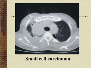
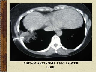
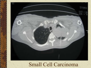
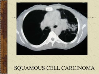
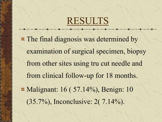
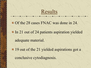
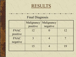
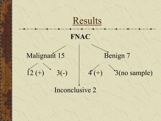
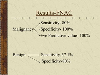
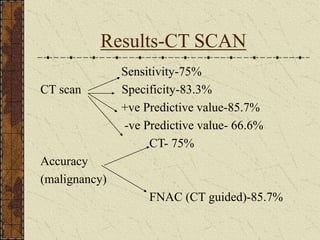
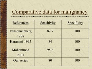
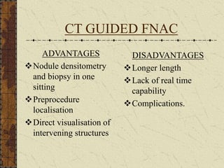
CT GUIDED FNAC : A
SIMPLE SOLUTION TO
COMPLEX PROBLEMS
Dr.Harish.N, Dr. Jayasree.L, Dr. Sara Ammu
Chacko, Dr. Harikumaran Nair
INSTITUTION: Medical College Hospital,
Thiruvananthapuram.
INTRODUCTION
History of medicine parallels the quest for correct
diagnosis by least invasive techniques.
The custodian of final diagnosis - pathologist.
How to produce the sample?
Less invasive – FNAC.
Guided FNAC - modality of choice – computed
tomography.
INTRODUCTION(cont)
Haaga and Alfidi - reported CT guided
biopsy for pulmonary nodules in 1976.
Geragthy et al - a diagnostic accuracy
benign-80% & malignant-90%.
VanSonnenberg et al - CT guidance
permits biopsy- regardless of size or
position.
AIMS OF THE STUDY
To evaluate the sensitivity and specificity of
computed tomography (CT) guided
aspiration cytology in lung lesions.
To characterise lung lesions by computed
tomography.
Materials and methods
Study period-March 2004 - March 2005.
Sample size-28.
Age – 20-70 yrs.
Males:22(78%), Females:6(22%).
Inclusion criteria: peripheral opacities in
chest X-ray.
Materials and methods(cont)
Exclusion criteria:
bleeding disorders.
chronic obstructive pulmonary disease
dyspnoea.
bullous disease.
unwilling patient.
contralateral pneumonectomy.
uncontrollable cough.
Materials and methods (contd)
The spiral CT scanner from Toshiba (X- vision)
was used.
Frontal scout view image of thorax.
Axial sections - 10mm,from apex to base.
Sagittal and coronal reconstructions - in selected
cases
Lesions analysed by thin sections(2-5mm).
Plain and CECT taken.
Lesions studied for their characteristics.
Chondroid hamartoma with calcification, no fat
density seen.
Technique of FNAC
Done as an OPD procedure.
Relevant investigations.
Informed consent.
After locating the lesion,best
approach(supine,prone and lateral
decubitus) was decided.
Site marked.
Local preparation done.
Technique of FNAC (cont)
Needle – disposable spinal needle – 22-23G
& 9cm in length.
Fixatives-wet fixation with 80% isopropyl
alcohol in coplin’s jar.
After anaesthesia needle introduced in
suspended respiration,perpendicularly.
Check CT slice taken.
TECHNIQUE OF FNAC
Squamous cell carcinoma
with calcification
Small cell carcinoma
ADENOCARCINOMA LEFT LOWER
LOBE
Small Cell Carcinoma
SQUAMOUS CELL CARCINOMA
RESULTS
The final diagnosis was determined by
examination of surgical specimen, biopsy
from other sites using tru cut needle and
from clinical follow-up for 18 months.
Malignant: 16 ( 57.14%), Benign: 10
(35.7%), Inconclusive: 2( 7.14%).
Results
Of the 28 cases FNAC was done in 24.
In 21 out of 24 patients aspiration yielded
adequate material.
19 out of the 21 yielded aspirations got a
conclusive cytodiagnosis.
RESULTS
Final Diagnosis
Malignancy
positive
Malignancy
negative
FNAC
positive
12 0 12
FNAC
negative
3 4 7
15 4 19
Results
FNAC
Malignant 15 Benign 7
12 (+) 3(-) 4 (+) 3(no sample)
Inconclusive 2
Results-FNAC
Sensitivity- 80%
Malignancy Specificity- 100%
+ve Predictive value- 100%
Benign Sensitivity-57.1%
Specificity-80%
Results-CT SCAN
Sensitivity-75%
CT scan Specificity-83.3%
+ve Predictive value-85.7%
-ve Predictive value- 66.6%
CT- 75%
Accuracy
(malignancy)
FNAC (CT guided)-85.7%
Complications
Total 3 cases : 12.5%
Pneumothorax: 2 cases(8%).
Patients were observed for 24hrs after which
chest X Ray taken and discharged accordingly.
Pulmonary haemorrhage: 1 case(4%).
As it is self limiting,patient was reassured and
made to lie with biopsy-site down.
All 3 resolved on conservative management.
Comparative data for malignancy
References Sensitivity Specificity
Vansonnenberg
1988
82.7 100
Haramati 1995 84 100
Mohammad
2001
95.6 100
Our series 80 100
CT GUIDED FNAC
ADVANTAGES
Nodule densitometry
and biopsy in one
sitting
Preprocedure
localisation
Direct visualisation of
intervening structures
DISADVANTAGES
Longer length
Lack of real time
capability
Complications.
CONCLUSION
CT scan and guided FNAC are effective
diagnostic modalities in patients with localised
pulmonary lesions.
It is a highly sensitive and specific technique with
a good diagnostic accuracy.
It can be used safely as an outpatient procedure in
the diagnosis of lung lesions.
FNAC lung.ppt

FNAC lung.ppt

  • 1.
    CT GUIDED FNAC: A SIMPLE SOLUTION TO COMPLEX PROBLEMS Dr.Harish.N, Dr. Jayasree.L, Dr. Sara Ammu Chacko, Dr. Harikumaran Nair INSTITUTION: Medical College Hospital, Thiruvananthapuram.
  • 2.
    INTRODUCTION History of medicineparallels the quest for correct diagnosis by least invasive techniques. The custodian of final diagnosis - pathologist. How to produce the sample? Less invasive – FNAC. Guided FNAC - modality of choice – computed tomography.
  • 3.
    INTRODUCTION(cont) Haaga and Alfidi- reported CT guided biopsy for pulmonary nodules in 1976. Geragthy et al - a diagnostic accuracy benign-80% & malignant-90%. VanSonnenberg et al - CT guidance permits biopsy- regardless of size or position.
  • 4.
    AIMS OF THESTUDY To evaluate the sensitivity and specificity of computed tomography (CT) guided aspiration cytology in lung lesions. To characterise lung lesions by computed tomography.
  • 5.
    Materials and methods Studyperiod-March 2004 - March 2005. Sample size-28. Age – 20-70 yrs. Males:22(78%), Females:6(22%). Inclusion criteria: peripheral opacities in chest X-ray.
  • 6.
    Materials and methods(cont) Exclusioncriteria: bleeding disorders. chronic obstructive pulmonary disease dyspnoea. bullous disease. unwilling patient. contralateral pneumonectomy. uncontrollable cough.
  • 7.
    Materials and methods(contd) The spiral CT scanner from Toshiba (X- vision) was used. Frontal scout view image of thorax. Axial sections - 10mm,from apex to base. Sagittal and coronal reconstructions - in selected cases Lesions analysed by thin sections(2-5mm). Plain and CECT taken. Lesions studied for their characteristics.
  • 8.
    Chondroid hamartoma withcalcification, no fat density seen.
  • 9.
    Technique of FNAC Doneas an OPD procedure. Relevant investigations. Informed consent. After locating the lesion,best approach(supine,prone and lateral decubitus) was decided. Site marked. Local preparation done.
  • 10.
    Technique of FNAC(cont) Needle – disposable spinal needle – 22-23G & 9cm in length. Fixatives-wet fixation with 80% isopropyl alcohol in coplin’s jar. After anaesthesia needle introduced in suspended respiration,perpendicularly. Check CT slice taken.
  • 11.
  • 12.
  • 13.
  • 14.
  • 15.
  • 16.
  • 17.
    RESULTS The final diagnosiswas determined by examination of surgical specimen, biopsy from other sites using tru cut needle and from clinical follow-up for 18 months. Malignant: 16 ( 57.14%), Benign: 10 (35.7%), Inconclusive: 2( 7.14%).
  • 18.
    Results Of the 28cases FNAC was done in 24. In 21 out of 24 patients aspiration yielded adequate material. 19 out of the 21 yielded aspirations got a conclusive cytodiagnosis.
  • 19.
  • 20.
    Results FNAC Malignant 15 Benign7 12 (+) 3(-) 4 (+) 3(no sample) Inconclusive 2
  • 21.
    Results-FNAC Sensitivity- 80% Malignancy Specificity-100% +ve Predictive value- 100% Benign Sensitivity-57.1% Specificity-80%
  • 22.
    Results-CT SCAN Sensitivity-75% CT scanSpecificity-83.3% +ve Predictive value-85.7% -ve Predictive value- 66.6% CT- 75% Accuracy (malignancy) FNAC (CT guided)-85.7%
  • 23.
    Complications Total 3 cases: 12.5% Pneumothorax: 2 cases(8%). Patients were observed for 24hrs after which chest X Ray taken and discharged accordingly. Pulmonary haemorrhage: 1 case(4%). As it is self limiting,patient was reassured and made to lie with biopsy-site down. All 3 resolved on conservative management.
  • 24.
    Comparative data formalignancy References Sensitivity Specificity Vansonnenberg 1988 82.7 100 Haramati 1995 84 100 Mohammad 2001 95.6 100 Our series 80 100
  • 25.
    CT GUIDED FNAC ADVANTAGES Noduledensitometry and biopsy in one sitting Preprocedure localisation Direct visualisation of intervening structures DISADVANTAGES Longer length Lack of real time capability Complications.
  • 26.
    CONCLUSION CT scan andguided FNAC are effective diagnostic modalities in patients with localised pulmonary lesions. It is a highly sensitive and specific technique with a good diagnostic accuracy. It can be used safely as an outpatient procedure in the diagnosis of lung lesions.