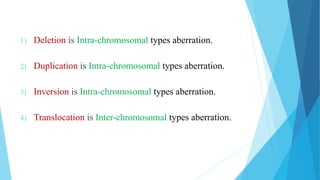
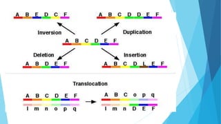
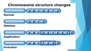
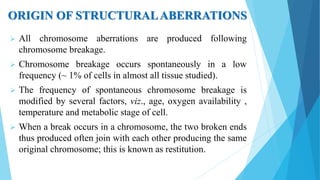
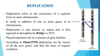
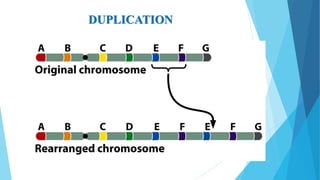
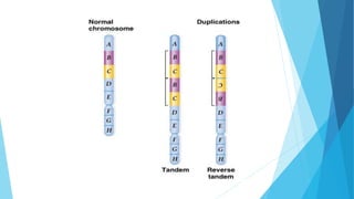
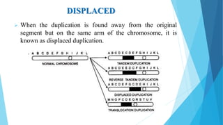
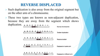
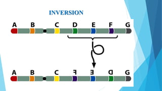
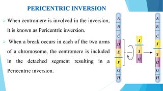
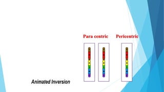
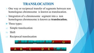
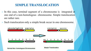
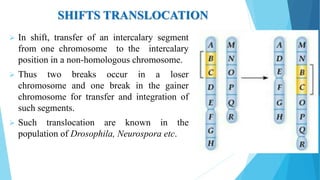
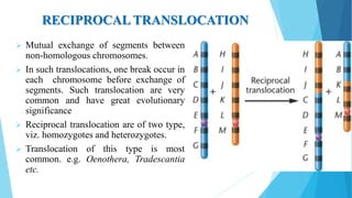
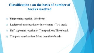
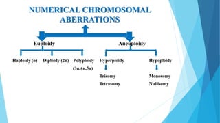
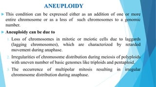
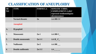
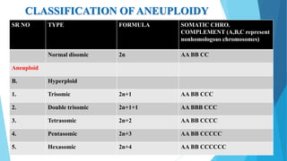
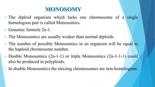
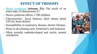
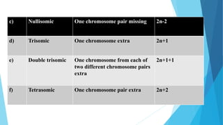
2. ASSIGNMENT PRESENTATION
(UNIT- 3)
Submitted By:-
1) Vaghela Gauravrajsinh K.
2) Patel Chinmaykumar G.
3) Patel Chiragkumar R.
4) Makwana Hitendrasinh B.
Submitted To:-
Dr. N.B.Patel sir,
Asso. Professor, Dept. of GPB,
C.P.College of Agriculture,
SDAU, SKNagar.
3. WHAT IS CHROMOSOME?
Chromosomes are the rod-shaped, filamentous bodies present in
the nucleus, which become visible during cell division.
They are the carriers of the gene or unit of heredity.
Chromosomes were first described by Strausberger (1875).
The term “Chromosome”, given by Waldeyer (1888).
Chromosome (Chroma means colour and Soma means body).
4. WHAT IS CHROMOSOMAL
ABERRATIONS ?
The somatic (2n) and gametic (n) chromosomes numbers of a
species ordinarily remain constant due to extremely precise
mitotic and meiotic cell divisions.
But occasionally, spontaneous variation in chromosome
number or structure do arise in nature, these are called
chromosomal aberrations.
Chromosomal aberrations are of two types:
1) Structural chromosomal aberrations
2) Numerical chromosomal aberrations
5. STRUCTURAL CHROMOSOMAL
ABERRATIONS
Structural chromosomal aberrations alter the chromosome
structure, i.e., the number, the sequence or the kind of genes
present in chromosome(s).
There are four common types of structural aberrations :-
1) Deletion or Deficiency
2) Duplication or Repeat
3) Inversion
4) Translocation
6. 1) Deletion is Intra-chromosomal types aberration.
2) Duplication is Intra-chromosomal types aberration.
3) Inversion is Intra-chromosomal types aberration.
4) Translocation is Inter-chromosomal types aberration.
7.
8.
9. ORIGIN OF STRUCTURALABERRATIONS
All chromosome aberrations are produced following
chromosome breakage.
Chromosome breakage occurs spontaneously in a low
frequency (~ 1% of cells in almost all tissue studied).
The frequency of spontaneous chromosome breakage is
modified by several factors, viz., age, oxygen availability ,
temperature and metabolic stage of cell.
When a break occurs in a chromosome, the two broken ends
thus produced often join with each other producing the same
original chromosome; this is known as restitution.
10. DELETION OR DEFICIENCY
Loss of a chromosome segment is known as deletion or deficiency.
The term deficiency was coined by Bridges in 1917. It involves the
one break.
Term deletion was coined by Painter and Muller in 1929. It involves
the two break.
Deletion was the first structural aberration detected by Bridges in 1917
from his genetic study on the X chromosome of Drosophila.
Types of Deletion or Deficiency :-
1) Terminal deletion:- Loss of either terminal segment of a
chromosome.
2) Interstitial deletion:- Loss of intercalary segment of a chromosome.
12. TERMINAL DELETION
A chromosome has two ends or terminals. Loss of either
terminal segment of a chromosome is known as terminal
deletion.
The deletion occurs in only in one chromosome of homozygous
pair, it is known as terminal heterozygous deletion.
The deletion occur in both the chromosome of a pair , it is
termed as homozygous deletion.
13. INTERSTITIAL DELETION
Some times there is loss of a segment of chromosome from
the intermediate portion or between telomere and centromere.
Loss of intercalary portion of chromosome is known as
interstitial deletion.
14. ORIGIN OF DELETION
Deficiency originates spontaneously or it may be induces
artificially like physical mutagen, chemical mutagen.
Terminal deficiency required a single break, Intercalary
deficiency required two break.
15. EFFECT OF DELETION
It could affect eye sight, smell, skin tone or loss of weight.
Small deletions are less likely to be fatal; large deletions are
usually fatal - there are always variations based on which
genes are lost. Some medium-sized deletions lead to
recognizable human disorders, e.g. Williams syndrome
Deletions are responsible for an array of genetic disorders,
including some cases of male infertility.
Deletion of part of the short arm of chromosome 5 results
in Cri du chat syndrome.
16. “Cri-du-chat” (cry-of-cat):- A specific deletion of a small portion
of chromosome 5; these children have severe mental retardation, a
small head with unusual facial features, and a cry that sounds like a
distressed cat.
“Philadelphia 22”:- Deletion of the chromosome 22.
Prader-Willi syndrome:- Deletion on long arm of chromosome 15.
Wolf- Hirschhorn syndrome:- Caused by partial deletion of the
short arm of chromosome 4.
17. USES OF DEFICIENCY
Deficiency may be used for the study of chromosome pairing
and its behavior during cells division.
They may be used for locating a gene on a particular
chromosomes.
Deficiency can be used to resolve special problems, such as,
the relationship between chiasma and crossing over.
18. DUPLICATION
Duplication refers to the occurrence of a segment
twice in same chromosome.
It result in addition of one or more genes in to
chromosome.
Duplication is also known as repeat and is first
reported in drosophila by Bridge in 1919.
Played important role in evolution of gene families.
According to Ohno(1970) duplication are the source
of all the new genes, and thus the basis of organic
evolution.
20. TYPES OF DUPLICATION
1) Interchromosomal duplication:- The duplicated segment of a
chromosome is present in another chromosome of the genome.
2) Intrachromosomal duplication:- The duplicated segment
remains in the same chromosome. It may be present at different
location. It has a two types:-
a) Direct tandem
b) Reverse tandem
21. DIRECT TANDEM DUPLICATION
In this case sequence of gene in the duplicated segment is
similar to the sequence of genes in the original segment of
a chromosome.
Here the sequence of gene in the duplicated segment is
reverse to the sequence of genes in the original segment
of a chromosome.
REVERSE TANDEM DUPLICATION
22.
23. DISPLACED
When the duplication is found away from the original
segment but on the same arm of the chromosome, it is
known as displaced duplication.
24. REVERSE DISPLACED
Such duplication is also away from the original segment but
on the other arm of a chromosome.
These two types are known as non-adjacent duplication,
because they are away from the segment which shows
duplication.
25. ORIGIN OF DUPLICATION
Origin of duplication involves chromosome breakage and
reunion of chromosome segment with its homologous
chromosome .
As a result , one of the two homologous involved in the
production of a duplication and up with a deficiency, while
the other has a duplication for the concerned segment.
26. EFFECT OF DUPLICATION
Duplication may produce specific effects when the phenotype is
affected due to a change in the position of a gene, it is called
position effect.
Duplication may lead to a more intense effect of a duplicated
gene.
Crossing over is suppressed in the duplicate region due to lack of
corresponding duplicated segment in the normal chromosome.
The gene number is increased in the chromosome having
duplication.
Presence of duplication leads to reduction in pollen fertility in
plant species.
27. USES OF DUPLICATION
Duplication can be used to study the chromosome behavior during meiosis,
such as, chromosome pairing, crossing over and their consequences.
Duplication offers number of possibilities in plant breeding. They can be
used to increase the dosage of certain desirable genes for increasing disease
or pest resistance, enzymatic activity or other characteristics.
Duplication has an advantage over polyploidy because the genetic
disbalance due to the duplication of chromosomal segments is lesser as
compared to polyploidy where the whole genome is duplicated.
In case where genes for resistance to diseases or pests are linked to some
undesirable genes, or the genes for resistance to various races are allelic, a
combination of resistance to different races can be obtained through
duplication.
28. USES OF DUPLICATION
Duplication can be used to study the chromosome behavior during meiosis,
such as, chromosome pairing, crossing over and their consequences.
Duplication offers number of possibilities in plant breeding. They can be
used to increase the dosage of certain desirable genes for increasing
disease or pest resistance, enzymatic activity or other characteristics.
Duplication has an advantage over polyploidy because the genetic
disbalance due to the duplication of chromosomal segments is lesser as
compared to polyploidy where the whole genome is duplicated.
In case where genes for resistance to diseases or pests are linked to some
undesirable genes, or the genes for resistance to various races are allelic, a
combination of resistance to different races can be obtained through
duplication.
29. INVERSION
When a segment of chromosome is oriented in the reverse direction,
such segment said to be inverted and the phenomenon is termed as
inversion.
The existence of inversion was first detected by Sturtevant and
Plunkett in 1926.
Inversion occur when part of chromosomes become detached, turn
through 180 ͦ and are reinserted in such way that are in reversed order.
When they rejoin, the wrong ends may become connected.
The part on one side of the loop connect with broken end different
from the one with which it was formerly connected.
This leaves the other two broken end to become attached.
31. TYPES OF INVERSION
Single Inversion
a) Pericentric inversion
b) Paracentric inversion
Complex Inversion
a) Independent inversion
b) Direct tandom inversion
c) Reverse tandom inversion
d) Included inversion
e) Overlapping inversion
32. SINGLE INVERSION
In this case, only one segment of the chromosome is
inverted. There are two types of single inversion.
It has a two types:-
a) Pericentric inversion
b) Paracentric inversion
33. PARACENTRIC INVERSION
The inversion in which centromere is not
involved is called Paracentric inversion. In this
type of inversion both breaks occur in one arm
of the chromosome.
When only the chromosome of a homologous
pair has inversion it is called inversion
heterozygote.
When both the members of homologous pair
have similar type of inversion, it is called
inversion homozygote.
34. PERICENTRIC INVERSION
When centromere is involved in the inversion,
it is known as Pericentric inversion.
When a break occurs in each of the two arms
of a chromosome, the centromere is included
in the detached segment resulting in a
Pericentric inversion.
36. COMPLEX INVERSION
Occurrence of more than one inversion in a chromosomes is
called complex inversion. Based on the mutual relationship
of a inverted regions.
It has a five types:-
a) Independent inversion
b) Direct tandom inversion
c) Reverse tandom inversion
d) Included inversion
e) Overlapping inversion
37. TRANSLOCATION
One way or reciprocal transfer of segments between non
homologous chromosome is known as translocation.
Integration of a chromosome segment into a non
homologous chromosome is known as translocation.
Three types:
a) Simple translocation
b) Shift
c) Reciprocal translocation
38. SIMPLE TRANSLOCATION
In this case, terminal segment of a chromosome is integrated at
one end of a non-homologous chromosome. Simple translocation
are rather rare.
Such translocation only a simple break occur in one chromosome.
39. SHIFTS TRANSLOCATION
In shift, transfer of an intercalary segment
from one chromosome to the intercalary
position in a non-homologous chromosome.
Thus two breaks occur in a loser
chromosome and one break in the gainer
chromosome for transfer and integration of
such segments.
Such translocation are known in the
population of Drosophila, Neurospora etc.
40. RECIPROCAL TRANSLOCATION
Mutual exchange of segments between
non-homologous chromosomes.
In such translocations, one break occur in
each chromosome before exchange of
segments. Such translocation are very
common and have great evolutionary
significance
Reciprocal translocation are of two type,
viz. homozygotes and heterozygotes.
Translocation of this type is most
common. e.g. Oenothera, Tradescantia
etc.
41. Classification : on the basis of number of
breaks involved
1) Simple translocation: One break
2) Reciprocal translocation or Interchange : Two break
3) Shift type translocation or Transposition: Three break
4) Complex translocation :More than three breaks
42. EFFECT OF TRANSLOCATION
In general, no phenotypic effects of translocations are visible,
but in case there is damage to the DNA during translocation,
recessive mutation may arise.
Translocation may also act as recessive lethal.
Position effect may be produced by certain translocation.
43. USES OF TRANSLOCATION
Study of chromosome behavior during meiosis.
Assignment of centromere position.
Interchange as genetic markers.
Determination of unknown locus of a gene and chromosome
mapping.
Association of linkage groups or genes to specific chromosomes.
Testing of the independent of linkage groups.
Association of different linkage groups.
44. Determination of the initiation of chromosome pairing.
Production of duplications
Evolution of allopolyploids.
Gamete selection.
Enlarging the genome.
Evolution of karyotype.
Interchange as a source of trisomic.
Interchange may be used for pest control.
USES OF TRANSLOCATION
45. NUMERICAL CHROMOSOMAL
ABERRATIONS
Numerical aberrations are those that cause a change in the number of
chromosomes and this chromosomes produced two types of cells or individuals.
1. Those whose somatic genome (chromosome complement) is the exact
multiple of the basic number characteristic of the species (EUPLOID).
2. Those in which the somatic number is an irregular multiplication of the basic
number. Occasionally there could be reduction in basic chromosomes
number also (ANEUPLOID).
47. EUPLOIDY
The change in chromosome number which involves entire set of
chromosomes.
Euploids have one or more complete genomes, which may be
identical with or distinct from each other.
The somatic chromosome number of a euploid individual is exact
multiple of basic chromosome number of that species.
Euploidy includes monoploids, diploids and polyploids.
48. MONOPLOIDS
Monoploids contain a single chromosome set and are
characteristically sterile.
In other words monoploid have the basic chromosome number (x) of
a species.
Monoploid (x) differ from haploids (n) which carry half or gametic
chromosome number.
In a true diploid species, both monoploid and haploid chromosome
number are same (i. e. x=n).
49. HAPLOID
Haploid is a general term used to designate the individuals
or tissues with a gametic chromosome number.
Single set of chromosome(denotes by “n”)
Differences between monoploids and haploids:
Monoploids Haploids
1) Represent gametic chromosome
number of a diploid species
Represent gametic chromosome
number of any species
2) Monoploids are always haploids Haploids cannot always be
monoploids
3) Contain single set of genome May contain one or more copies of
genome.
50. ORIGIN OF HAPLOID
Parthenogenesis and Apogamy.
Somatic reduction and chromosome elimination.
Anther, Pollen and Ovule culture.
DOUBLE HAPLOID:-Double haploid plants are
developed from haploids by doubling the chromosome
number of haploid plants by colchicine treatment.
51. CHARACTERISTIC FEATURES OF
HAPLOID PLANTS
Haploids are smaller, less vigorous than their diploid phenotypes.
Haploids are sterile, as the chromosomes have no regular pairing
partner homologous chromosomes during meiosis and they are
found as univalent at metaphase I of meiosis.
The meiotic products are deficient in one or more chromosomes.
Haploids can be produced through anther culture, parthenocarpy,
delayed pollination etc.
52. DIPLOID
Diploidy is characterized by presence of two genomes in each
somatic cell of the diploid organism.
Most animals and plants are diploids.
The Diploidy is related with fertility, balanced growth, vigour,
adaptability and survival of diploid organisms.
53. POLYPLOIDY
The organisms with more than two genomes are called polyploids.
Among plants, polyploidy occurs in multiple series of 3, 4, 5, 6, 7,
8 etc. of the basic chromosome number and thus resulting in
triploids, tetraploid, pentaploid, hexaploid, heptaploid, octaploid
etc., respectively.
Generally ploidy levels higher than tetraploid are not commonly
encountered in the natural population.
However there are some exceptions. E.g. hexaploid (6x) wheat,
octaploids (8x) straw berries, many commercial fruits and
ornamental plants, liver cells of man etc.
54. KINDS OF POLYPLOIDS
Polyploids are distinguished on the basis of source of
chromosomes into three main kinds.
1) Auto polyploids
2) Allopolyploids
3) Segmental allopolyploids
55. TYPES OF POLYPLOIDY
Autopolyploid:- Multiple copies of identical chromosome
sets; usually develop normally; cells are proportionately larger
than diploid
Alloploidy:- multiple copies of non-identical (homologous)
chromosome sets; includes genomes of two different species;
usually display “hybrid” characteristics
Segmental allopolyploids: genome present in an individual
are partially homologous
56. AUTOPOLYPLOIDS
In a plant, when same set of chromosomes of a genome are
increased in number, autopolyploid are obtained.
The prefix “auto” indicates that the ploidy involves
homologous chromosome sets.
Genetical and morphological characters expressed by
autopolyploid depend on the genetic constitution of parent
plant.
In many species, autopolyploids show an increase invigour
and size this phenomenon is known as gigantism.
However, autopolyploid occur rarely in natural populations.
57. MORPHOLOGICAL FEATURES OF
AUTOPOLYPLOIDS
Large cell size than diploid (larger stomata but freq. lower
than normal).
Larger pollen grains.
Slower growth and late flowering.
Larger leaves, flower and fruits but generally less in number.
Reduced fertility due to meiotic irregularities.
Increase vigour and vegetative growth but yield is lower.
Lower dry matter.
58. AUTOTRIPLOIDS
Autotriploid have three complete sets of genomes of the same
species in somatic cell.
Generally, in nature they originate by the fusion of a haploid
gamete with a diploid gamete.
Triploids are generally highly sterile due to defective gamete
formation. But triploid of some species are highly fertile e.g.
spinach
The triploid plants do not produce true seeds.
Triploids are useful only in those plant species which propagate
asexually like banana, sugarcane, apple, sugar beet.
59. AUTOTETRAPLOIDS:
In autotetraploid, four copies of the genome of same species
(AAAA or BBBB) are present.
They may arise spontaneously or can be induced artificially
by doubling the chromosomes of a diploid pieces with
colchicine treatment.
Autotetraploid are usually larger and more vigorous than the
diploid species.
E.g. Rye, alfalfa, grasses, groundnut, potato, coffee.
60. ALLOPOLYPLOIDS
A polyploids containing genetically different chromosome sets from two
or more species is known as allopolyploid.
Natural allopolyploids most likely originate through chromosome
doubling of F1 hybrid produced by chance through natural
hybridization between two distinct species of the same genus or from
different genera.
Amphidiploid: It is an allopolyploid (allotetraploid) which arises by
combining genomes of two different species.
61. Natural allopolyploids:- Inter-specific crossing followed by
chromosome doubling in nature have resulted in origin of
some natural allopolyploid crops like cotton, tobacco,
mustard, wheat etc.
Artificial allopolyploids:- Artificial allopolyploids have
been synthesized in some crops either to study the origin of
naturally available allopolyploids or to explore the
possibilities of creating new species. Some examples of
artificial allopolyploids are Triticale, Raphanobrassica.
62. ANEUPLOIDY
This condition can be expressed either as an addition of one or more
entire chromosome or as a loss of such chromosomes to a genomic
number.
Aneuploidy can be due to
1. Loss of chromosomes in mitotic or meiotic cells due to laggards
(lagging chromosomes), which are characterized by retarded
movement during anaphase.
2. Irregularities of chromosome distribution during meiosis of polyploids
with uneven number of basic genomes like triploids and pentaploid.
3. The occurrence of multipolar mitosis resulting in irregular
chromosome distribution during anaphase.
63. CLASSIFICATION OF ANEUPLOIDY
SR
NO
TYPE FORMULA SOMATIC CHRO.
COMPLEMENT (A,B,C
represent nonhomologous
chromosomes)
Normal disomic 2n AA BB CC
Aneuploid
A. Hypoploid
1. Monosomic 2n-1 AA BB C_
2. Double monosomic 2n-1-1 AA B_ C_
3. Nullisomic 2n-2 AA BB _ _
4. Double nullisomic 2n-2-2 AA _ _ _ _
64. CLASSIFICATION OF ANEUPLOIDY
SR NO TYPE FORMULA SOMATIC CHRO.
COMPLEMENT (A,B,C represent
nonhomologous chromosomes)
Normal disomic 2n AA BB CC
Aneuploid
B. Hyperploid
1. Trisomic 2n+1 AA BB CCC
2. Double trisomic 2n+1+1 AA BBB CCC
3. Tetrasomic 2n+2 AA BB CCCC
4. Pentasomic 2n+3 AA BB CCCCC
5. Hexasomic 2n+4 AA BB CCCCCC
66. MONOSOMY
The diploid organism which lacks one chromosome of a single
homologous pair is called Monosomics.
Genomic formula 2n-1.
The Monosomics are usually weaker than normal diploids.
The number of possible Monosomics in an organism will be equal to
the haploid chromosome number.
Double Monosomics (2n-1-1) or triple Monosomics (2n-1-1-1) could
also be produced in polyploids.
In double Monosomics the missing chromosomes are non-homologous.
67. NULLISOMY
Diploid organisms which have lost a pair of homologous
chromosomes are called Nullisomics.
Genomic formula 2n-2.
Nullisomics are not usually found in natural populations, but have to
be obtained by intercrossing or selfing of Monosomics (2n-1).
Nullisomics series are not of great agronomic importance, but used
for genetic studies.
They exhibit reduced vigour, fertility and survival.
Double Nullisomy (2n-2-2) involves loss of two pairs of homologous
chromosomes.
68. TRISOMY
Trisomics are those organisms which have one extra chromosome
(2n+1).
Extra chromosome may belong to any one of the different
chromosome pairs, the number of possible Trisomic in an
organism will be equal to the haploid chromosome number.
An individual having two extra chromosomes each belonging to a
different chromosome pair is called double trisomic (2n + 1 + 1).
Depending on the nature of extra chromosome, simple Trisomics
are of three types.
69. Primary Trisomics:- The additional chromosome is normal
one in primary Trisomics.
Secondary Trisomics:- Trisomics having isochromosome as
additional chromosome.
Tertiary Trisomics:- When additional chromosome in a
trisomic is translocate one, it is known as tertiary trisomic.
70. EFFECT OF TRISOMY
Down syndrome (trisomy 21): The result of an
extra copy of chromosome 21.
Down syndrome affects 1:700 children
Characteristic:- facial features, short stature about
120 cm; heart defects.
Susceptibility to respiratory disease, shorter lifespan.
Prone to developing early Alzheimer's and leukemia
Often sexually underdeveloped and sterile, mental
retardation.
71. TETRASOMY
Tetrasomics have a particular chromosome represented four times.
Tetrasomics are those organisms which have two extra chromosome
(2n+2).
Therefore the general chromosome formula for Tetrasomics is 2n+2.
Trisomics and Tetrasomics are together known as hyperploid or
polysomic, which refers to addition of one or two chromosomes to a
single or two different homologous pairs.
72. NUMERICAL CHANGE IN CHROMOSOME
AND THEIR SYMBOLS
SR NO. Term Type of change Symbol
Heteroploid A change from diploid
A. Euploid Number of genomes or copies of a genome is more or
less than two
a) Monoploid One copy of a single genome x
b) Haploid Gametic chromosome complement n
c) Diploid Two copies of genome 2x
d) Polyploidy More than two copies of one genome or two copies
each of two or more genomes
73. 1. Autoployploid Genomes are identical with each other
i. Autotriploid Three copies of one genome 3x
ii. Autotetraploid Four copies of one genome 4x
iii. Autopentaploid Five copies of one genome 5x
Iv. Autohexaploid Six copies of one genome 6x
v. Autoheptaploid Seven copies of one gnome 7x
vi. Autooctaploid Eight copies of one genome 8x
74. 2. Allopolyploid Two or more distinct genomes
I Allotetraploid Two copies each of two distinct
genomes
(2x1 + 2x2)
ii Allohexaploid Two copies each of three
distinct genomes
(2x1 + 2x2 + 2x3)
iii Allooctaploid Two copies each of four
distinct genomes
(2x1 + 2x2 + 2x3 +
2x4)
B. Aneuploid One or few chromosomes extra or
missing from 2n
2n+ few
a) Monosomic One chromosome missing 2n-1
b) Double monosomic One chromosome from each of
two different chromosome pairs
missing
2n-1-1
75. c) Nullisomic One chromosome pair missing 2n-2
d) Trisomic One chromosome extra 2n+1
e) Double trisomic One chromosome from each of
two different chromosome pairs
extra
2n+1+1
f) Tetrasomic One chromosome pair extra 2n+2
77. USE OF ANEUPLOID FOR LOCATING
GENES
Aneuploidy
Loss or gain of one or few chromosomes as compared to
normal somatic chromosome complement.
Generally as a view of locating the genes we use
nullisomic, monosomic or Trisomics as per convenience.
78. TRISOMIC ANALYSIS
Use of Trisomics for locating the genes and preparing
chromosome mapping called trisomic analysis.
BRIDGES (1921) was first to use Trisomics to locate the gene ey
(eyeless) has its locus in the fourth chromosome of Drosophila
melanogaster.
Similar identifications followed shortly in Datura: the gene “
White ” was identified with the trisomic type “ Poinsettia ” (
BLAKESLEE and FARNHAM 1923).
79. ANALYSIS
Assigning linkage groups to specific chromosome using primary
trisomic
Mapping of genes on specific chromosome arms using secondary,
tertiary and telocentric Trisomics,
1. Location of genes on specific chromosome arm
2. Location of centromere and orientation of linkage group
3. Conversion of recombination values into map distances
80. ASSIGNING LINKAGE GROUPS TO
CHROMOSOMES
Complete set of trisomic Strain carrying gene on
particular chromosome
Trisomic Disomic
Selfed or testcross Discard
Ratio analysed
81. Genotype of
trisomic or
triploid
Chromosomal
Segregation
Max. eq.
segregation
50% 25% 50% 25% 50%
Aaa 17:1 11:1 14:1 41:4 67:5
Aaa 2:1 11:7 29:16 3:2 23:13
Theoretical trisomic F2 ratios obtained in duplex and simplex
genotypes due to three.
Different modes of segregation.
Segregation types and transmission of X+1 gametes
Normal F2 ratio 3:1
82. Theoretical trisomic test ratios obtained in duplex (AAa) and
simplex (Aaa) genotype due to three different modes of
segregation.
Test cross Chromoso
mal
Segregati
on
Max. eq.
segregatio
n
50% 25% 50% 25% 50% 25%
AAa X aa 5:1 3:1 4:1 11:4 19:5 35:13
aa X AAa 2:1 2:1 2:1 2:1 2:1 2:1
Aaa X aa 1:1 5:7 7:8 2:3 11:13 19:29
aa X Aaa 1:2 1:2 1:2 1:2 1:2 1:2
Normal test ratio 1:1
Segregation types and transmission of X+1 gametes
83. LOCATING GENE ON SPECIFIC
CHROMOSOME ARM
Secondary, tertiary or
telocentric
Strain carrying gene on particular
chromosome
Trisomic Disomic
Discard
Selfed or testcross
Ratio analysed
If dominant allele located on extra chromosome arm
then in all:0 ratio observed… if not normal disomic ratio observed
85. MONOSOMIC & NULLISOMIC ANALYSIS
Monosomic analysis
Used for locating genes in Polyploid species and in maize (can
tolerate monosomic
Monosomic analysis
For monogenic trait For digenic trait Using intervarietal
Ch. substitution
Locating on ch. arms
Analysis of F1
For dominant gene
Analysis of F2
For recessive genes
Analysis of F3
For hemizygous
ineffective genes
86. LOCATING DOMINANT GENE BY ABSENCE
OF EXPRESSION IN NULLISOMICS
When dominant gene located on chromosome.
Nullisomic for that chromosome will not show that character.
E.g., genes for red seed colour-on chromosome 3D.
Gene for awn suppression- on chromosome 4B and 6B both
for wheat.
87. LOCATING DOMINANT GENE BY
ANALYSIS OF F1
Cross whole set of monosomic using as female containing
dominant marker with normal male containing recessive for
that trait.
Locating recessive gene by analysis in F2
Cross between whole set of monosomic containing recessive
trait with normal male carrying homozygous dominant for that
gene.
In F1 all plants show dominant phenotype for both critical and
non critical lines.
88. LOCATING HEMIZYGOUS INEFFECTIVE
GENE THROUGH ANALYSIS OF F3
In some cases effect of aa differs from abut AA and Aa
show dominance.
aa shows recessive character a- is known as hemizygous
ineffective.
Ex, spheroccocum character in wheat.
It shows same ratios in F2 for critical and non-critical lines.
But in F3 study the ratio of progeny of plants which
showed dominance in F2.
89. FOR DIGENIC TRAIT
When character controlled by two genes, it may have
various types of interactions.
Critical lines have AAB- , A-BB, aab- , a-bb.
Non-critical lines have AaBb, AABB, aabb.
90. LOCATING GENES USING INTERVARIETAL
CHROMOSOMAL SUBSTITUTIONS
Use of whole chromosome substitutions
1) Monosomic series crossed with donor variety.
2) F1monosomics selected cytologically.
3) Selfed to get disomic for the univalent chromosome of donor.
4) Then disomics are back-crossed to recipient to recover recipient.
5) If this substitution leads to major morphological changes then we can
conclude that gene located on that chromosome.
They use intervarietal chromosome substitutions for locating characters
like awning, earliness, lodging, plant height, protein content, 1000 kernel
weight
They used recipient variety: Chinese spring
Donor varieties: Thatcher, Hope and Timstein
91. LOCATING GENES ON CHROMOSOME
ARMS
Locating gene on one of two arms of chromosomes by using
telocentric chromosomes in form of
Monotelosomics - 20II + 1 tI
Monoisosomics - 20II + 1 iI
Ditelosomics- 20II + 1 tII
Monotelodisomics- 20II+ 1 heteroII
• Example : In Chinese spring wheat monotelosomics for
chromosome arm 6BS are awned (Chinese spring is awnless), so it
concluded that awn inhibitor gene B2 is present on 6BL (Sears, 1962)
Use of chromosome banding techniques
92. SOMATIC SEGREGATION AND CHIMERAS
SOMATIC SEGREGATION: When an organism produces several types of
offspring the nor mal moment of segregation is at the reduction division, in
which case segregation usually obeys Mendel's laws.
But other types of segregation may occur.
Much less is known about them than about the normal type, but a distinction
may be made between three types of event, mutation, break-up of a chimaera,
and plastid inheritance.
Somatic segregation is more important in plants than in animals, because in the
latter the germ cells are usually early differentiated from the soma, and whereas
in a plant with white and green branches the gametes will of ten be different on
the two branches, an insect whose right and left wings differ will commonly
produce only one type of gamete.
93. CHIMERAS
Definition:
“A plant composed of tissues of two or more genotypes or
ideotypes; as a consequences of mutation, fusion of
different zygotes or grafting.”
94. TYPES OF CHIMERAS
a)According to their structure:
I) Sectorial Chimera:
It is a plant in which a part of one cell layer has a different
genotypes .
crimson sectorial-chimera
95. b) Periclinal Chimera: It is a plant in which whole of
one cell layer has different genotypes.
Sandwich Periclinal chimera Pale leaf edge Periclinal
chimera
96. ORIGIN
There are often authentic accounts of the origin of
chimeras:
by spontaneous or induced mutations.
by the sorting-out from variegated seedlings after
plastid mutation.
by grafting, and by the layering of mixed populations of cells
within callus tissue cultures.
by somatic hybridization through protoplast fusion.
97. ENDOMITOSIS
Definition:
“It is the phenomenon of replication of chromosomes
without cell or nuclear division leading to polyploidy”
98. SOME IMPORTANT FEATURES
The process can be induced in isolated tissues by treatment with
colchicine, which prevents spindle formation so the centromeres of the
daughter chromosomes are unable to move apart into separate nuclei.
It may occur as an error in part of a plant, producing, for example, a
tetraploid branch on a diploid plant.
It occurs as a normal feature in some tissues of higher plants, e.g. the
phloem cells of some leguminous plants are Polyploid.
This type of polyploidy, where some of the cells of a plant have more than
the normal complement of chromosomes for the species, is known as
endopolyploidy.
If endomitosis occurs in cells in the germ line or during the second
division of meiosis then unreduced gametes may result.
99. Definition:-
“ It is the phenomenon which results in the reduction of
somatic chromosome complements and involves the
segregation of whole genome.”
100. IMPORTANT FEATURES
Its mechanism may involves the abnormalities of spindle such as
multipolar spindle formations.
It was first described in insects and later it was found to occur
spontaneously in plants and plant tissues.
It can be induced by certain chemicals in different plants, e.g.,
by chloramphenicol in barley root tip and by
paraflouorphenylalanine in grape seedling.
The most completely analysed case of somatic reduction in
plants is that recorded recently by Brown in cotton.
101. EVOLUTIONARY SIGNIFICANCE OF
CHROMOSOMALABERRATIONS
Polyploidy played a significant role in the origin and evolution of many plant
species.
The primary phylogenetic effect of polyploidy is to stabilize selected hybrid
genotypes.
It also provides a mechanism by which daughter and parental populations
become immediately isolated from each other.
Polyploidy also buffers genotypes against the shock of absorbing foreign
genomes, making hybridization possible between species that are otherwise
genetically isolated from each other.
Polyploidy, frequently termed ‘‘whole genome duplication’’, is a major force in
the evolution of many eukaryotes.
102. ALLOPOLYPLOIDY
Origin:
Interspecific hybridization followed by chromosome doubling
Meiotic irregularities – unreduced gamets
Effect of Allopolyploid on plant:
Allopolyploids are highly vigorous than diploids with some
exceptional.
Apomictic
Adaptability differs from parents
Ex: Raphanobrassica and triticale
104. GENETICS OF ALLOPOLYPLOIDS:
Allopolyploid speciation often results in more individual genetic variation than in
a diploid species.
Increased heterozygosity, the generation of novel heteromeric enzymes, and the
formation of new gene combinations.
Alloploids arise from the combination and subsequent doubling of different
genomes, a cytological event is called Alloploidy.
The genomes that are combined differ in degrees of homology, some being close
enough to pair with each other, whereas others are too divergent to pair.
Some of the chromosome of one genome may share a function in common with
some chromosomes in a different genome.
Wheat - pairing between homologous chromosome is prevented by Ph locus
Allopolyploids shows meiotic behaviour as that of diploids – formation of
bivalents & produce disomic ratios
105. ROLE OF ALLOPOLYPLOID IN
EVOLUTION
Wheat
The bread wheat (Triticum aestivum) is an allopolyploid.
It is believed that A genome of wheat has come from Triticum monococcum
(2n=14), D genome from Triticum tauschi (2n=14) and B genome from unknown
source probably from an extinct species (2n=14).
Thus hexaploid wheat has two copies of the genomes from three species.
First allotetetraploid Triticum turgidum developed from a cross between Triticum
monococcum and unknown species of B genome.
Then cross between T. turgidum and T. tauschi resulted in in the development of
hexaploid wheat T. aestivum.
106.
107. BRASSICA
Three basic species:-
1) Brassica nigra (BB, n=8)
2) B. oleracea (CC, n=9) and
3) B. campestris (AA, n=10).
The cross between Brassica nigra and B. oleracea gave rise to B.
carinata.
Cross between B. campestris and B. oleracea led to the
development of B. napus, and cross between B. campestris and B.
nigra resulted in the development of B. juncea.
All the resulting species are amphidiploids.
109. APPLICATIONS OFALLOPOLYPLOIDY IN
CROP IMPROVEMENT:
1. Bridging cross: Nicotiana digluta
2. Creation of new crop species
3. Interspecific Gene Transfer
4. Tracing the origin of crop species
110. LIMITATIONS OF ALLOPLOIDY
1) It is not possible to predict the effect of alloploids.
2) There may have defects in the newly synthesized alloploids.
3) There are only a small proportion of alloploids are
promising.
4) Chances of developing new species are very low.
111. BALANCED LETHALAND CHROMOSOME
COMPLEXES
Balanced Lethal Systems
Lethal genes
Some genes in an organism are known to cause the death of the
organism. These genes can be either dominant or recessive, but the
organism must be homozygous for them.
These genes are essential, and are therefore, important for survival.
Hence, alleles of a lethal gene show a deviation from the normal
Mendelian inheritance.
For instance, the Mendelian inheritance shows a phenotypic ratio of 3:1,
whereas in the case of expression of lethal alleles there is a deviation
from this ratio.
112. Lethal alleles are produced when mutation in a usual allele
distorts the function of an essential gene.
This in turn results in a phenotype, which when expressed, is
fatal to the organism carrying them.
An example is the gene expressing the coat color in mouse. A
French geneticist found that yellow mouse was dominant over
the brown mouse, and the color was determined by a single gene
“C”.
He also found that yellow mouse was never homozygous for the
“C” gene. Lethal alleles are either dominant or recessive.
When the allele is fully dominant it can cause the death of the
organism in both heterozygous and homozygous conditions.
But when the allele is recessive in nature the organism dies only
when the lethal allele is in the homozygous condition.
113. TYPES OF LETHALALLELES
1) Recessive lethals,
2) Dominant lethals,
3) Conditional lethals,
4) Balanced lethals, and
5) Gametic lethals.
114. BALANCED LETHALS
Two different lethal systems can operate in an organism, and
balance each other's effect.
Both genes are tightly linked in the repulsion phase of linkage
where a dominant allele of one gene is linked to a recessive allele
of another gene. In such cases, one parent contributes the
dominant allele of gene one and recessive allele of gene two,
while the other parent contributes the recessive allele of gene one
and dominant allele of gene two. This arrangement of lethal
alleles maintains a heterozygous combination, and ensures that
homozygotes for lethal chromosomes do not occur.
115. The SMC (Structure Maintenance of Chromosome)
complexes are the key components of higher-order chromatin
fibers and play important roles in genome stability.
Three SMC complexes are present in most eukaryotic cells:
cohesin (SMC1/3), condensin (SMC2/4) and SMC5/6 complex.
Cohesin can make internal loops or embrace two sister
chromatids (feature essential for proper chromosome
segregation);
condensin interconnects loops to condense chromatin during
mitosis.
The SMC5/6 complex is involved in the homologous
recombination-based DNA repair, in replication fork stability and
processing, and in cohesin regulation.
116. Transcription factors control cell-specific gene expression programs by binding
regulatory elements and recruiting cofactors and the transcription apparatus to the
initiation sites of active genes.
One of these cofactors is cohesin, a structural maintenance of chromosomes (SMC)
complex that is necessary for proper gene expression. We report that a second
SMC complex, condensin II, is also present at transcriptional regulatory elements
of active genes during interphase and is necessary for normal gene activity.
Both cohesin and condensin II are associated with genes in euchromatin and not
heterochromatin.
The two SMC complexes and the SMC loading factor NIPBL are particularly
enriched at super-enhancers, and the genes associated with these regulatory elements
are especially sensitive to reduced levels of these complexes.
Thus, in addition to their well-established functions in chromosome maintenance
during mitosis, both cohesin and condensin II make important contributions to the
functions of the key transcriptional regulatory elements during interphase.