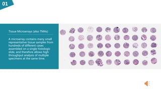
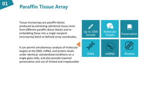
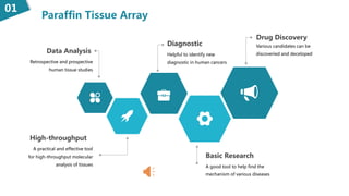
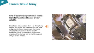
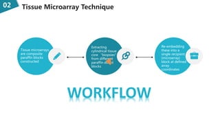
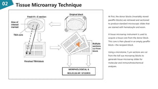
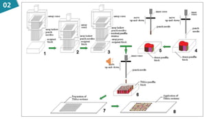
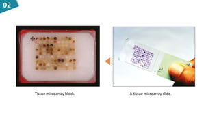
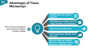
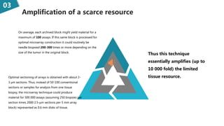
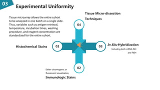
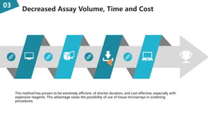
The Use of
Tissue Mircoarray
Creative Bioarray
What is Tissue Mircoarray?01
01
A microarray contains many small
representative tissue samples from
hundreds of different cases
assembled on a single histologic
slide, and therefore allows high
throughput analysis of multiple
specimens at the same time.
Tissue Microarrays (also TMAs)
According to the different making methods,tissue
array can be divided into two categories:
01
Frozen
Tissue Array
Paraffin
Tissue Array
 Frozen tissue array
consists of fresh
frozen tissues.
 The DNA, RNA, and
protein in frozen
tissues retain their
native
characteristics.
 Paraffin tissue array
was first developed.
 Most of the arrays
can be easily made
by microarraying
instrument.
Paraffin Tissue Array
01
Tissue microarrays are paraffin blocks
produced by extracting cylindrical tissue cores
from different paraffin donor blocks and re-
embedding these into a single recipient
(microarray) block at defined array coordinates.
It can permit simultaneous analysis of molecular
targets at the DNA, mRNA, and protein levels
under identical, standardized conditions on a
single glass slide, and also provide maximal
preservation and use of limited and irreplaceable.
Up to 1000
Sample
DNA mRNA
Preservation
Molecular
Targets
Protein
Paraffin Tissue Array
01
Data Analysis
Retrospective and prospective
human tissue studies
High-throughput
A practical and effective tool
for high-throughput molecular
analysis of tissues
Diagnostic
Helpful to identify new
diagnostic in human cancers
Basic Research
A good tool to help find the
mechanism of various diseases
Various candidates can be
discoveried and deceloped
Drug Discovery
Frozen Tissue Array
01
Lots of scientific experimental results
from formalin fixed tissues are not
reliable.
Since frozen tissue sections don’t go through any
fixation procedures, and therefore, the DNA, RNA,
and protein in frozen tissues retain their native
characteristics much more than in paraffin
embedded tissues. Consequently frozen tissue
array should be the best tool for high throughput
analysis on this purpose.
Tissue Microarray
Technique02
Tissue Microarray Technique02
Tissue microarrays
are composite
paraffin blocks
constructed
Extracting
cylindrical tissue
core “biopsies”
from different
paraffin donor
blocks
Re-embedding
these into a
single recipient
(microarray)
block at defined
array
coordinates
WORKFLOW
02 Tissue Microarray Technique
At first, the donor blocks (invariably stored
paraffin blocks) are retrieved and sectioned
to produce standard microscopic slides that
are stained with hematoxylin and eosin.
A tissue microarray instrument is used to
acquire a tissue core from the donor block.
This core is then placed in an empty paraffin
block—the recipient block.
Using a microtome, 5 μm sections are cut
from the tisll sue microarray blocks to
generate tissue microarray slides for
molecular and immunohistochemical
analyses.
研究过程及方法02
02
Tissue microarray block. A tissue microarray slide.
02
15 24 60 15036 72
Advantages of
Tissue Microarrays03
Advantages of Tissue
Microarrays
03
There are numerous advantages of
tissue microarray over standard
techniques, including:
Amplification of a scarce
resource
Simultaneous analysis of very
large numbers of specimens
Experimental uniformity
Decreased assay volume, time
and cost
Does not destroy original
block for diagnosis and thus
conserves valuable tissue
Amplification of a scarce resource
03
Thus this technique
essentially amplifies (up to
10 000 fold) the limited
tissue resource.
On average, each archived block might yield material for a
maximum of 100 assays. If this same block is processed for
optimal microarray construction it could routinely be
needle biopsied 200-300 times or more depending on the
size of the tumor in the original block.
Optimal sectioning of arrays is obtained with about 2-
3 μm sections. Thus, instead of 50-100 conventional
sections or samples for analysis from one tissue
biopsy, the microarray technique could produce
material for 500 000 assays (assuming 250 biopsies per
section times 2000 2.5-μm sections per 5 mm array
block) represented as 0.6 mm disks of tissue.
添加标题
Experimental Uniformity
03
04
01 03
02
Tissue Micro-dissection
Techniques
In Situ Hybridization
Including both mRNA ISH
and FISH
Histochemical Stains
Immunologic Stains
Either chromogenic or
fluorescent visualization,
Tissue microarray allows the entire cohort
to be analyzed in one batch on a single slide.
Thus, variables such as antigen retrieval,
temperature, incubation times, washing
procedure, and reagent concentration are
standardized for the entire cohort.
Decreased Assay Volume, Time and Cost
03
This method has proven to be extremely efficient, of shorter duration, and cost effective, especially with
expensive reagents. This advantage raises the possibility of use of tissue microarrays in screening
procedures.
Where you can get
the tissue array?04
Creative Bioarray is an innovative
biotechnology company providing a
comprehensive range of pre-made and
custom tissue arrays.
Creative Bioarray
Our in-house repository with a comprehensive
archive of over 500,000 human tissue samples
and 30,000 animal samples.
1
All TMAs include slide layout and tissue
pathology information.2
We will be able to offer the research
community sets of large and high quality
arrays linked to clinical information.
2
In addition to TMA products, we also provide
comprehensive pathological services based on
the TMA.
3
High-quality
Abundant
Information
Samples
Library
Related
Services
Custom
Design
GLP
Non-GLP
Tissue Array
at
Creative Bioarray
Contect Us
https://www.creative-bioarray.com
info@creative-bioarray.com

The use of tissue array

  • 1.
    The Use of TissueMircoarray Creative Bioarray
  • 2.
    What is TissueMircoarray?01
  • 3.
    01 A microarray containsmany small representative tissue samples from hundreds of different cases assembled on a single histologic slide, and therefore allows high throughput analysis of multiple specimens at the same time. Tissue Microarrays (also TMAs)
  • 4.
    According to thedifferent making methods,tissue array can be divided into two categories: 01 Frozen Tissue Array Paraffin Tissue Array  Frozen tissue array consists of fresh frozen tissues.  The DNA, RNA, and protein in frozen tissues retain their native characteristics.  Paraffin tissue array was first developed.  Most of the arrays can be easily made by microarraying instrument.
  • 5.
    Paraffin Tissue Array 01 Tissuemicroarrays are paraffin blocks produced by extracting cylindrical tissue cores from different paraffin donor blocks and re- embedding these into a single recipient (microarray) block at defined array coordinates. It can permit simultaneous analysis of molecular targets at the DNA, mRNA, and protein levels under identical, standardized conditions on a single glass slide, and also provide maximal preservation and use of limited and irreplaceable. Up to 1000 Sample DNA mRNA Preservation Molecular Targets Protein
  • 6.
    Paraffin Tissue Array 01 DataAnalysis Retrospective and prospective human tissue studies High-throughput A practical and effective tool for high-throughput molecular analysis of tissues Diagnostic Helpful to identify new diagnostic in human cancers Basic Research A good tool to help find the mechanism of various diseases Various candidates can be discoveried and deceloped Drug Discovery
  • 7.
    Frozen Tissue Array 01 Lotsof scientific experimental results from formalin fixed tissues are not reliable. Since frozen tissue sections don’t go through any fixation procedures, and therefore, the DNA, RNA, and protein in frozen tissues retain their native characteristics much more than in paraffin embedded tissues. Consequently frozen tissue array should be the best tool for high throughput analysis on this purpose.
  • 8.
  • 9.
    Tissue Microarray Technique02 Tissuemicroarrays are composite paraffin blocks constructed Extracting cylindrical tissue core “biopsies” from different paraffin donor blocks Re-embedding these into a single recipient (microarray) block at defined array coordinates WORKFLOW
  • 10.
    02 Tissue MicroarrayTechnique At first, the donor blocks (invariably stored paraffin blocks) are retrieved and sectioned to produce standard microscopic slides that are stained with hematoxylin and eosin. A tissue microarray instrument is used to acquire a tissue core from the donor block. This core is then placed in an empty paraffin block—the recipient block. Using a microtome, 5 μm sections are cut from the tisll sue microarray blocks to generate tissue microarray slides for molecular and immunohistochemical analyses.
  • 11.
  • 12.
    02 Tissue microarray block.A tissue microarray slide.
  • 13.
    02 15 24 6015036 72
  • 14.
  • 15.
    Advantages of Tissue Microarrays 03 Thereare numerous advantages of tissue microarray over standard techniques, including: Amplification of a scarce resource Simultaneous analysis of very large numbers of specimens Experimental uniformity Decreased assay volume, time and cost Does not destroy original block for diagnosis and thus conserves valuable tissue
  • 16.
    Amplification of ascarce resource 03 Thus this technique essentially amplifies (up to 10 000 fold) the limited tissue resource. On average, each archived block might yield material for a maximum of 100 assays. If this same block is processed for optimal microarray construction it could routinely be needle biopsied 200-300 times or more depending on the size of the tumor in the original block. Optimal sectioning of arrays is obtained with about 2- 3 μm sections. Thus, instead of 50-100 conventional sections or samples for analysis from one tissue biopsy, the microarray technique could produce material for 500 000 assays (assuming 250 biopsies per section times 2000 2.5-μm sections per 5 mm array block) represented as 0.6 mm disks of tissue. 添加标题
  • 17.
    Experimental Uniformity 03 04 01 03 02 TissueMicro-dissection Techniques In Situ Hybridization Including both mRNA ISH and FISH Histochemical Stains Immunologic Stains Either chromogenic or fluorescent visualization, Tissue microarray allows the entire cohort to be analyzed in one batch on a single slide. Thus, variables such as antigen retrieval, temperature, incubation times, washing procedure, and reagent concentration are standardized for the entire cohort.
  • 18.
    Decreased Assay Volume,Time and Cost 03 This method has proven to be extremely efficient, of shorter duration, and cost effective, especially with expensive reagents. This advantage raises the possibility of use of tissue microarrays in screening procedures.
  • 19.
    Where you canget the tissue array?04
  • 20.
    Creative Bioarray isan innovative biotechnology company providing a comprehensive range of pre-made and custom tissue arrays. Creative Bioarray
  • 21.
    Our in-house repositorywith a comprehensive archive of over 500,000 human tissue samples and 30,000 animal samples. 1 All TMAs include slide layout and tissue pathology information.2 We will be able to offer the research community sets of large and high quality arrays linked to clinical information. 2 In addition to TMA products, we also provide comprehensive pathological services based on the TMA. 3
  • 22.
  • 23.

Editor's Notes

  • #2 The tissue microarrays together with gene chips,prothein chips and cell chips, constituting a series of biochips, which enable researchers to effectively learn about hundreds of tissue samples at the same time.
  • #3 So what is tissue microarray?
  • #4 Tissue microarray is a recent innovation in the field of pathology. They consist of paraffin blocks in which up to 1000 separate tissue cores are assembled in array fashion to allow multiplex histological analysis.
  • #5 Frozen tissue array consists of fresh frozen tissues. Since frozen tissue sections don’t go through any fixation procedures, and therefore, the DNA, RNA, and protein in frozen tissues retain their native characteristics. Paraffin tissue array was first developed, and it can help scientists high throughput analyze gene and protein expressions in multiple tissue samples, especially analyze different protein levels with antibodies by immunohistochemistry
  • #6 Tissue microarrays are paraffin blocks produced by extracting cylindrical tissue cores from different paraffin donor blocks and re-embedding these into a single recipient (microarray) block at defined array coordinates. It can permit simultaneous analysis of molecular targets at the DNA, mRNA, and protein levels under identical, standardized conditions on a single glass slide, and also provide maximal preservation and use of limited and irreplaceable archival tissue samples.
  • #7 This versatile technique, in which data analysis is automated facilitates retrospective and prospective human tissue studies. It is a practical and effective tool for high-throughput molecular analysis of tissues that is helping to identify new diagnostic and prognostic markers and targets in human cancers, and has a range of potential applications in basic research, prognostic oncology and drug discovery.
  • #8 Though most of the arrays can be easily made by microarraying instrument, paraffin-embedded tissues have limitations. Buffered formalin solutions cross-link proteins and nucleic acids when they are used for fix tissues. The DNA, RNA, and protein within the tissues are damaged at various levels during the fixation. Therefore, lots of scientific experimental results from formalin fixed tissues are not reliable. Since frozen tissue sections don’t go through any fixation procedures, and therefore, the DNA, RNA, and protein in frozen tissues retain their native characteristics much more than in paraffin embedded tissues. Consequently frozen tissue array should be the best tool for high throughput analysis on this purpose.
  • #10 Microarray is a technique for organizing minute amounts of biological samples on a solid support. Tissue microarrays are composite paraffin blocks constructed by extracting cylindrical tissue core “biopsies” from different paraffin donor blocks and re-embedding these into a single recipient (microarray) block at defined array coordinates.
  • #11  At first, the donor blocks (invariably stored paraffin blocks) are retrieved and sectioned to produce standard microscopic slides that are stained with hematoxylin and eosin. An experienced pathologist examines the slides to mark the area of interest, which is commonly an area of cancer, after which the samples can be arrayed. A tissue microarray instrument is used to acquire a tissue core from the donor block.16 This core is then placed in an empty paraffin block—the recipient block. Then, using a microtome, 5 μm sections are cut from the tisll sue microarray blocks to generate tissue microarray slides for molecular and immunohistochemical analyses.
  • #12 Here is a procere of tissue array.Here is a procedure of tissue array. The current Beecher Instruments arraying device is designed to produce sample circular spots that are 0.6 mm in diameter at a spacing of 0.7-0.8 mm. The number of spots on a single slide is variable depending on the array design; the current comfortable maximum with the 0.6 mm needle is about 600 spots per standard glass microscope slide.The core is placed at a specifically assigned coordinate (X-Y guide), which is accurately recorded, typically on a spreadsheet, such as Microsoft Excel. The sampling process can then be repeated many times from different donor blocks until hundreds, or even thousands, of cores are placed into one recipient block, producing the final tissue microarray block.
  • #13 The left is the final tissue microarray block. After using a microtome, we will get tissue microarray slides for molecular and immunohistochemical analyses like the photo in the right.
  • #14 New technologies are under development that may allow as many as 2000 or more sections per slide. Using this method, an entire cohort of cases can be analyzed by staining just one or two master array slides, instead of staining hundreds of conventional slides.
  • #16 There are numerous advantages of tissue microarray over standard techniques.
  • #17 On average, each archived block might yield material for a maximum of 100 assays. If this same block is processed for optimal microarray construction it could routinely be needle biopsied 200-300 times or more depending on the size of the tumor in the original block. Thus this technique essentially amplifies (up to 10 000 fold) the limited tissue resource.
  • #18  With this technology, each tissue sample is treated in an identical manner and microarrays are amenable to a wide range of techniques, including histochemical stains, immunologic stains with either chromogenic or fluorescent visualization, in situ hybridization (including both mRNA ISH and FISH), and even tissue micro-dissection techniques.
  • #19 As only a very small amount of reagent (a few μL) is required to analyze an entire cohort, less laboratory personnel are required to perform the experiments. This method has proven to be extremely efficient, of shorter duration, and cost effective, especially with expensive reagents.4 This advantage raises the possibility of use of tissue microarrays in screening procedures.
  • #21 Creative Bioarray is one of the most reliable providers with the highest quality tissue arrays in the industry and maintains the most stringent standards on quality controls. We provide a comprehensive range of pre-made and custom tissue arrays.
  • #22 Creative Bioarray carries a variety of TMAs representing normal and cancer human tissue slides. All TMAs include slide layout, tissue pathology information, and clinical information. In addition to TMA products, we also provide comprehensive pathological services based on the TMA.
  • #23 Creative Bioarray has ability to help you build your research. For more infomation, please contact us.