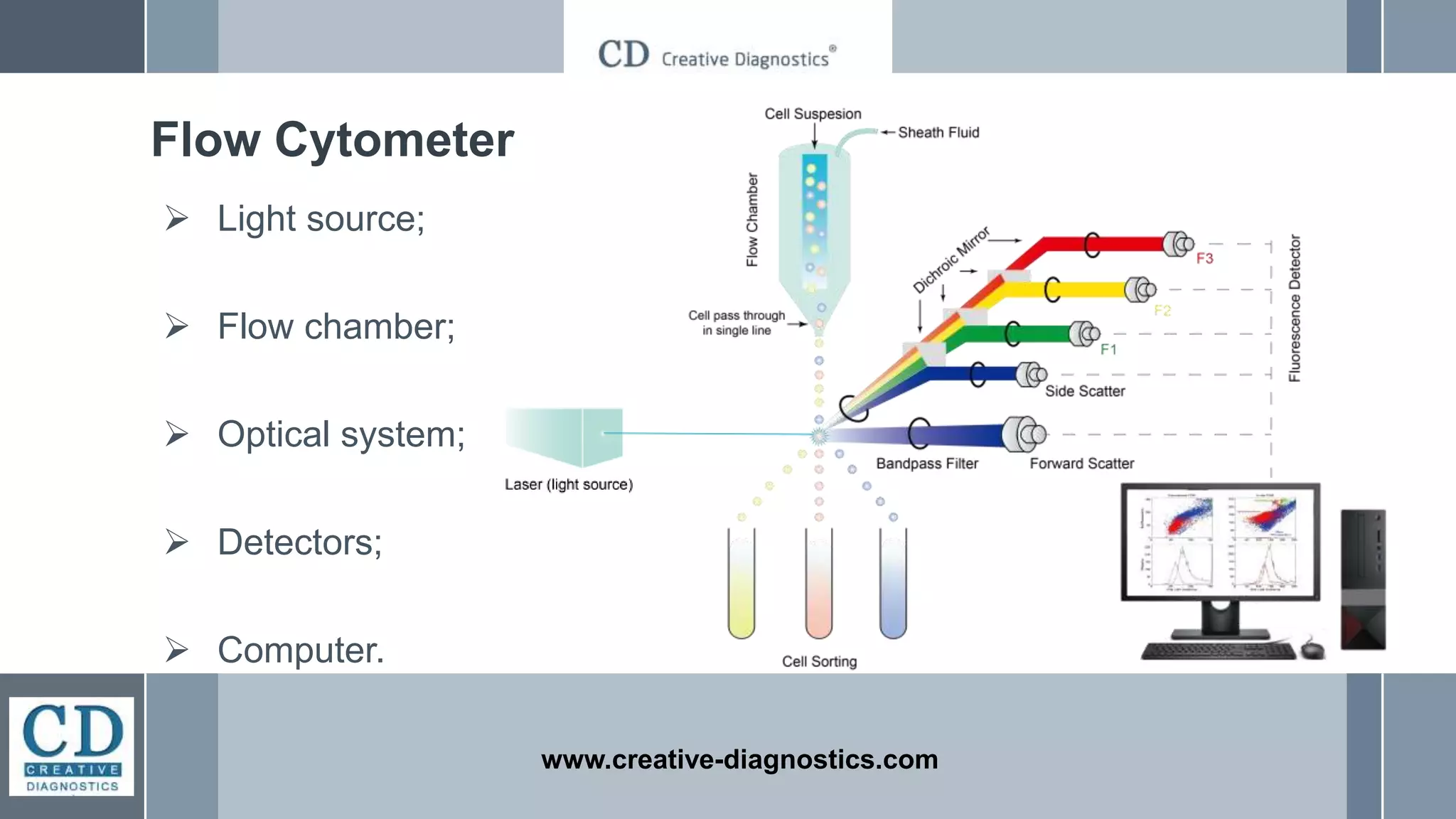
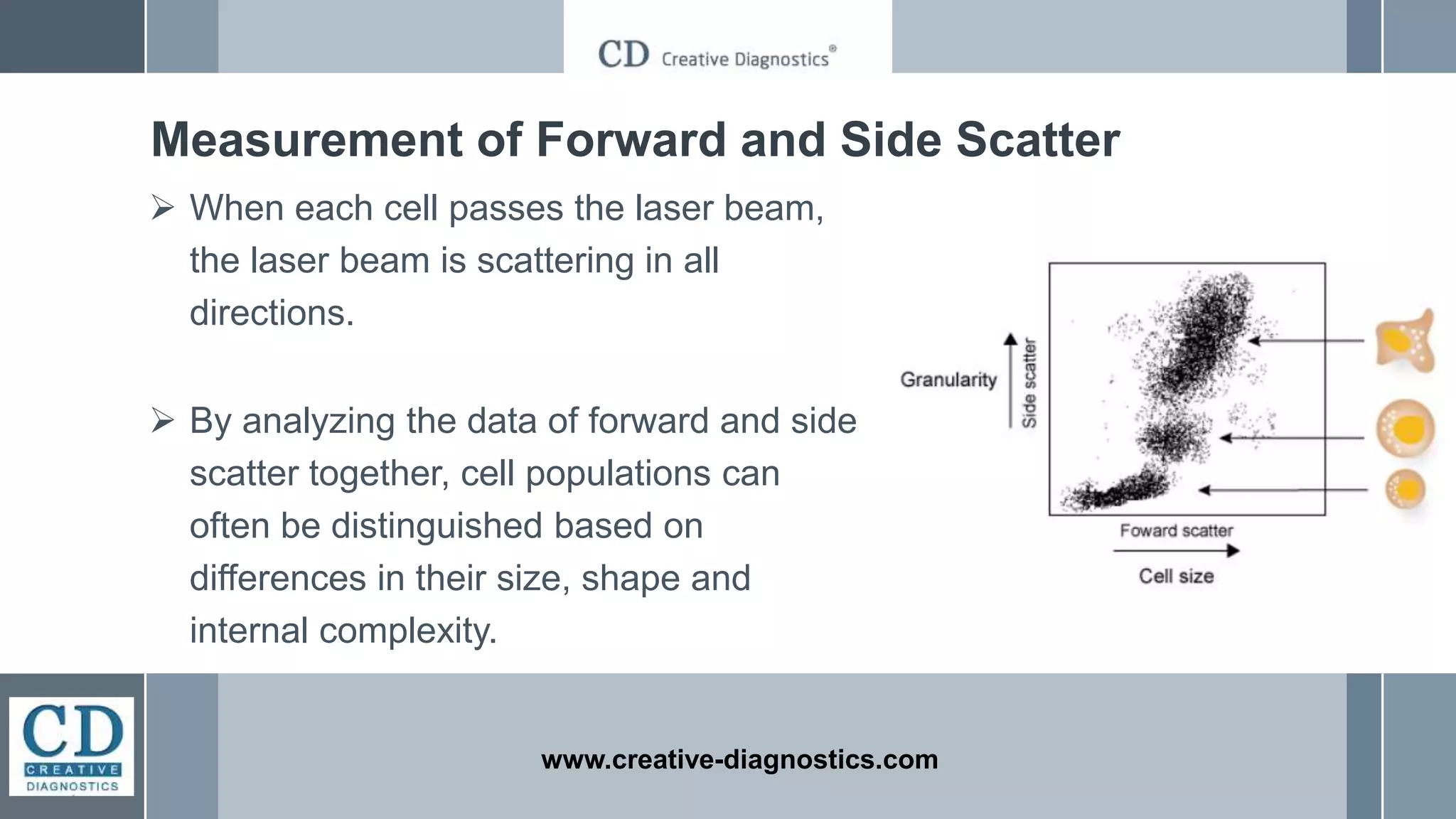
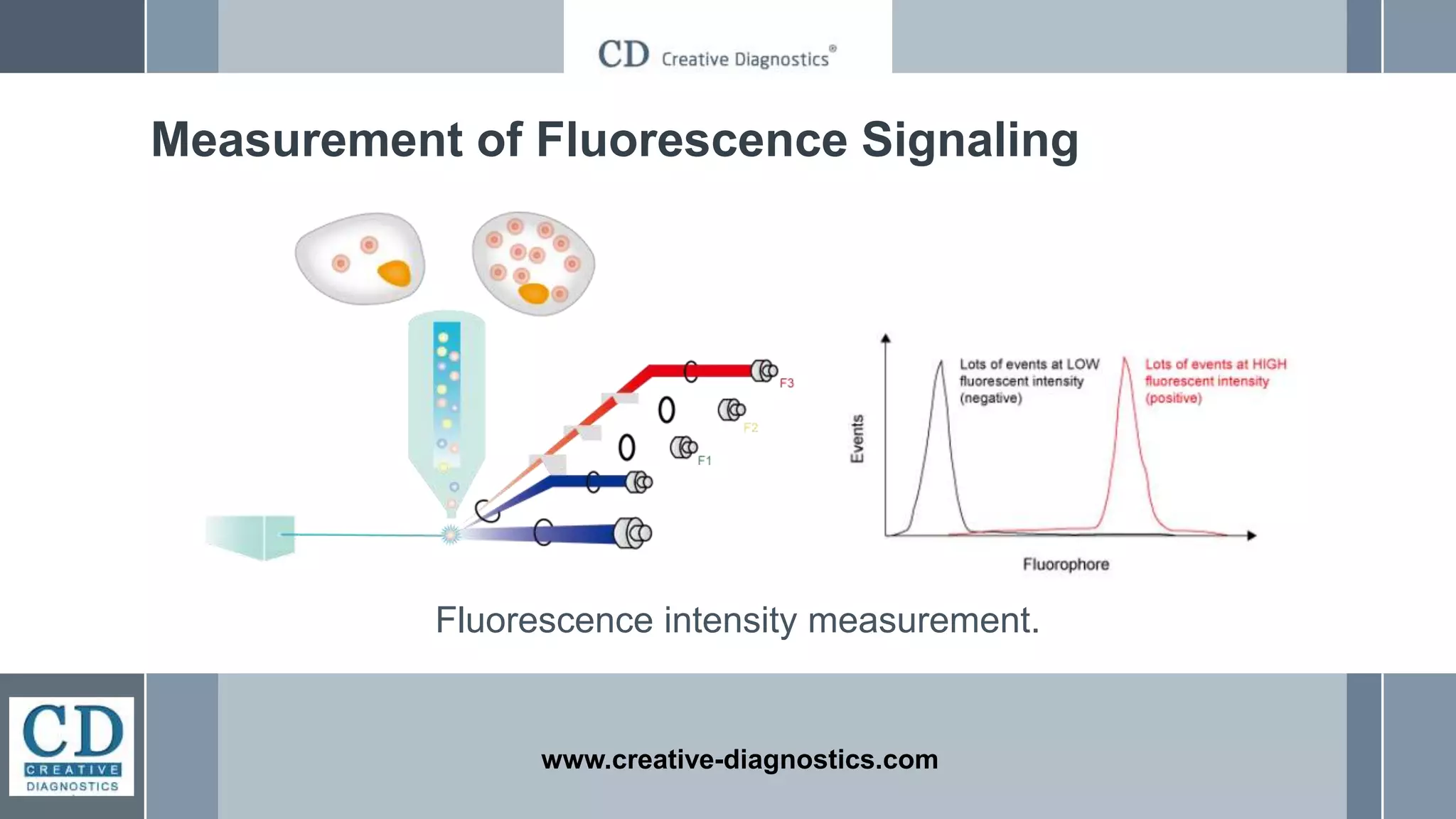
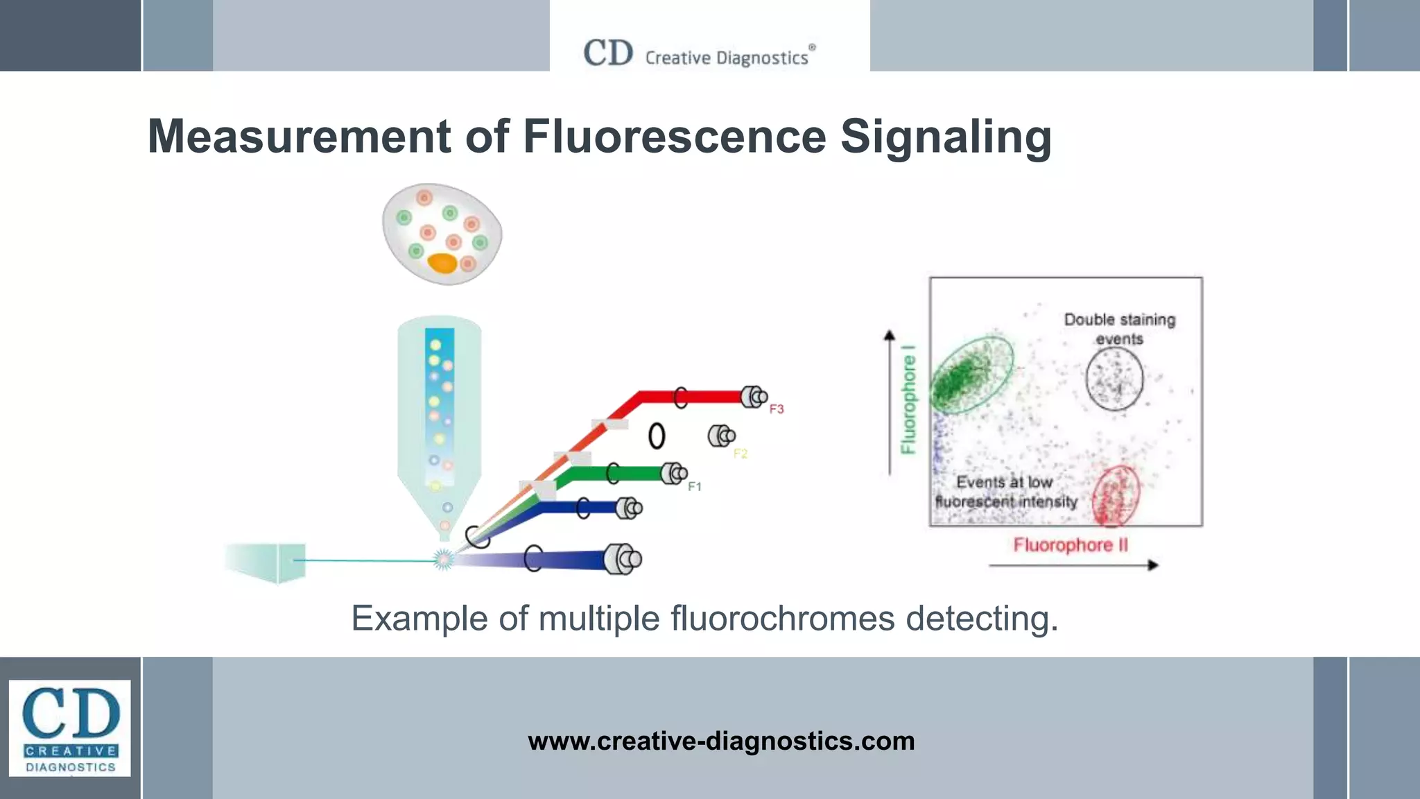
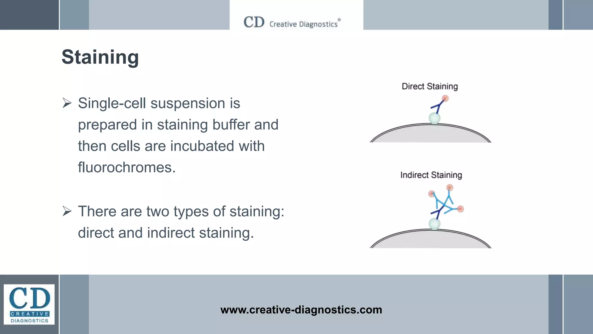
Flow cytometry is a laser-based technique that quantitatively analyzes individual cells in a heterogeneous population, allowing for rapid, simultaneous multi-parameter assessments. It involves sorting cells using fluorescence-activated cell sorting (FACS) and requires a flow cytometer composed of key components such as a light source and optical system. The technique employs fluorescent-labeled antibodies for cell phenotyping and intracellular analysis, with two types of staining methods: direct and indirect.


![FlowBasics2[1]](https://cdn.slidesharecdn.com/ss_thumbnails/7f56678c-0f61-43d6-bbfe-d51ebe159eed-160219222349-thumbnail.jpg?width=640&height=640&fit=bounds)


