This document provides information about syndactyly, including its definition, embryology, etiology, types, evaluation, management, surgical techniques, complications, and its association with certain genetic syndromes. Syndactyly is a fusion of soft tissue or skeletal elements of adjacent digits. It occurs when normal digital separation fails during development. Surgical correction aims to separate the digits and reconstruct the intervening skin and tissues. Timing, flap design, and postoperative care require consideration to optimize outcomes and prevent contractures. Syndactyly can be an isolated anomaly or part of genetic syndromes like Apert syndrome or Poland syndrome.
Dr Bipin Ghanghurde, hand surgeon, Mumbai, +917738729068,
Bipinghanghurde@gmail.com
Syndactyly is a common hereditary digit malformation where
adjacent fingers are webbed due to a failure to separate during limb development. Surgical release is indicated in almost every case except for a mild, incomplete syndactyly without functional impairment.
References
Braun TL, Trost JG, Pederson WC: Syndactyly Release. SeminPlastSurg 30:162-70, 2016.
2. Cronin TD. Syndactylism: results of zig-zag incision to prevent postoperative contracture. PlastReconstrSurg (1946) 1956;18(6): 460–468.
3. Dao KD, Shin AY, Billings A, Oberg KC, Wood VE: Surgical treatment of congenital syndactyly of the hand. J Am AcadOrthopSurg 12:39-48, 2004.
4. Malik S: Syndactyly: phenotypes, genetics and current classification. Eur J Hum Genet 20:817-24, 2012.
5. Tonkin MA: Failure of differentiation part I: Syndactyly. Hand Clin 25:171-93, 2009.
6. Withey SJ, Kangesu T, Carver N, Sommerlad BC. The open finger technique for the release of syndactyly. J Hand Surg [Br] 2001; 26(1):4–7.
1. The document discusses various congenital hand anomalies classified using the Swanson and modified classifications.
2. Key classifications include failures of formation (transverse arrest, longitudinal arrest), differentiation (syndactyly, contractures), duplication (polydactyly), overgrowth (macrodactyly), undergrowth (hypoplastic thumb), and generalized skeletal abnormalities (Apert syndrome, Poland syndrome).
3. The modified classification organizes anomalies by malformations involving the entire limb, hand plate, or unspecified structures, as well as deformities and dysplasias. Specific conditions like symbrachydactyly, triphalangeal thumb, and brachydacty
A fingertip injury is defined as any soft tissue, nail or bony injury distal to the dorsal and volar skin creases at the distal interphalangeal joint and insertions of long flexor and extensor tendons of a finger or thumb.
The fingertips are exposed to all aspects of daily living,
recreation and work and it is perhaps no surprise they
are the most commonly injured part of the hand
Syndactyly is a condition where two or more digits, such as fingers or toes, are fused together. It can be classified as simple or complex based on whether the soft tissue or bones are fused. Syndactyly is usually congenital and occurs when the webbing between digits fails to dissolve during early fetal development. While syndactyly surgery is generally indicated, risks include hypertrophic scarring and skin flap sloughing, especially in lower limb procedures on young children. Post-operative scar management may include massage, splinting, ultrasound therapy, or laser treatment.
The document discusses ulnar nerve palsy and tendon transfers used to treat it. It begins by describing the anatomy of the ulnar nerve and its motor and sensory functions. It then discusses clinical findings associated with ulnar nerve injuries at different locations. Various tendon transfers are summarized that aim to restore small and ring finger flexion, key pinch, correct clawing, and improve grip strength for patients with ulnar nerve palsy. These include transferring forearm muscles like the ECRB to restore key pinch or correct clawing. The modified Stiles-Bunnell procedure is also summarized, which uses the middle finger superficialis tendon to dynamically correct clawing during finger flexion.
The document discusses the use of the Snow and Littler procedure for managing cleft hand deformities. It provides background on classifications of cleft hands and discusses goals of surgical management, which include releasing syndactyly and thumb adduction deformities. The Red Cross Hospital experience from 2003-2007 is described, where 20 cleft hands were treated, including 6 with the Snow and Littler procedure. Complications included finger flexion contractures and rotation of the transferred index metacarpal bone, but patients reported high satisfaction overall.
The document discusses the Position of Safe Immobilisation (POSI), which is a position used to rest the hand during periods of immobilization. It involves flexing the MCP joints 60-90 degrees, extending the PIP joints fully, extending the DIP joints fully, and extending the wrist 10-45 degrees. This position is important because it minimizes the risk of joint stiffness and contractures when immobilizing the hand. The POSI can be used for fractures, tendon injuries, nerve injuries, burns, and other hand injuries or conditions. It is important to splint the hand in the POSI as soon as possible to prevent stiffness and contractures from developing.
External fixation is a method of stabilizing fractures using pins connected to an external frame. It is indicated for open fractures with soft tissue injury, long bone and pelvic fractures, and in multiple trauma patients. The basic components are pins inserted through bones, clamps connecting pins to rods or rings, and external rods or rings. Proper pin placement and construct are important for stability. Complications can include pin site infections, loosening, and breakage. Care includes daily inspection and cleaning of pins.
Finger tip injuries are common, especially in working adults and children, and require prompt treatment to restore function, sensibility, and a viable length. Injuries are classified based on the structures involved and treatments include revision amputation, primary closure, skin grafting, or flap reconstruction depending on the severity and location of the injury. Various flap techniques like V-Y advancement flaps or cross finger flaps are used to reconstruct the pulp and cover bone depending on the zone and site of injury on the finger or thumb.
The document discusses tendon injuries of the hand, including the anatomy, morphology, nutrition, and zones of injury of both flexor and extensor tendons. It covers the etiology, signs and symptoms, examination, detection, and treatment including surgical repair and postoperative rehabilitation of tendon injuries. The key aspects are meticulous surgical technique, appropriate postoperative mobilization, and supervision through rehabilitation to minimize complications of tendon injuries.
This document provides an overview of common injuries around the knee joint. It describes the anatomy of the knee including bones and ligaments. Common mechanisms of injury are discussed for fractures around the knee like condylar fractures of the femur, patella fractures, and tibial plateau fractures. Injuries to the ligaments including ACL, PCL, MCL and LCL are also summarized. Treatment approaches for many of these injuries including nonsurgical and surgical options are highlighted. Other topics covered include meniscal injuries, knee dislocations, and patella dislocations. Complications of various knee injuries are also mentioned.
Apply gentle pressure proximally
Surgeon: Check distal pulses and capillary refill
If no improvement:
Consider temporary arteriotomy or venous shunt
Delay closure and observe
Flap or graft may be needed
This document discusses flexor tendon injuries and repairs in the hand. It covers tendon anatomy, zones of injury, types of repairs, and postoperative rehabilitation protocols. Flexor tendon injuries most commonly occur in males aged 15-30 years old. There are two main postoperative protocols - passive flexion protocol with splinting for 6-9 weeks or early active tension protocol with splinting for 4 weeks and early active exercises. Precise surgical technique and strict adherence to rehabilitation are needed for successful results.
The document discusses the embryology and development of the upper limb. It covers topics like:
- The formation of the limb bud and structures involved in outgrowth like the apical ectodermal ridge and progress zone.
- How the limb bud develops and differentiates over time into the hand plate and cartilage models of bones.
- Rotation and ossification of the bones during the 7th week of development.
- Common congenital anomalies of the upper limb like syndactyly and polydactyly.
- Classification systems for various upper limb anomalies.
- Principles and timing of surgical treatment for different anomalies.
This document discusses polydactyly, which is the congenital duplication of fingers. It begins by describing the embryology and timeline of upper limb development. It then classifies polydactyly into preaxial, central, and postaxial types based on the duplicated digit. Preaxial polydactyly, or thumb duplication, is discussed in depth, including genetics, classification systems like the Wassel classification, clinical assessment, treatment goals and surgical techniques for different types of thumb duplication. Potential complications are also mentioned.
This document provides an overview of Monteggia fracture dislocations, beginning with definitions, history, epidemiology, classification, mechanisms of injury, clinical features, management, complications, and recent updates. Monteggia fractures, first described in 1814, constitute 1-2% of forearm fractures. Bado's 1958 classification divides them into four types based on the direction of radial head dislocation and location of the ulna fracture. Type I is the most common, involving anterior radial head dislocation and ulna fracture. Nonoperative treatment typically involves closed reduction and casting, while surgery is indicated for failed reductions. Complications can include nerve injuries, ossification, and compartment syndrome.
Femoral shaft fractures occur in the diaphysis of the femur between 5 cm distal to the lesser trochanter and 5 cm proximal to the adductor tubercle. They are commonly caused by high-energy trauma in young adults and falls in the elderly. Clinical evaluation involves assessing neurovascular status, associated injuries, and deformity or shortening of the leg. X-rays are used to confirm the diagnosis and classify the fracture. Treatment options include traction, casting, intramedullary nailing, plate fixation, or external fixation depending on the patient's age and the fracture pattern. Complications can include blood loss, nerve injuries, infections, and non-union.
The document discusses various local flap options for reconstructing fingertip injuries. It describes the anatomy of the fingertip and goals of reconstruction which are to close wounds, maximize sensation, preserve length and function. Common local flaps include the volar V-Y flap, bilateral V-Y flaps, cross-finger flap, thenar flap and lateral island flaps. Choice depends on wound orientation and configuration.
This document provides an overview of the management of hand fractures. It discusses the goals of treatment which include restoring anatomy, reducing malrotation and angulation, maintaining reduction with minimal surgery, and rapid mobilization. Most closed hand fractures can be treated with closed reduction and splinting, while unstable or intra-articular fractures often require operative fixation techniques like K-wiring, tension band wiring, plating, or external fixation. Common fractures of the hand including metacarpals, phalanges, and thumb are described along with appropriate treatment options and techniques. Potential complications of treatment are also outlined.
This document provides information on finger tip and soft tissue reconstruction of the hand. It begins with an introduction to finger tip injuries, including common causes. It then discusses the anatomy of the finger tip and classifications of fingertip amputations. Several local flap techniques for reconstructing finger tip defects are described in detail, including composite tip grafts, free pulp grafts, V-Y advancement flaps, homodigital flaps, and cross finger flaps. Goals of management and assessment of injuries are also covered. The document concludes with a discussion of local flaps that can be used for covering defects on the dorsum of the hand.
This document discusses various congenital hand anomalies classified by Swanson into categories such as failure of formation, differentiation, duplication, overgrowth, undergrowth, and constriction syndromes. Specific conditions covered include macrodactyly (overgrowth of digits), undergrowth conditions like hypoplastic thumb, and syndromes such as Madelung deformity. Evaluation, classification, etiology, clinical features, and treatment options are provided for each condition.
This document provides information about Dupuytren's contracture, including its history, definition, epidemiology, associated conditions, pathogenesis, treatment options, and complications. Specifically, it describes how Dupuytren's contracture results from abnormal proliferation of fibroblasts in the palmar fascia, which can cause fingers to bend into the palm. Treatment involves nonsurgical options like collagenase injections or surgery to release contracted tissues through techniques like fasciectomy. Postoperative rehabilitation with splinting and range of motion exercises aims to prevent recurrence of contractures.
Humeral shaft fractures can often be treated nonoperatively with a brace, though operative options include plating, flexible nailing, or locked intramedullary nailing. Plates and nails have similar union rates but nails have more complications so plates are generally preferable. Flexible nails are also an effective option. Radial nerve palsy is a risk, especially with distal fractures, and may require exploration. Most humeral shaft fractures heal well with either operative or nonoperative treatment depending on the specific situation and patient factors.
This document discusses neck of femur fractures (NOF), also known as hip fractures. It covers the epidemiology, risk factors, anatomy, classification, diagnosis, and treatment of NOF fractures. NOF fractures typically occur in elderly patients from low-energy falls and are associated with osteoporosis, while in younger patients they usually result from high-energy injuries. Treatment depends on factors like the patient's age, health, and fracture classification, and may involve closed or open reduction, fixation with screws or nails, or arthroplasty. Complications can include nonunion, osteonecrosis, fixation failure, dislocation, and increased mortality risk especially in older or less healthy patients.
This document discusses syndactyly, a congenital hand anomaly where adjacent digits are fused. It describes the typical surgical techniques used to correct syndactyly, including the use of local flaps and skin grafts. Recently, some reconstructive techniques have been developed that do not require skin grafts. The document outlines the steps of syndactyly release surgery, including flap design and elevation, separation of digits, and use of skin grafts if needed. Postoperative complications are also discussed, such as web creep caused by scar contracture.
This document provides information about syndactyly, including:
- It is the most common congenital hand malformation, occurring in about 1 in 2,000 live births.
- It can range from simple incomplete fusions to complex fusions involving bones and other structures.
- Treatment involves carefully separating the fingers while reconstructing skin and other tissues to form new web spaces. Multiple flap techniques may be used depending on the specifics of each case.
- Proper postoperative immobilization and splinting is important to maintain the results long term and prevent web creep.
The objective in the management of soft-tissue injuries of the hand is to achieve primary wound healing.
The choice of treatment of fingertip is based on the
mechanism of injury ,
the size of the defect,
location and status of the wound
injuries to other parts of the hand
other factors(patient’s age, sex, general health, and occupation)
This document discusses the principles and management of various congenital hand anomalies. It covers topics such as:
- Surgical management should aim to improve function and appearance with minimal risk. Hand function depends on thumb mobility and pinch grip.
- Timing of surgery depends on technical difficulty and cortical plasticity. Surgery may be delayed to treat other conditions.
- Transverse deficiencies are often managed non-surgically with prosthetics. Longitudinal deficiencies often benefit from surgery.
- Specific procedures are described for conditions like symbrachydactyly, radial hypoplasia, central ray deficiency, thumb and finger hypoplasia/aplasia. Syndactyly and poly
This document discusses reconstructive options for fingertip injuries. The goals of reconstruction are to close wounds, maximize sensory return, preserve length and joint function, and obtain a cosmetic appearance. Options include healing by secondary intention, skin grafting, and local flap reconstruction using flaps such as volar V-Y flaps, bilateral V-Y flaps, cross-finger flaps, and thenar flaps. Major complications are hypersensitivity and cold intolerance, which usually resolve after 1-2 years.
The document discusses various reconstructive options for fingertip injuries, including healing by secondary intention, skin grafting, and local flap reconstruction techniques such as volar V-Y flaps, bilateral V-Y flaps, and cross-finger flaps. The goals of fingertip reconstruction are to close the wound, maximize sensory return, preserve length and joint function, and obtain a satisfactory cosmetic appearance. Selection of the reconstructive method depends on factors like the level and depth of the injury, amount of exposed tissue, and characteristics of the individual patient.
This document discusses various options for non-microsurgical coverage of soft tissue defects in the hand, including healing by secondary intention, vacuum-assisted closure, skin grafting, and various local, regional, and distant flaps. It provides details on techniques such as the Moberg flap, Tranquilli-Leali flap, cross-finger flap, and their indications. It also discusses the vascular anatomy of the hand and design principles for different types of flaps used in hand reconstruction.
This document discusses various techniques for pollicization, which is a hand surgery to create a functioning thumb from another finger. It begins by defining pollicization and describing the anatomy and function of the thumb. It then discusses different conditions that may require pollicization, including thumb hypoplasia, absence due to trauma, macrodactyly, multifinger hands, and mirror hands. For each condition, it provides details on the surgical procedure for pollicization, including steps like isolating nerves/vessels, shortening bones, transferring muscles/tendons, and positioning the new thumb. Post-operative casting is also addressed. The document contains many diagrams illustrating thumb anatomy and different pollicization procedures.
The document provides an overview of hand anatomy including terminology, skin, fascia, muscles, tendons, bones, joints, nerves and vasculature. Key points include descriptions of various hand incisions and their purposes, fascial layers and spaces of the hand, flexor tendon anatomy and zones of injury, and anatomy of important muscles like the thenar and hypothenar muscles. The document covers the detailed functional anatomy of the hand.
1. Entropion refers to inward rolling and rotation of the eyelid margin toward the eyeball. There are several types including congenital, cicatricial, senile/involutional, and mechanical.
2. Senile/involutional entropion is the most common type and affects only the lower eyelid in older people. It is caused by horizontal laxity of the eyelid due to weakening of the orbicularis muscle and vertical lid instability due to weakening of the lower eyelid retractors.
3. Surgical techniques for correcting senile entropion include transverse everting sutures, Wies operation, and Jones plication. The Wies
This document discusses reconstruction of the thumb. It begins by describing the anatomy of the thumb bones and joints. It then discusses various soft tissue reconstruction options for different types and sizes of thumb defects, including local flaps, cross-finger flaps, and free flaps. It also covers osteoplastic reconstruction using a bone graft and flap in multiple stages to reconstruct large bony defects of the thumb. The goals of reconstruction are to restore length, stability, mobility, and sensate soft tissue coverage to the thumb.
This document discusses various flap techniques used in periodontal surgery. It defines flaps as sections of gingiva and mucosa surgically separated from underlying tissues to provide access to bone and roots. Full and partial thickness flaps are classified based on the depth of tissue reflection. Techniques include the modified Widman flap, undisplaced flap, apically displaced flap, papilla preservation flap, and techniques for distal molar surgery. Healing after flap surgery is described in stages from initial clot formation to establishment of new connective tissue attachment after 4 weeks.
1) Macrodactyly is an overgrowth of one or more fingers, most often the index finger. It can involve enlargement of skin, bone, nerves, and other tissues.
2) The cause is unknown but may involve abnormal nerve or blood supply. Recent evidence suggests a genetic mutation can cause abnormal growth regulation.
3) Treatment involves surgical procedures like debulking excess tissue or shortening the enlarged bones to improve appearance and function.
The document discusses different techniques for periodontal pocket therapy using periodontal flaps. It describes the Modified Widman flap, Undisplaced flap, and Apically displaced flap. The Modified Widman flap involves reflecting the gingiva to access root surfaces while allowing reattachment with minimal recession. The Undisplaced flap eliminates the pocket wall with the initial incision. The Apically displaced flap aims to eliminate pockets while retaining attached gingiva by displacing the entire mucogingival unit apically. Each technique involves specific incisions and steps to access, debride, and close the surgical site.
This document discusses thumb hypoplasia, including its epidemiology, pathophysiology, classification, clinical examination, investigations, and management. It notes that thumb hypoplasia is considered part of radial longitudinal deficiency. The classification system discussed is the Blauth classification, which includes types I-V. Management depends on the type, and may include observation, opposition tendon transfer, web space release and stabilization, pollicization, or toe transfer. Pollicization of the index finger remains the preferred treatment for more severe types when possible.
The document discusses various local flap options for reconstructing fingertip injuries. It describes the anatomy of the fingertip and goals of reconstruction which are to close wounds, maximize sensation, preserve length and function. Local flap options mentioned include volar V-Y flaps, bilateral V-Y flaps, cross-finger flaps, thenar flaps and lateral island flaps. Choice of flap depends on wound orientation and configuration.
Gingivectomy is the surgical excision of gum tissue to eliminate periodontal pockets and create a favorable environment for gum healing. It involves marking pockets, making internal bevel incisions, removing gum tissue and granulation, and root planing. Healing occurs through clot formation, granulation tissue growth, and epithelialization over 2-7 weeks. While it effectively eliminates pockets, gingivectomy risks reducing attached gum and exposing root surfaces.
This document discusses periodontal flaps, which are sections of gingiva surgically separated from underlying tissues to provide access to bone and roots. It defines different types of flaps classified by bone exposure, placement, and papilla design. Indications and contraindications for various flaps are outlined. Procedures for modified Widman, undisplaced, apically displaced, and regenerative flaps are described. Distal molar surgery techniques and use of periodontal packs are also summarized.
This document provides definitions and details regarding periodontal flap surgery. It defines a periodontal flap as a section of gingiva and/or mucosa surgically separated from underlying tissues to provide access to bone and roots. It discusses indications, contraindications, classifications of flaps based on bone exposure and placement, and procedures for various flap types like modified Widman flap and apically displaced flap. Healing after flap surgery and use of periodontal packs are also summarized. The document aims to comprehensively cover periodontal flap surgery planning and techniques.
These lecture slides, by Dr Sidra Arshad, offer a simplified look into the mechanisms involved in the regulation of respiration:
Learning objectives:
1. Describe the organisation of respiratory center
2. Describe the nervous control of inspiration and respiratory rhythm
3. Describe the functions of the dorsal and respiratory groups of neurons
4. Describe the influences of the Pneumotaxic and Apneustic centers
5. Explain the role of Hering-Breur inflation reflex in regulation of inspiration
6. Explain the role of central chemoreceptors in regulation of respiration
7. Explain the role of peripheral chemoreceptors in regulation of respiration
8. Explain the regulation of respiration during exercise
9. Integrate the respiratory regulatory mechanisms
10. Describe the Cheyne-Stokes breathing
Study Resources:
1. Chapter 42, Guyton and Hall Textbook of Medical Physiology, 14th edition
2. Chapter 36, Ganong’s Review of Medical Physiology, 26th edition
3. Chapter 13, Human Physiology by Lauralee Sherwood, 9th edition
NAVIGATING THE HORIZONS OF TIME LAPSE EMBRYO MONITORING.pdfRahul Sen
Time-lapse embryo monitoring is an advanced imaging technique used in IVF to continuously observe embryo development. It captures high-resolution images at regular intervals, allowing embryologists to select the most viable embryos for transfer based on detailed growth patterns. This technology enhances embryo selection, potentially increasing pregnancy success rates.
Kosmoderma Academy, a leading institution in the field of dermatology and aesthetics, offers comprehensive courses in cosmetology and trichology. Our specialized courses on PRP (Hair), DR+Growth Factor, GFC, and Qr678 are designed to equip practitioners with advanced skills and knowledge to excel in hair restoration and growth treatments.
10 Benefits an EPCR Software should Bring to EMS Organizations Traumasoft LLC
The benefits of an ePCR solution should extend to the whole EMS organization, not just certain groups of people or certain departments. It should provide more than just a form for entering and a database for storing information. It should also include a workflow of how information is communicated, used and stored across the entire organization.
Know the difference between Endodontics and Orthodontics.Gokuldas Hospital
Your smile is beautiful.
Let’s be honest. Maintaining that beautiful smile is not an easy task. It is more than brushing and flossing. Sometimes, you might encounter dental issues that need special dental care. These issues can range anywhere from misalignment of the jaw to pain in the root of teeth.
- Video recording of this lecture in English language: https://youtu.be/Pt1nA32sdHQ
- Video recording of this lecture in Arabic language: https://youtu.be/uFdc9F0rlP0
- Link to download the book free: https://nephrotube.blogspot.com/p/nephrotube-nephrology-books.html
- Link to NephroTube website: www.NephroTube.com
- Link to NephroTube social media accounts: https://nephrotube.blogspot.com/p/join-nephrotube-on-social-media.html
How to Control Your Asthma Tips by gokuldas hospital.Gokuldas Hospital
Respiratory issues like asthma are the most sensitive issue that is affecting millions worldwide. It hampers the daily activities leaving the body tired and breathless.
The key to a good grip on asthma is proper knowledge and management strategies. Understanding the patient-specific symptoms and carving out an effective treatment likewise is the best way to keep asthma under control.
DECLARATION OF HELSINKI - History and principlesanaghabharat01
This SlideShare presentation provides a comprehensive overview of the Declaration of Helsinki, a foundational document outlining ethical guidelines for conducting medical research involving human subjects.
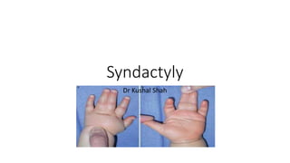
5. Definition and embryology
• Its a variable fusion of the soft tissue or skeletal elements or both
of adjacent digits, and it occurs when the normal processes of
digital separation and web space formation fail to some degree.(
figure 1)
• Normally digits form as condensations of mesoderm within the
terminal paddle of the embryonic upper limb. Spaces form between
the fingers in a distal to proximal direction to the level of the normal
we space by a process of regulated apoptosis which is dependent
on the apical ectodermal ridge ( AER) and the molecular
signaling
• The normal web space slopes 45 degrees in a dorsal to palmar
direction from the metacarpal heads to the midproximal phalanx
(Figure 2).
• The second and fourth webs are wider than the third web, allowing
greater abduction of the index and small fingers.
• The first web space is a broader diamond-shaped expanse of skin
composed of the glabrous skin of the palm and thinner mobile skin
dorsally
6. Etiology
• Are fairly common and often run in families
• Occur in about one out of every 2,500-3,000 newborns
• Affect boys more often than girls( 2:1)
• Affect whites more often than blacks or Asians
• Bilateral about 50 % of the time
• Can occur alone or as part of a genetic syndrome, such as
Apert syndrome
• Can sometimes be seen prior to birth by ultrasound
9. Pre op evaluation
• which web space(s) is involved
• the extent of the syndactyly
• the involvement of the nail
• and the presence of other anomalies.
• Lack of differential motion between the digits may indicate bony fusion or an
extra digit, or both, concealed within the conjoined digits.
• Examine entire upper limb, the contralateral hand, the chest wall, and the feet.
• Radiographs may reveal skeletal fusion, a concealed extra digit
(synpolydactyly), or other bony or articular deformities.
• Further imaging with ultrasound or magnetic resonance imaging can be useful
in determining the flexor tendon and Vascular anatomy in complex cases
10. Management
• Syndactyly can have cosmetic, functional, or
developmental impacts on the growing child.
• The appearance of the hand is altered, more so
with complete complicated forms of syndactyly.
• Syndactyly of the first web space hampers grasp
and the development of pinch.
• Syndactyly of the second, third, and fourth web
spaces inhibits independent digital motion,
particularly abduction, and therefore reduces the
span of the hand.
• Syndactyly between digits of unequal length
causes tethering of the longer digit, which
deviates toward the shorter digits and may also
cause a flexion contracture at the proximal
interphalangeal joint (PIP) that progresses with
11. Surgical contra indication
• include mild incomplete
syndactyly without functional
impairment
• medical conditions that preclude
surgery, or complex
• syndactylies that risk further
functional impairment with
attempted separation.
• In complicated complex – there
are insufficient components in the
fused mass to produce
independent, stable, and mobile
digits .
• This situation typically arises in
central brachysyndactyly or
synpolydactyly, and separation
risks reducing function.
12. Surgical factors to be taken into account
• timing of the procedure or procedures
• staging the releases of multiple web space
syndactylies
• creation of a commissure
• techniques of separation and resurfacing of the
digits
• postoperative dressing and aftercare.
13. Timing of surgery
• Syndactyly release has been performed in the neonatal period or
during infancy, or it has been delayed until childhood.
• Longterm reviews by Flatt and Ger have shown better outcomes
with release after 18 months, although early surgery may be
dictated by progressive skeletal deviation or deformity.
• The goal is to complete all the releases by school age
• . In multiple staged release - the first procedure can be combined
with isolated release of the fingertips and distal phalangeal fusions
of all the digits to reduce the tethering effect between surgical
procedures
14. Surgical anatomy
• cleeland's ligament:
- coalesce in interdigital space
forming a dorsal roof over digital vessels and
nerves as well as forming a septum between
them;
- digital nerves and arteries may not be
available for both digits;
- vessels may be entwined, or
absent w/ in the bridge;
- aberent anatomy is more common
w/ more complex deformities;
- nerves should be teased apart
using magnification;
15. Surgical steps
• (1) separation of the digits
• (2) commissure reconstruction
• (3) resurfacing of the intervening borders of
the digits.
• (4) Paronyhcial fold formation
16. Seperation of digits
• Release of syndactyly requires
careful planning to optimize use of
the available skin and to allow
surgical exposure for separation of
digits and structures.
• Separation of the digits requires
division or excision of fascial
interconnections between the digits,
with care taken to identify and
preserve the individual
neurovascular bundles and the
venous plexus on the dorsum of the
digits and of the commissure flap
• . Bifurcation of the common digital
nerve and artery may be distal to
the planned position of the web
space.
• In this situation, the digital artery
can be ligated provided the other
side of the digit is unoperated or the
contralateral digital artery is known
to be intact
Cronin and Skoog
–dorsal and volar
triangular flap
with matched zig
zag incision
Somarlad open
finger technique
18. Commissure reconstruction
• A basic tenet of syndactyly
release is reconstruction of the
interdigital commissure with a
local skin flap.
• Incision design must be placed
such that inevitable scar
contraction will avoid joint or
web space contracture
• For 1st web space: Other
options include a transposition
flap from the index finger, a
combination of transposition
flaps from the radial and ulnar
borders of the index and thumb,
respectively, or a “V-to-Y”
advancement of the central
web.
Butterfly flap for
web deepening
4 flap Z
plasty
for1stweb
space
19. Resurfacing of the digit
• Resurfacing the digits is achieved with the
palmar and dorsal flaps raised from the
conjoined digits supplemented with skin
grafts.
• Full-thickness skin grafts are preferred
over split thickness skin grafts to lessen
secondary graft contracture
• Resurfacing the digits without skin graft
may require some reduction of digital
diameter by excising the subcutaneous fat
of the digit while preserving the dorsal
venous system
21. Paronychial fold formation
• Release of a complete
syndactyly, particularly when
associated with distal
phalangeal fusion, requires
the formation of a paronychial
fold.
• The distal phalangeal tufts
may be covered using the
technique described by
Buck-Gramcko.
• Laterally based long narrow
triangular flaps are raised
from the hyponychium of the
conjoined digital mass and
folded around to form the
lateral nail fold
22. Post operative dressing
• The dressings must apply gentle compression across the
skin graft sites and protect the separated digits.
• Nonadherent dressings and moist cotton are placed into the
web spaces and reinforced with large amounts of soft gauze.
• In young children, the compressive hand dressing is
reinforced by above-the elbow plaster or a soft cast to
prevent inadvertent removal.
• The elbow is positioned in at least 90 degrees of flexion to
minimize the chances of the cast sliding off the arm.
• The dressings are removed 3 weeks after surgery, and then
gentle washing and wound care are needed. The wounds are
protected until they are dry and healed.
• Normal hand use is allowed after the dressing has been
removed.
• Once healing has taken place, an elasticized compression
glove may be fitted and worn for up to 3 months for scar
management.
• Scar massage by oil/gel , silicone gel sheets, or elastomere
products can be used to treat areas of hypertrophic scarring.
28. Syndactyly : and its associated syndrome
• Acrosyndactyly
Poland's syndrome:
- hypoplasia of hand and simple syndactyly of fingers on the same side as
the absent pectoral muscles (and other chest wall muscles);
- Apert's Syndrome:
- when all digits are joined, as is common in spoon hand of
Apert's syndrome (acrocephalosyndactyly), it is important to release border
digits-thumb and small finger-first;
- remaining 3 joined fingers can be managed by removing middle digit, thus
creating a three-fingered hand with a thumb and sufficient
skin for closure;
- Chromosomal Syndromes:
- trisomy of 13, 18, or 21;
- deletion of short arm of chromsome 5;
- Craniofacial Syndromes:
- Aglossia adactylia
- Mobius Syndrome
- Oculomandibulofacial syndrome
29. Acrosyndactyly:
• Classification:
- Type I:
- conjoined finger tips with well formed webs w/ normal
depth;
- treatment involves separation and contouring of the tips
of the digits;
- partial digit ablation may be required;
- Type II:
- tips of digits are joined and web formation is incomplete;
- treatment involves separation of tips of digits and
deepening of web space;
- Type III:
- absent web spaces, sinus tracts, joined digit clefts;
- as in simple syndactyly border digits are reconstructed
first
•
30. Apert syndrome
• Acrocephalosyndactyly – craniosynostosis with acrosyndactyly and
symphalangism and clinodactyly of thumb
• M:F – 1.5:1
• AD/AR
• 1:2,00,000
• Single gene mutation
34. Radial thumb clinodactyly
• Dao recommended a release of the
abnormal abductor pollicis brevis
tendon insertion into the distal
phalanx and reinsertion the proximal
phalanx, excision of the metacarpal
head ulnar prominence, and pinning
of the interphalangeal and
metacarpophalangeal joints.
• Oishi and Ezaki proposed that the
thumb be reconstructed by releasing
the abnormal abductor pollicis
brevis insertion, opening or closing
wedge osteotomy of the proximal
phalanx, and a V-Y advancement
flap on the radial side of the thumb.